A modular approach to the synthesis of N-glycans for attachment to an aluminum oxide-coated glass slide (ACG slide) as a glycan microarray has been developed and its use for the profiling of an HIV broadly neutralizing antibody has been demonstrated.
We present a highly efficient way for the rapid preparation of a wide range of N-linked oligosaccharides (estimated to exceed 20,000 structures) that are commonly found on human glycoproteins. To achieve the desired structural diversity, the strategy began with the chemo-enzymatic synthesis of three kinds of oligosaccharyl fluoride modules, followed by their stepwise α-selective glycosylations at the 3-O and 6-O positions of the mannose residue of the common core trisaccharide having a crucial β-mannoside linkage. We further attached the N-glycans to the surface of an aluminum oxide-coated glass (ACG) slide to create a covalent mixed array for the analysis of hetero-ligand interaction with an HIV antibody. In particular, the binding behavior of a newly isolated HIV-1 broadly neutralizing antibody (bNAb), PG9, to the mixture of closely spaced Man5GlcNAc2 (Man5) and 2,6-di-sialylated bi-antennary complex type N-glycan (SCT) on an ACG array, opens a new avenue to guide the effective immunogen design for HIV vaccine development. In addition, our ACG array embodies a powerful tool to study other HIV antibodies for hetero-ligand binding behavior.
N-glycans on glycoproteins are covalently linked to the asparagine (Asn) residue of the consensus Asn-Xxx-Ser/Thr sequon, which affect several biological processes such as protein conformation, antigenicity, solubility, and lectin recognition1,2. The chemical synthesis of N-linked oligosaccharides represents a significant synthetic challenge because of their huge structural micro heterogeneity and highly branched architecture. Careful selection of protecting groups to tune reactivity of building blocks, achieving selectivity at anomeric centers, and proper use of promoter/activator(s) are key elements in synthesis of complex oligosaccharides. To solve this problem of complexity, a great amount of work to advance N-glycan synthesis was reported recently3,4. In spite of these robust approaches, finding an effective method for the preparation of a wide range of N-glycans (~20,000) remains a major challenge.
The rapid mutation rate of HIV-1 to achieve the extensive genetic diversity and its ability to escape from neutralizing antibody response, is among the greatest challenges to develop a safe and prophylactic vaccine against HIV-15,6,7. One effective tactic that HIV uses to avoid the host immune response is the post-translational glycosylation of envelope glycoprotein gp120 with a diverse N-linked glycans derived from the host glycosylation machinery8,9. A recent report regarding the precise analysis of recombinant monomeric HIV-1 gp120 glycosylation from human embryonic kidney (HEK) 293T cells suggests the occurrence of structural microheterogeneity with a characteristic cell-specific pattern10,11,12. Therefore, understanding the glycan specificities of HIV-1 bNAbs requires well characterized gp120 related N-glycan structures in a quantity sufficient for analysis.
The discovery of glycan microarray technology provided high throughput-based exploration of specificities of a diverse range of carbohydrate-binding proteins, viruses/bacterial adhesins, toxins, antibodies, and lectines13,14. The systematic glycans arrangement in an arrayed chip-based format could determine problematic low affinity protein-glycan interactions through multivalent presentation15,16,17,18. This chip-based glycan arrangement conveniently appears to effectively mimic cell-cell interfaces. To enrich the technology and overcome the uneven issue associated with conventional array formats, our group recently developed a glycan array on an aluminum oxide-coated glass (ACG) slide using phosphonic acid-ended glycans to enhance the signal intensity, homogeneity, and sensitivity19,20.
To improve the current understanding about glycan epitopes of newly isolated HIV-1 broadly neutralizing antibodies (bNAbs), we have developed a highly efficient modular strategy for the preparation of a broad array of N-linked glycans21,22 to be printed on an ACG slide (see Figure 1). Specificity profiling studies of HIV-1 bNAbs on an ACG array offered the unusual detection of hetero-glycan binding behavior of highly potent bNAb PG9 that was isolated from HIV infected individuals23,24,25.
1. Preparation of D1/D2 Arm Modules22
2. Preparation of Glycan 10
3. Preparation of Glycans with Phosphonic Acid Tail19,22
4. Glycan Array
A modular chemo-enzymatic strategy for the synthesis of a wide array of N-glycans is presented in Figure 1. The strategy is based on the fact that diversity can be created at beginning by chemo-enzymatic synthesis of the three important modules, followed by the α-specific mannosylation at the 3-O and/or 6-O position of the mannose residue of the common core trisaccharide of N-glycans. Considering the structural diversity of bi-, tri-, and tetra-antennary complex type N-glycan structures, we believed that a set of oligosaccharyl donors and the core trisaccharides acceptor with preinstalled alkyl handlecould be used as starting materials to generate the desired structural diversity (Figure 1). The applicability of glycosyl fluoride donors in building a complex glycan library has been proven previously21.
To demonstrate the effectiveness of our strategy, a bi-antennary isomeric structure (10, Figure 3) was selected for synthesis. The D1 arm antennae 4 and D2 arm antennae 5 of glycan 10 were generated on large scales by chemo-enzymatic methods. In particular, the disaccharide acceptor 1 was enzymatically galactosylated by using β-1, 4-galactosyltransferase and uridine 5'-diphosphogalactose (UDP-Gal) to form trisaccharide 2. Next, the intermediate 2 was fucosylated at GlcNAc 3-O position in presence of α-1, 3-fucosyltransferase from Helicobacter pylori (Hpα1,3FT) to afford the desired tetrasaccharide 3. For chemical ligation to core, building blocks 2 and 3 were first peracetylated, and the reducing end p-methoxy phenol group was removed. Finally, fluoride was installed in the presence of DAST to get the desired modules 4 and 5, respectively (Figure 2).
Having the desired modules in hand, we next proceed to the stereoselective 3-O glycosylation of 4 to the core trisaccharide 6 under catalysis of silver triflate and hafnocene dichloride to provide the respective hexasaccharide 7 (Figure 3). The benzylidene protection that masking 4, 6-OH was removed using catalytic p-toluene sulfonic acid (p-TSA). Taking advantage of its reactivity, primary 6-OH of 8 was reacted with fluoride module 5 under similar experimental conditions to achieve the required decasaccharide 9. At last, global deprotection was performed to get glycan 10, which was further characterized using NMR and mass spectroscopy (See the Supplementary Data File).
Glycans containing a pentyl amine tail at the reducing end were modified with phosphonic acid linkers and attached to ACG surface through phosphonate chemistry (Figure 4). At last, HIV-1 bNAb PG9 was screened for its glycan specificity using homo- and hetero-glycans arrays to demonstrate for the first time that PG9 interacted with adjacent heteroglycans in the V1/V2 loop of HIV-1 gp120 surface (Figure 5).
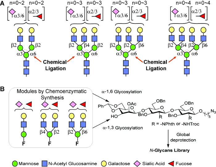
Figure 1: A general modular strategy for the preparation of N-glycans. (A) The number of N-glycans generated by this strategy that commonly occur on human glycoproteins is estimated to exceed 20,000. (B) Three types of modules prepared by this chemo-enzymatic approach that can be used for α-seletive glycosylations. Please click here to view a larger version of this figure.
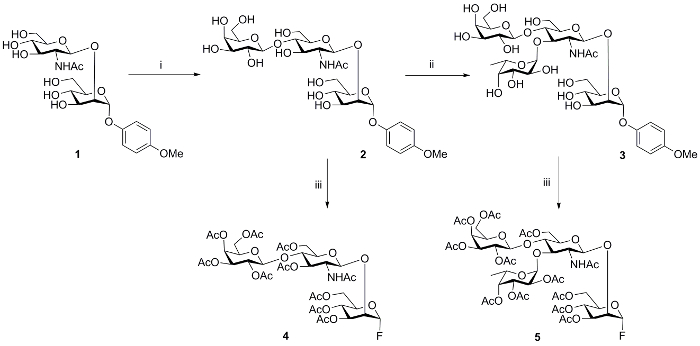
Figure 2: Chemoenzymatic synthesis of modules. i, UDP-galactose, β 1, 4-GalT, 15 h, 86%; ii, GDP-fucose, α 1, 3-FucT, 15 h, 84%; iii, (1) Ac2O, pyridine, RT, 12 h; (1) CAN, ACN: toluene: H2O, (3) DAST, CH2Cl2, -30 oC. CAN: Cerium ammonium nitrate; DAST: Diethylaminosulfur trifluoride.
Product nomenclature : 1, p-methoxyphenyl-O-2-acetamido-2-deoxy-β-D- glucopyranosyl-(1→ 2)-α-D-mannopyranoside; 2, p-methoxyphenyl-O-β-D-galactopyranosyl-(1→4)-2-acetamido-2- deoxy-β-D-glucopyranosyl-(1→2)-α-D-mannopyranoside; 3, p-methoxyphenyl-O-β-D- galactopyranosyl-(1→4)-[α-L-fucopyranosyl-(1→3)-2-acetamido-2-deoxy-β-D-glucopyranosyl]-(1→2)-α-D-mannopyranoside; 4, [2,3,4,6-O-tetraacetyl-β-D-galactopyranosyl]-(1→4)-[3,6-O- diacetyl-2-acetamido-2-deoxy-β-D-glucopyranosyl]-(1→2)-3,4,6-O-triacetyl-α-D-mannopyranosyl fluoride; 5, [2,3,4,6-O-tetraacetyl-β-D-galactopyranosyl]-(1→4)-[2,3,4-O-triacetyl-α-L- fucopyranosyl(1→3)-3,6-O-diacetyl-2-acetamido-2-deoxy-β-D-glucopyranosyl]-(1→2)-3,4,6-O-triacetyl-α-D-mannopyranosyl fluoride Please click here to view a larger version of this figure.
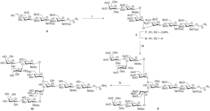
Figure 3: Chemical glycosylation of D1/D2 arm modules to core acceptor. i, 4, AgOTf, Cp2HfCl2, toluene, 4 Å MS, 0 oC to RT, 70%; ii, p-TSA, acetonitrile, RT, 57%; iii, 5, AgOTf, Cp2HfCl2, toluene, 4 Å MS, 0 oC to RT, 34%; iv, (1) LiOH, 1,4-dioxane: H2O; 90 oC, 12 h; (2) Ac2O, pyridine, 12 h; (3) NaOMe, MeOH, 12 h; (4) Pd(OH)2, MeOH : H2O : HCOOH (5:3:2), H2, 36%. AgOTf: Silver trifluromethanesulfonate; Cp2HfCl2: Bis(cyclopentadienyl)hafnium dichloride, MS: molecular sieves, Product nomenclature : 7, 5-Azidopentyl-O-{[2,3,4,6-O-tetraacetyl-β-D-galactopyranosyl]-(1→4)-[3,6-O-diacetyl-2- acetamido-2-deoxy-β-D-glucopyranosyl]-(1→2)-[3,4,6-O-triacetyl-α-D-mannopyranosyl]}-(1→3)-[2-O-acetyl-4,6-O-benzylidine-β-D-mannopyranosyl-(1→4)-O-(3,6-di-O-benzyl-2-deoxy-2-(2,2,2-trichloroethoxy)carbonylamino-β-D-glucopyranosyl)-(1→4)-O-3,6-di-O-benzyl-2-deoxy-2-(2,2,2-trichloroethoxy)carbonylamino-β-D-glucopyranosid. 8, 5-Azidopentyl-O-(2-O- acetyl-3,4,6-tri-O -benzyl-α-D-mannopyranosyl-(1→3)-2-O-acetyl-4,6-O-benzylidine-β-D-mannopyranosyl-(1→4)-O-(3,6-di-O-benzyl-2-deoxy-2-phthalimido-β-D-glucopyranosyl)-(1→4)-O-3,6-di-O-benzyl-2-deoxy-2-phthalimido-β-D-glucopyranoside. 9, 5-Azidopentyl-O-{[2,3,4,6-O-tetraacetyl-β-D- galactopyranosyl]-(1→4)-[3,6-O-diacetyl-2-acetamido-2-deoxy-β-D-glucopyranosyl]-(1→2)-[3,4,6-O-triacetyl-α-D-mannopyranosyl]}-(1→3)-{2,3,4,6-O-tetraacetyl-β-D-galactopyranosyl]-(1→4)-[2,3,4-O-triacetyl-α-LS160fucopyranosyl-(1→3)-3,6-O-diacetyl-2-acetamido-2-deoxy-β-D-glucopyranosyl]-(1→2)-3,4,6-O-triacetyl-α-D-mannopyranosyl}-(1→6)-[2-O-acetyl-β-D-mannopyranosyl-(1→4)-O-(3,6-di-O-benzyl-2-deoxy-2-(2,2,2-trichloroethoxy)carbonylamino-β-D-glucopyranosyl)-(1→4)-O-3,6-di-O-benzyl-2-deoxy-2-(2,2,2-trichloroethoxy)carbonylamino-β-Dglucopyranoside 10, 5-Aminopentyl-β-D-galactopyranosyl-(1→4)-2-acetamido-2-deoxy-β-D-glucopyranosyl- (1→2)-α-D-mannopyranosyl]-(1→3),-[β-D-galactopyranosyl-(1→4)(α-L-fucopyranosyl-(1→3)-2-2-acetamido-2-deoxy-β-D-glucopyranosyl)-(1→2)-α-D-mannopyranosyl]-(1→6)-β-D-mannopyranosyl-(1→4)-2-acetamido-2-deoxy-β-D-glucopyranosyl-(1→4)-2-acetamido-2-deoxy-β-D-glucopyranoside Please click here to view a larger version of this figure.
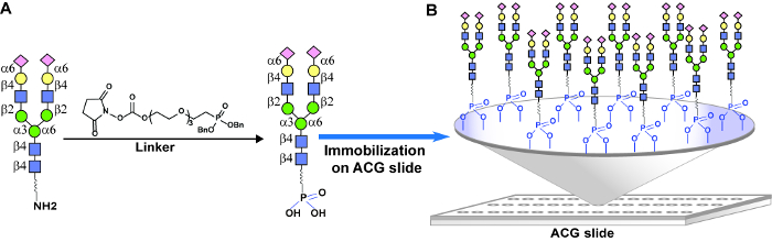
Figure 4: Glycan immobilization on an ACG array. (A) Chemical modification of glycan with amino tail into a phosphonic acid tail for covalent attachment to the ACG slide through phosphonate chemistry. (B) Distribution of glycans on an ACG surface. Please click here to view a larger version of this figure.
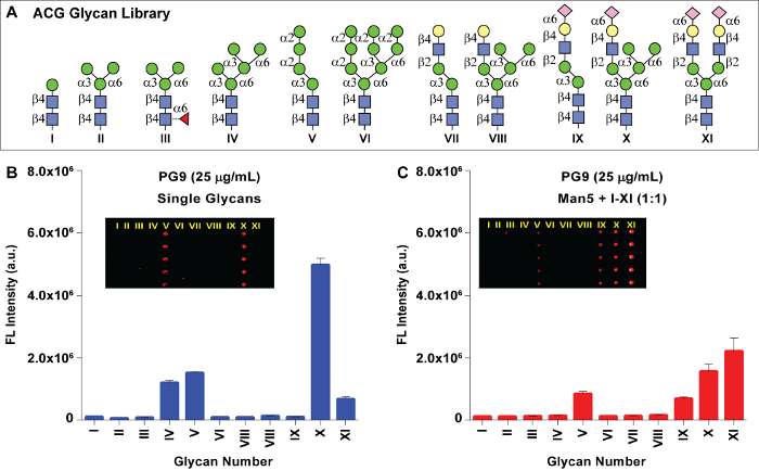
Figure 5: Glyan array analysis. (A) Structures of synthetic N-glycans that are printed on an ACG array. (B) Binding analysis of PG9 to individual glycans I-XI printed on ACG array (left panel) and to glycan mixtures of Man5 mixed with glycans I-XI (right panel) with 100 µM concentration. The molar concentrations in µM for PG9 are given in the legend. The mean signal intensities and standard error calculated for five independent replicates on the array are shown. Insets show fluorescence images. Please click here to view a larger version of this figure.
A class of HIV-1 bNAbs including PG9, PG16, and PGTs 128, 141 – 145 were reported to be highly potent in neutralizing 70 – 80% of circulating HIV-1 isolates. The epitopes of these bNAbs are highly conserved among the variants of the entire HIV-1 group M, therefore they may guide the effective immunogen design for an HIV vaccine to elicit neutralizing antibodies23,24,25. As a part of our ongoing efforts to identify the glycan epitopes of HIV-1 broadly neutralizing antibodies, we reported the chemical and enzymatic synthesis of a panel of high mannose, hybrid, and complex type oligosaccharides to form a glycan array21,22. However, the commonly used glycan arrays on N-hydroxysuccinimide-coated (NHS) glass slides are unable to detect low affinity carbohydrate-protein interactions, probably due to heterogeneous glycan distribution resulting from hydrolysis of reactive N-hydroxysuccimide functional groups on its surface. To overcome the limitations of conventional array formats, we have developed a glycan array on an aluminum oxide coated-glass (ACG) slide. We further performed a homogeneity comparison between the ACG array and the commonly used NHS array to prove that the ACG array offered a more homogeneous glycan presentation on its surface22. Our preliminary binding analysis was not able to observe binding of PG9 to any of the glycans on NHS array (data not shown). Therefore, to define the binding specificity of PG9, the glycans I-XI were attached to a phosphonic acid tail and printed on the ACG slide with 100 µM of individual glycans (Figure 4). The glycan immobilization on an ACG slide through phosphonate chemistry offered a more stable and homogenous distribution. Each of the glycans was printed with five replicates and slide images were obtained from a fluorescence scan after DyLight649-conjugated donkey anti-Human IgG antibody incubation. The binding analysis of PG9 towards individual glycans printed on ACG array (Figure 5, left panel) suggests that PG9 interacted strongly with hybrid-type glycan (X). The interactions were also detected for high mannose type Man5 (glycan IV) and the complex-type glycan (XI).
The molecular level interactions between PG9 and hybrid-type glycan (X) are difficult to determine due to the lack of their co-crystal structure information. The hybrid-type glycan X is composed of a complex-type arm at the 3-O position of the core and a trimannose arm linked to the 6-O position of the central trisaccharide. Binding of PG9 to hybrid glycan X suggests that PG9 requires both mannose and sialic acid residues in close vicinity for high affinity interactions. To identify the glycan combination that best fits into the PG9 binding pocket, we performed a mixed-glycan array study. Glycan IV was mixed with every glycan from I-XI in a 1:1 mole ratio and spotted on an ACG array. The binding analysis of PG9 to each of the mixtures indicated that a combination of Man5 and a SCT (IV+XI) resulted in the highest affinity towards PG9 compared to IV or XI alone. In addition, mixtures containing Man5 and those containing sialylated antennae such as glycans VIII and X were also detected22. For the first time, these results offered an array based proof for hetero-glycans' binding behavior of PG9 that was suggested by crystal structure studies of PG9 with the HIV-1 gp120 V1/V2 domain23.
In conclusion, we have demonstrated an efficient modular chemo-enzymatic strategy for the preparation of highly diverse N-linked oligosaccharides that occur on human glycoproteins. In addition, the development of an ACG array provides an effective means to determine extremely weak protein-carbohydrate interactions and, more importantly, the interactions that occur through hetero-glycans.
The authors have nothing to disclose.
The authors thank the Thin Film Technology Division, Instrument Technology Research Center (ITRC) and National Applied Research Laboratories, Hsinchu Science Park, Taiwan. This work was supported by the National Science Council (grant no. MOST 105-0210-01-13-01) and Academia Sinica.
Acetic acid | Sigma Aldrich | 64197 | |
Acetonitrile | Sigma Aldrich | 75058 | |
Acetic anhydride | Sigma Aldrich | 108247 | |
Anhydrous magnesium sulfate | Sigma Aldrich | 7487889 | |
Boron trifluoride ethyl etherate | Sigma Aldrich | 109637 | |
Bovine serum albumin | Sigma Aldrich | 9048468 | |
Bio-Gel P2 polyacrylamide | Bio-Rad | 1504118 | |
Bis(cyclopentadienyl)hafnium(IV) dichloride | Sigma Aldrich | 12116664 | |
β-1, 4 Galactosyl transferases from bovine milk | Sigma Aldrich | 48279 | |
BioDot Cartesion technology with robotic pin SMP3 (Stealth Micro Spotting Pins) | Arrayit | ||
Cerium ammonium molybdate | TCI | C1794 | |
Cerium ammonium nitrate | Sigma Aldrich | 16774213 | |
Clean glass slide | Schott | ||
Cytidine-5′-monophospho-N-acetylneuraminic acid | Sigma Aldrich | 3063716 | |
Deuterated chloroform | Sigma Aldrich | 865496 | |
Donkey Anti-Human IgG (Alexa Fluor647 conjugated | Jackson Immuno Research, USA | 709605098 | |
Dichloromethane | Sigma Aldrich | 75092 | |
Diethylaminosulfur trifluoride | Sigma Aldrich | 38078090 | |
Dimethylformamide | Sigma Aldrich | 68122 | |
Ethyl acetate | Sigma Aldrich | 141786 | |
Ethylene glycol | Acros Organic | 107211 | |
FAST frame slide incubation chambers | Sigma Aldrich | ||
Guanosine 5'-diphospho-b-L-fucose disodium salt | Sigma Aldrich | 15839700 | |
Lab tracer 2.0 software | Section 4 of the Protocol | ||
GenePix Pro 4300A reader (microarray image analysis) | moleculardevices | www.moleculardevices.com | |
GraphPad Prism Software (Image processing ) | GraphPad Software, Inc | http://www.graphpad.com/guides/prism/6/user-guide/ | |
Lithium hydroxide | Sigma Aldrich | 1310652 | |
Manganese chloride | Sigma Aldrich | 7773015 | |
Methanol | Sigma Aldrich | 67561 | |
N-butanol | Sigma Aldrich | 71363 | |
Oxalic acid | Acros Organic | 144627 | |
Palladium hydroxide | Sigma Aldrich | 12135227 | |
Phosphate Buffered Saline | Thermo Fisher Scientific | 10010023 | |
Pyridine | Sigma Aldrich | 110861 | |
P-Toluene sulfonic acid monohydrate | Sigma Aldrich | 773476 | |
Silver triflate | Sigma Aldrich | 2923286 | |
Sodium bicarbonate | Sigma Aldrich | 144558 | |
Sodium chloride | Sigma Aldrich | 7647145 | |
Sodium hydrogen carbonate | Sigma Aldrich | 144558 | |
Sodium methoxide | Sigma Aldrich | 124414 | |
Sodium sulfate | Sigma Aldrich | 7757826 | |
Toluene | Sigma Aldrich | 108883 | |
Tris buffer | Amresco | N/A | Ultra-pure grade |
Tween-20 | Amresco | 9005645 | |
Uridine diphosphate galactose (UDP-galactose) | Sigma Aldrich | 137868521 |