感应加热小磁球周围水近红外测温技术
Summary
提出了一种利用1150年和 1412 nm 波长测量感应加热小磁球周围水温度的技术。
Abstract
介绍了一种测量感应加热小磁球周围水和非混浊水介质温度的方法。这个技术利用1150年和1412毫微米的波长, 水的吸收系数取决于温度。水或非混浊水凝胶含有2.0 毫米或0.5 毫米直径的磁性球体被辐照1150毫微米或 1412 nm 入射光, 选择使用一个狭窄的带通滤波器;此外, 二维吸光度图像, 这是横向预测的吸收系数, 是通过近红外相机获取。当温度的三维分布可以假设为球状对称时, 将逆阿贝尔变换应用于吸光度剖面估计。根据时间和感应加热功率, 观察到温度的变化。
Introduction
在许多科研领域和应用中, 需要一种测量介质中小热源附近温度的技术。例如, 在磁性热疗的研究中, 这是一种利用磁粒子电磁感应或小磁片进行癌症治疗的方法, 对于准确预测磁性产生的温度分布至关重要。粒子1,2。但是,虽然微波3,4, 超声波 5,6,7,8, 光声 9, 拉曼 10, 和磁共振 11,对12基于的温度测量技术进行了研究和开发, 目前无法准确测量出这种内部温度分布。到目前为止, 在一些位置的单位置温度或温度是通过温度传感器测量的, 在感应加热的情况下, 是非磁性光纤温度传感器13,14。或者, 通过红外辐射温度计远程测量介质的表面温度, 以估计内部温度14。然而, 当含有小热源的介质是水层或非浑浊的水介质时, 我们已经证明, 近红外吸收技术对于测量温度15、16是有用的, 17,18,19。本文介绍了该技术的详细协议和代表性结果。
近红外吸收技术是基于近红外区域水吸收带温度依赖性的原理。如图 1a所示, ν1 + ν2 + ν3的水的吸收带在 1100 nm 到 1250 nm 波长 (λ) 范围内, 并随着温度的变化而变短波长。增加19。这里, ν1 + ν2 + ν3表示此波段对应于三基本的 O H 振动模式的组合: 对称拉伸 (ν1), 弯曲 (ν 2)和求解反对称拉伸(ν 3)20, 21。频谱的这种变化表明, 波段中最温度敏感的波长是λ ≈ 1150 nm。其他吸收的水带也表现出类似的行为, 有关温度15,16,17,18,20,21。ν1 + ν3在范围λ = 1350−1500 nm 中观察到的水带, 其温度依赖性在图 1b中显示。在ν1 + ν3带水中, 1412 nm 是温度敏感度最高的波长。因此, 有可能获得二维 (2D) 温度图像, 使用近红外相机捕捉2D 吸光度图像在λ = 1150 或 1412 nm。由于水的吸收系数在λ = 1150 毫微米小于在λ = 1412 毫微米, 前波长为大约10毫米厚实的水介质是适当的, 而后者适合大约1毫米厚实部分。最近, 使用λ = 1150 nm, 我们得到的温度分布在一个10毫米厚的水层包含感应加热1毫米直径钢球19。此外, 使用λ = 1412 nm15,17, 测量了0.5 毫米厚水层中的温度分布。
基于近红外测温技术的一个优点是, 它简单的设置和实现, 因为它是一种透射吸收测量技术, 不需要荧光, 荧光粉, 或其他热探针。此外, 其温度分辨率小于 0.2 K15,17,19。这种良好的温度分辨率不能通过基于干涉测量的其他传输技术来实现, 这种方法经常用于热和传质研究22,23,24。然而, 我们注意到, 近红外温度成像技术在局部温度变化较大的情况下并不适用, 因为由大温度梯度引起的光偏转成为占主导地位的19。本文从实际使用的角度介绍了这一问题。
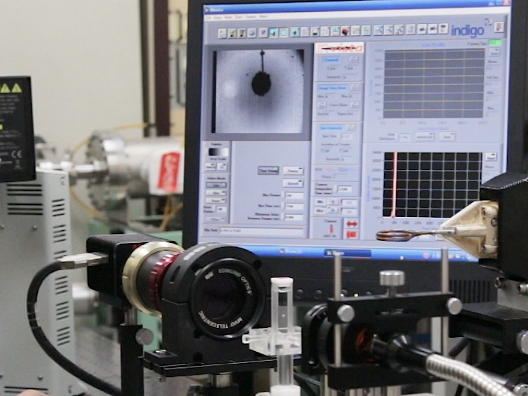
本文介绍了基于近红外测温技术的小磁球感应加热的实验设置和步骤;此外, 它还提供了两个代表性的2D 吸光度图像的结果。一个图像是一个2.0 毫米直径的钢球在10.0 毫米厚的水层, 捕获在λ = 1150 nm。第二个图像是一个0.5 毫米直径的钢球在2.0 毫米厚的麦芽糖糖浆层, 捕获在λ = 1412 nm。本文还提出了将逆阿贝尔变换 (IAT) 应用于2D 吸光度图像的三维 (3D) 径向分布的计算方法和结果。当3D 温度分布假定为球状对称时, IAT 是有效的, 如加热球体 (图 2)19中的情况。对于 IAT 计算, 在这里采用了一种多高斯函数拟合方法, 这是因为可以通过分析25、26、27、28、29 (IATs) 来获得高斯函数的可解析性.并适合于单调地减少数据;这包括使用来自单一热源的热传导的实验。
Protocol
Representative Results
Discussion
本文所提出的技术是利用近红外吸收水的温度依赖性, 在建立必要的设备和实施方面没有很大困难的一种新方法。使用卤素灯和 NBPF 可以很容易地产生入射光。然而, 激光不能使用, 因为相干干扰模式将出现在图像上。可用于可见光使用的普通光学透镜和玻璃细胞, 因为它们在λ上传输足够的光 (1150 毫微米和 1412 nm)。此外, InGaAs 相机现在可以以相对低廉的价格购买。
NBPFs …
Divulgazioni
The authors have nothing to disclose.
Acknowledgements
作者感谢健太山田先生、Ryota 藤冈先生和水木许田先生对实验和数据分析的支持。这项工作得到了 jsp KAKENHI 赠款25630069号, 铃木基金会和日本精密测量技术促进基金会的支持。
Materials
| Induction heating system | CEIA, Italy | SPW900/56 | 780 kHz, 5.6 kW (max). |
| Coil | SA-Japan | custom | Water-cooled copper tube; two-turn; outer dia. 28 mm. |
| Water chiller | Matsumoto Kikai, Japan | MP-401CT | |
| Halogen lamp | Hayashi Watch-Works, Japan | LA-150UE-A | |
| Narrow bandpass filter for λ = 1150 nm | Andover | 115FS10-25 | Full width at half-maximum (FWHM): 10 nm. |
| Narrow bandpass filter for λ = 1412 nm | Andover | semi-custom | Full width at half-maximum (FWHM): 10 nm. |
| Bandpass filter for λ = 850−1300 nm | Spectrogon | SP-1300 | |
| Bandpass filter for λ = 1100−2000 nm | Spectrogon | SP-2000 | |
| NIR camera | FLIR Systems | Alpha NIR | InGaAs |
| Image acquisition software | FLIR Systems | IRvista | |
| Image processing software | NIH | ImageJ | ver. 1.51r |
| Image processing software | MathWorks | Matlab | ver. 2016a |
| Telecentric lens | Edmond Optics | 55350-L | X1 |
| Steel sphere (0.5 mm dia.) | Kobe Steel, Japan | Fe-1.5Cr-1.0C-0.4Mn (wt %) | |
| Steel sphere (2.0 mm dia.) | Kobe Steel, Japan | Fe-1.5Cr-1.0C-0.4Mn (wt %) | |
| Maltose syrup as aqueous gel | Sonton, Japan | Mizuame | Food product |
Riferimenti
- Moros, E. G. . Physics of Thermal Therapy. , (2012).
- Périgo, E. G., Hemery, G., Sandre, O., Ortega, D., Garaio, E., Plazaola, F., Teran, F. J. Fundamentals and advances in magnetic hyperthermia. Appl Phys Rev. 2, 041302 (2015).
- Bardati, F., Marrocco, G., Tognolatti, P. Time-dependent microwave radiometry for the measurement of temperature in medical applications. IEEE Trans Microwave Theo Tech. 52, 1917-1924 (2004).
- Levick, A., Land, D., Hand, J. Validation of microwave radiometry for measuring the internal temperature profile of human tissue. Meas Sci Technol. 22, 065801 (2011).
- Daniels, M. J., Varghese, T., Madsen, E. L., Zagzebski, J. A. Non-invasive ultrasound-based temperature imaging for monitoring radiofrequency heating-phantom results. Phys Med Biol. 52, 4827 (2007).
- Daniels, M. J., Varghese, T. Dynamic frame selection for in vivo ultrasound temperature estimation during radiofrequency ablation. Phys Med Biol. 55, 4735 (2010).
- Seo, C. H., Shi, Y., Huang, S. -. W., Kim, K., O’Donnell, M. Thermal strain imaging: A review. Interface Focus. 1, 649-664 (2011).
- Bayat, M., Ballard, J. R., Ebbini, E. S. Ultrasound thermography: A new temperature reconstruction model and in vivo results. AIP Conf Proc. 1821, 060004 (2017).
- Petrova, E., Liopo, A., Nadvoretskiy, V., Ermilov, S. Imaging technique for real-time temperature monitoring during cryotherapy of lesions. J Biomed Opt. 21, 116007 (2016).
- Gardner, B., Matousek, P., Stone, N. Temperature spatially offset Raman spectroscopy (T-SORS): Subsurface chemically specific measurement of temperature in turbid media using anti-Stokes spatially offset Raman spectroscopy. Anal Chem. 88, 832-837 (2016).
- Yoshioka, Y., Oikawa, H., Ehara, S., Inoue, T., Ogawa, A., Kanbara, Y., Kubokawa, M. Noninvasive measurement of temperature and fractional dissociation of imidazole in human lower leg muscles using 1H-nuclear magnetic resonance spectroscopy. J Appl Physiol. 98, 282-287 (2004).
- Galiana, G., Branca, R. T., Jenista, E. R., Warren, W. S. Accurate temperature imaging based on intermolecular coherences in magnetic resonance. Science. 322, 421-424 (2008).
- Rapoport, E., Pleshivtseva, Y. . Optimal Control of Induction Heating Processes. , (2006).
- Lucía, O., Maussion, P., Dede, E. J., Burdío, J. M. Induction heating technology and its applications: Past developments, current technology, and future challenges. IEEE Trans Ind Electron. 61, 2509-2520 (2014).
- Kakuta, N., Kondo, K., Ozaki, A., Arimoto, H., Yamada, Y. Temperature imaging of sub-millimeter-thick water using a near-infrared camera. Int J Heat Mass Trans. 52, 4221-4228 (2009).
- Kakuta, N., Fukuhara, Y., Kondo, K., Arimoto, H., Yamada, Y. Temperature imaging of water in a microchannel using thermal sensitivity of near-infrared absorption. Lab Chip. 11, 3479-3486 (2011).
- Kakuta, N., Kondo, K., Arimoto, H., Yamada, Y. Reconstruction of cross-sectional temperature distributions of water around a thin heating wire by inverse Abel transform of near-infrared absorption images. Int J Heat Mass Trans. 77, 852-859 (2014).
- Kakuta, N., Yamashita, H., Kawashima, D., Kondo, K., Arimoto, H., Yamada, Y. Simultaneous imaging of temperature and concentration of ethanol-water mixtures in microchannel using near-infrared dual-wavelength absorption technique. Meas Sci Technol. 27, 115401 (2016).
- Kakuta, N., Nishijima, K., Kondo, K., Yamada, Y. Near-infrared measurement of water temperature near a 1-mm-diameter magnetic sphere and its heat generation rate under induction heating. J Appl Phys. 122, 044901 (2017).
- Libnau, F. O., Kvalheim, O. M., Christy, A. A., Toft, J. Spectra of water in the near- and mid-infrared region. Vib Spectrosc. 7, 243-254 (1994).
- Siesler, H. W., Ozaki, Y., Kawata, S., Heise, H. M. . Near-Infared Spectroscopy. , (2002).
- Shakher, C., Nirala, A. K. A review on refractive index and temperature profile measurements using laser-based interferometric techniques. Opt Laser Eng. 31, 455-491 (1999).
- Assebana, A., Lallemanda, M., Saulniera, J. -. B., Fominb, N., Lavinskaja, E., Merzkirchc, W., Vitkinc, D. Digital speckle photography and speckle tomography in heat transfer studies. Opt Laser Technol. 32, 583-592 (2000).
- Ambrosini, D., Paoletti, D., Spagnolo, S. G. Study of free-convective onset on a horizontal wire using speckle pattern interferometry. Int J Heat Mass Trans. 46, 4145-4155 (2003).
- Bracewell, R. N. . The Fourier Transform and Its Applications. , (2000).
- Yoder, L. M., Barker, J. R., Lorenz, K. T., Chandler, D. W. Ion imaging the recoil energy distribution following vibrational predissociation of triplet state pyrazine-Ar van der Waals clusters. Chem Phys Lett. 302, 602-608 (1999).
- De Colle, F., de Burgo, C., Raga, A. C. Diagnostics of inhomogeneous stellar jets: convolution effects and data reconstruction. Astron Astrophys. 485, 765-772 (2008).
- Green, K. M., Borrás, M. C., Woskov, P. P., Flores, G. J., Hadidi, K., Thomas, P. Electronic excitation temperature profiles in an air microwave plasma torch. IEEE Trans Plasma Sci. 29, 399-406 (2001).
- Bendinelli, O. Abel integral equation inversion and deconvolution by multi-Gaussian approximation. Astrophys J. 366, 599-604 (1991).
- Dorband, B., Muller, H., Gross, H., Gross, H. Vol. 5 Metrology of Optical Components and Systems. Handbook of Optical System. , (2012).
- Sheikholeslami, M., Rokni, H. B. Simulation of nanofluid heat transfer in presence of magnetic field: A review. Int J Heat Mass Trans. 115, 1203-1233 (2017).
- Häfeli, U., Schütt, W., Teller, J., Zborowski, M. . Scientific and Clinical Applications of Magnetic Carriers. , (2013).