Here, we present a protocol to produce a large number of GMP-grade exosomes from synovial fluid mesenchymal stem cells using a 3D bioreactor.
Exosomes secreted by mesenchymal stem cells (MSCs) have been suggested as promising candidates for cartilage injuries and osteoarthritis treatment. Exosomes for clinical application require large-scale production. To this aim, human synovial fluid MSCs (hSF-MSCs) were grown on microcarrier beads, and then cultured in a dynamic three-dimension (3D) culture system. Through utilizing 3D dynamic culture, this protocol successfully obtained large-scale exosomes from SF-MSC culture supernatants. Exosomes were harvested by ultracentrifugation and verified by a transmission electron microscope, nanoparticle transmission assay, and western blotting. Also, the microbiological safety of exosomes was detected. Results of exosome detection suggest that this approach can produce a large number of good manufacturing practices (GMP)-grade exosomes. These exosomes could be utilized in exosome biology research and clinical osteoarthritis treatment.
Osteoarthritis (OA), resulting from joint cartilage and underlying bone breakdown, remains a severe challenge leading to disability1,2. Without blood and nerve supply, cartilage self-healing ability is minimal once being injured3,4. In the past decades, therapies based on autologous chondrocyte implantation (ACI) have made some progress in OA treatment5. For chondrocyte isolation and expansion, harvesting small cartilage from the OA joint's non-weight bearing area is necessary, causing injuries to the cartilage. Also, the procedure will require a second operation to implant the expanded chondrocytes6. Thus, one-step therapies for OA treatment without cartilage injuries are under extensive exploration.
Mesenchymal stem cells (MSCs) have been suggested as promising alternatives for OA treatment7,8. Originating from multiple tissues, MSCs can differentiate into chondrocytes with specific stimulation. Importantly, MSCs can modulate immune responses via anti-inflammation9. Therefore, MSCs hold significant advantages in OA treatment by repairing cartilage defects and modulating the immune response, especially in the inflammation milieu. For OA treatment, MSCs from synovial fluid (SF-MSCs) have recently attracted much attention due to their stronger chondrocyte differentiation ability than other MSC sources10,11. Notably, at the orthopedic clinic, the extraction of inflammatory SF from the joint cavity is a routine therapy to relieve the pain symptom of OA patients. Extracted inflammatory SF usually is disposed of as medical waste. Both patients and doctors are ready to consider autologous MSCs isolated from the inflammatory SF as OA treatment with very few ethical conflicts. However, SF-MSC therapy is compromised due to tumorigenic risks, long-time storage, and distant shipment barriers.
Exosomes, secreted by many cell types, including MSCs, carry most of the parent cell bio-information. It has been investigated in-depth as a cell-free therapy12,13. According to the updated resources available on the clinical trial government (ClinicalTrials.gov) website, more extensive exosome clinical studies are initiated and undertaken in the research fields of cancer, hypertension, and neuro-degenerative diseases. SF-MSC exosome treatment could be an exciting and challenging trial to cope with OA. Good manufacturing practice (GMP)-grade and large-scale exosome production are essential for clinical translation. Small-scale exosome isolation has been widely performed based on two-dimensional (2D) cell culture. However, large-scale exosome production strategies need optimization. A large-scale exosome manufacturing method was developed in this study, based on massive SF-MSC culture in xeno-free conditions. After ultracentrifugation from cell culture supernatants, exosome safety and function were validated.
This study was approved by the Human Ethics Committee of Shenzhen Second People's Hospital. A schematic diagram of exosomes isolated from hSF-MSCs in vitro protocol is shown in Figure 1.
1. Human SF-MSCs culture and identification
2. 3D bioreactor cell culture
3. Exosome identification and safety detection
4. In vitro exosome function detection
5. Statistical analysis
Flow cytometry was used to identify the surface markers of SF-MSCs, according to the minimal criteria to define human MSCs recommended by the International Society for Cellular Therapy14,15. Flow cytometry analysis revealed that SF-MSCs cultured in this study met the identification criteria of MSCs. They were negative for CD34, CD45, and HLA-DR (below 3%) and positive for CD73, CD90, and CD105 (above 95%) (Figure 2).
Under inverted microscopy, it was noticed that the SF-MSCs proliferate on microcarriers (Figure 3A). After cells were digested and washed from the 2D culture plate and 3D culture microcarriers, the cell number was counted. Compared to 2D culture, 3D culture induced the SF-MSC to proliferate more quickly from 6 days onwards (Figure 3B). Results of three independent experiments were presented.
To identify the exosomes (Exos) from SF-MSCs of 2D and 3D culture, the exosome-associated proteins (CD63, CD9, and CD81) and negative protein (calnexin) were detected using western blotting. Results revealed that the 2D-Exos and 3D-Exos express CD63, CD9, and CD81, while they are negative for calnexin (Figure 4A). Also, the exosome diameter and morphology were assayed using NTA and TEM. Nanosight analysis demonstrated that the diameter of 2D-Exos (Figure 4B) and 3D-Exos (Figure 4D) is approximately 120 nm. Transmission electronic microscope analysis revealed the morphology of 2D-Exos and 3D-Exos, showing roughly spheroidal vesicles (Figure 4C,E).
After 3D culture, the particle concentration of 30-160 nm sized particles is 4.0 x 10⁶ per mL analyzed using NTA. However, after the 2D culture, the particle concentration of 30-160 nm particles is 2.5 x 10⁶ per mL (Figure 5A). When calculating the exosome protein yield in the medium, 3D culture produced more exosome protein than 2D culture (Figure 5B). Thus, compared to 2D culture, 3D culture significantly enhanced exosome yield.
The exosomes were first labeled with Dil, and then incubated at a concentration of 10 µg/mL with the chondrocytes for 1 h, 2 h, and 3 h to examine whether exosomes can enter primary chondrocytes in vitro. Dil-labeled exosomes entered primary chondrocytes, with the peak seen at 3 h (Figure 6).
To detect the function of exosomes as nanocarriers, exosomes were loaded with Cy3-labeled miR-140 through electroporation, and then treated with chondrocytes at a concentration of 10 µg/mL for 3 h. Results demonstrated that exosomes could deliver miR-140 to chondrocytes (Figure 7). All the results met the specifications; all samples were sterile and negative for Mycoplasma.
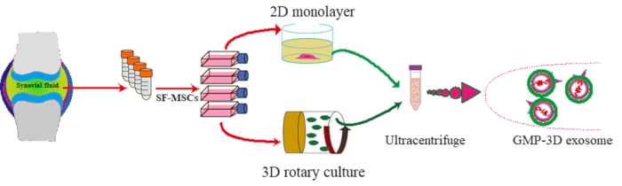
Figure 1: Schematic diagram of exosomes isolated from hSF-MSCs in vitro. Please click here to view a larger version of this figure.
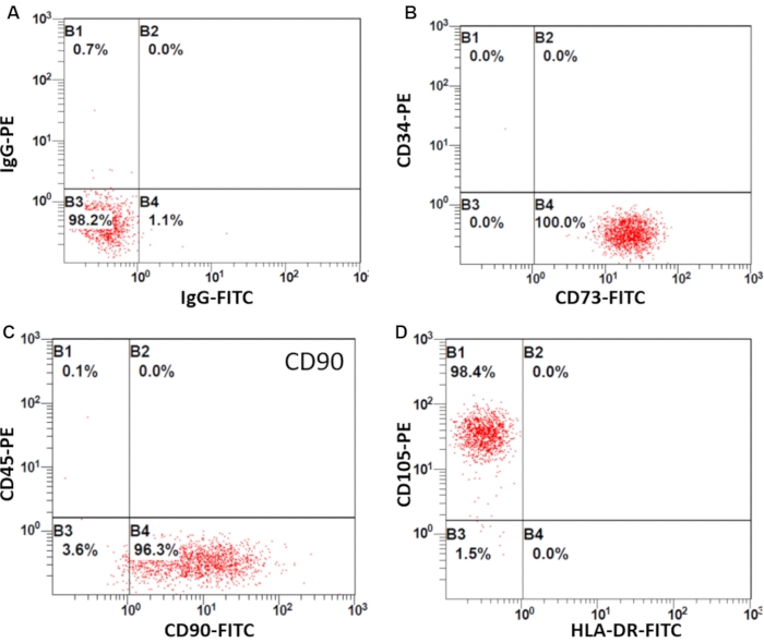
Figure 2: Identification of SF-MSCs by flow cytometry. Flow cytometry shows the positive or negative immunophenotype of hSF-MSCs. (A) Labeling with an IgG1 isotype control antibody. (B) CD73 is positive, and CD34 is negative. (C) CD90 is positive, and CD45 is negative. (D) CD105 is positive, and HLA-DR is negative, known as MSC markers. Please click here to view a larger version of this figure.
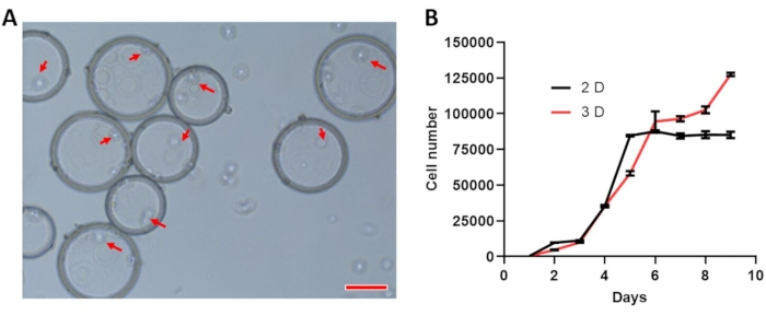
Figure 3: SF-MSC growth curve. (A) Representative images showing SF-MSCs (red arrows) on microcarriers under inverted microscopy (scale bar =100 µm). (B) The growth curve of SF-MSC under 2D and 3D culture. Please click here to view a larger version of this figure.
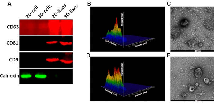
Figure 4: Identification of exosomes. (A) Western blotting results of the 2D-Exos and 3D-Exos. (B) Nanosight analysis of the diameter of 2D-Exos and 3D-Exos. (C) TEM detection of the morphology of 2D-Exos and 3D-Exos. Please click here to view a larger version of this figure.
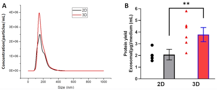
Figure 5: The enhanced yield of exosome production by 3D bioreactor culture. (A) Representative results of exosome size analyzed by NTA. (B) Protein yield = exosomal protein (µg)/conditioned medium (mL). Plots show yield for each method and the mean ± SD of all measurements (** p < 0.01). Statistical comparisons were performed by one-way ANOVA with post-hoc Bonferroni's correction and by Student's t-test. ** p < 0.01 was considered to be a significant difference. Please click here to view a larger version of this figure.
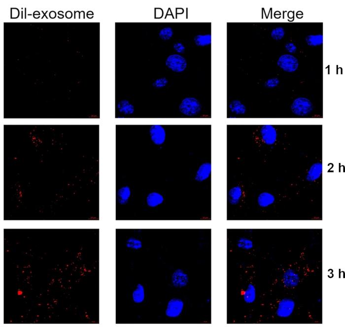
Figure 6 Representative images showing the internalization of Dil-labeled exosomes by primary chondrocytes. Chondrocytes were incubated with Dil-labeled exosomes for 1 h, 2 h, and 3 h. Exosomes were labeled with Dil (red), and nuclei were labeled with Hoechst (blue). Samples were detected at 60x magnification. Scale bar = 10 µm. Please click here to view a larger version of this figure.
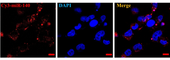
Figure 7: In vitro delivery of miR-140 by MSC exosomes. After up-taking Cy3-labeled miR-140 that was encapsulated in SF-MSCs-derived exosomes, chondrocytes were imaged. Scale bar = 10 µm. Please click here to view a larger version of this figure.
The mesenchymal stem cells have been widely used in regenerative medicine due to their self-renewal, differentiated into tissue cells with specialized functions, and paracrine effects16,17. Notably, the paracrine effects exerted by exosomes have attracted much attention18. Exosomes carry the bio-information of MSCs and perform their biological function and overcome MSC shortcomings, such as troublesome storage and shipment. Thus, exosomes derived from MSCs have been used for therapeutic interventions, which have attracted the most attention in OA therapy.
Currently, there are two methods to propagate MSCs, 2D culture and three-dimensional (3D) culture19. 2D culture is a conventional way to culture MSCs for in vitro studies with low costs. However, it is time-consuming and limited in scale potential. Also, MSC propagation in the 2D microenvironment quickly deduces the stemness20,21, one of the most critical characteristics of MSCs. Thus, 2D culture cannot fulfill the requirements for MSC therapy. In this study, to the aim of SF-MSCs in OA therapy, we endeavor to maintain SF-MSC characteristics using the culture of a 3D bioreactor, which can more accurately mimic the biological microenvironment. Recently, other researchers have used scaffolds or microcarrier-based 3D to culture MSCs. We found that the 3D collagen scaffolds allowed more concentrated exosomes produced by human bone marrow-derived MSCs than 2D culture23.
As exosomes can perform a large part of MSC function while avoiding some shortcomings of MSCs, we aim to produce exosomes for OA therapy. This study further detected the exosome production and delivery function using this system based on the large quantity and high quality of MSC propagation of 3D culture. The Rotary Cell Culture System (RCCS) was used for 3D culture to produce exosomes from large-scale MSC propagation. Compared to traditional 2D flask culture, this 3D culture system may avoid contamination from repeated medium change and cell passage. More importantly, this study showed that a 3D bioreactor could enhance exosome production to meet the clinical study requirements. Of note, exosomes isolated from 3D culture supernatants can deliver miRNAs into cells, suggesting that 3D culture does not interfere with exosome function. Microbial and endotoxin detection results further support that this study has established a protocol that could produce exosomes, which are biologically safe and promising for OA therapy. At present, the exosome quantity produced in this study cannot satisfy commercial needs. Hence, strategies for an enormous amount of exosome production need to be developed.
The authors have nothing to disclose.
National Natural Science Foundation of China (No. 81972116, No. 81972085, No. 81772394); Key Program of Natural Science Foundation of Guangdong Province (No.2018B0303110003); Guangdong International Cooperation Project (No.2021A0505030011); Shenzhen Science and Technology Projects (No. GJHZ20200731095606019, No. JCYJ20170817172023838, No. JCYJ20170306092215436, No. JCYJ20170413161649437); China Postdoctoral Science Foundation (No.2020M682907); Guangdong Basic and Applied Basic Research Foundation (No.2021A1515010985); Sanming Project of Medicine in Shenzhen (SZSM201612079); Special Funds for the Construction of High-level Hospitals in Guangdong Province.
BCA assay kit | ThermoFisher | 23227 | Protein concentration assay |
Blood agar plate | Nanjing Yiji Biochemical Technology Co. , Ltd. | P0903 | Bacteria culture |
CD105 antibody | Elabscience | E-AB-F1243C | Flow cytometry |
CD34 antibody | Elabscience | E-AB-F1143C | Flow cytometry |
CD45 antibody | BD Bioscience | 555483 | Flow cytometry |
CD63 antibody | Abclonal | A5271 | Western blotting |
CD73 antibody | Elabscience | E-AB-F1242C | Flow cytometry |
CD81 antibody | ABclonal | A5270 | Western blotting |
CD9 antibody | Abclonal | A1703 | Western blotting |
CD90 antibody | Elabscience | E-AB-F1167C | Flow cytometry |
Centrifuge | Eppendorf | Centrifuge 5810R | |
CO2 incubator | Thermo | Cell culture | |
Confocal laser scanning fluorescence microscopy | ZEISS | LSM 800 | |
Cytodex | GE Healthcare | Microcarrier | |
Dil | ThermoFisher | D1556 | Exosome label |
EZ-PCR Mycoplasma detection kit | BI | 20-700-20 | Mycoplasma detection |
Flowcytometry | Beckman | MSC identification | |
Gene Pulser II System | Bio-Rad Laboratories | 1652660 | Gene transfection |
GraphPad Prism 8.0.2 | GraphPad Software, Inc. | Version 8.0.2 | |
HLA-DR antibody | Elabscience | E-AB-F1111C | Flow cytometry |
Lowenstein-Jensen culture medium | Nanjing Yiji Biochemical Technology Co. , Ltd. | T0573 | Mycobacterium tuberculosis culture |
MesenGro | StemRD | MGro-500 | MSC culture |
Nanosight NS300 | Malvern | Nanosight NS300 | Nanoparticle tracking analysis |
NTA 2.3 software | Malvern | Data analysis | |
Odyssey FC | Gene Company Limited | Fluorescent western blotting | |
OptiPrep electroporation buffer | Sigma | D3911 | Gene transfection |
Protease inhibitors cocktail | Sigma | P8340 | Proteinase inhibitor |
RNase A | Qiagen | 158924 | Removal of RNA |
Sabouraud agar plate | Nanjing Yiji Biochemical Technology Co., Ltd. | P0919 | Fungi culture |
TEM | JEM-1200EX | ||
The Rotary Cell Culture System (RCCS) | Synthecon | RCCS-4HD | 3D culture |
Ultracentrifuge | Beckman | Optima XPN-100 | Exosome centrifuge |