This study presents an easy-to-use, complete, and simple set of methods to label and analyze glomeruli from CUBIC-cleared mouse kidneys. Data such as glomerulus number and volume can be obtained easily and reliably using fluorescein isothiocyanate (FITC)-Dextran, light sheet fluorescence microscopy (LSFM), or common confocal microscopy and software such as Imaris.
The glomeruli are fundamental units in the kidney; hence, studying the glomeruli is pivotal for understanding renal function and pathology. Biological imaging provides intuitive information; thus, it is of great significance to label and observe the glomeruli. However, the glomeruli observation methods currently in use require complicated operations, and the results may lose label details or three-dimensional (3D) information. The clear, unobstructed brain imaging cocktails and computational analysis (CUBIC) tissue clearing technology has been widely used in renal research, allowing for more accurate detection and deeper detection depth. We found that mouse glomeruli can be rapidly and effectively labeled by tail vein injection of medium molecular weight FITC-Dextran followed by the CUBIC clearing method. The cleared mouse kidney could be scanned by a light-sheet microscope (or a confocal microscope when sliced) to obtain three-dimensional image stacks of all the glomeruli in the entire kidney. Processed with appropriate software, the glomeruli signals could be easily digitized and further analyzed to measure the number, volume, and frequency of the glomeruli.
The number and volume of glomeruli are very important for the diagnosis and treatment of various kidney diseases1,2,3,4,5. The golden standard of glomeruli number estimation is the physical dissector/fractionator combination. However, this method requires special reagents and equipment, making it slow and expensive6,7,8,9. Biopsy provides a wealth of information, but obviously, this method is only suitable for rough estimations10,11. Medical imaging technologies, including magnetic resonance imaging (MRI), computed tomography (CT), and X-ray, are also widely used in glomerular detection12,13,14,15, but such technologies require bulky instruments. New methods, such as matrix-assisted laser desorption/ionization (MALDI) imaging mass spectrometer16 or the thick and thin section method17, have also been used in glomerular detection, though they remain tedious and laborious.
With the help of transparency technologies, it is possible to observe deeper depths and obtain richer and more complete information from thick tissues or even whole organs18,19,20,21,22,23. Therefore, transparency technologies have been widely used in kidney research24. The observation and detection of glomeruli in the cleared kidneys are also involved. However, these published articles either only briefly referred to glomerular detection25 or used difficult-to-achieve labeling methods such as transgenic animals26, self-produced dyes13, or high-concentration antibody incubation27 to label the glomeruli. In addition, although studies had analyzed glomeruli in cleared kidneys, the analyses were always limited13 or relied on analysis algorithms established by the authors themselves26.
We have previously demonstrated a more convenient way to label the glomeruli in mice kidneys28. By using Imaris, we found that glomeruli count, frequency, and volume could be quickly obtained. Thus, here we present a more accessible, comprehensive, and simplified set of methods to label and analyze the glomeruli of mice kidneys.
Adult C57BL/6 mice (6 weeks of age, 25-30 g) were used in this study. All procedures were performed in compliance with local regulations of animal welfare and experimental ethics. The study was approved by the West China Hospital of Sichuan University Biomedical Research Ethics Committee.
1. Glomeruli labeling and tissue preparation
2. Clearing
3. Image acquisition
4. Data processing and quantification
NOTE: Process the image stacks with Imaris (image analysis) software, using the Surface function to label the glomeruli and perform analysis.
This study provides a simple and efficient method for labeling and analyzing the glomeruli in mice kidneys.
Glomeruli (blood vessels) can be well labeled by intravascularly injected FITC-Dextran. After the clearing process, the kidney became transparent (Figure 1A), and the glomeruli could be clearly observed by using light-sheet microscopy (Figure 1B) or confocal microscopy (Figure 1C). Confocal microscopy has a limited scanning depth, so kidneys should be cut into approximately 1 mm-thick slices. If a light-sheet microscope is used, the whole kidney can be scanned directly.
With the clear signals, it was easy to count the number of glomeruli. In fact, the labeling was so effective that the glomeruli could be counted with the naked eye. The volume of glomeruli could also be directly measured. Of course, software such as Imaris greatly accelerated this process. Using the Surface function, all the glomeruli in a kidney slice (Figure 2), in an entire kidney (Figure 3), or in a strip (Figure 4) could be selected, and the number and volume of the glomeruli could be obtained directly (Figure 2C, Figure 3C, Figure 4C). The volume of the selected region could also be measured (Figure 2B, Figure 3B, Figure 4B), so the glomeruli volume ratio and frequency in a certain region or in the whole kidney could be calculated (Figure 2D, Figure 3D, Figure 4D). An area of the kidney could be selected to perform calculations in specific regions. We presented a strip similar to a biopsy. The number, volume, and frequency of the glomeruli in the strip could also be obtained easily (Figure 4).
As shown in the figures, the results presented in this study are (N = number, F = frequency, V = volume, V(a) = average volume, V(t) = total volume):
Nglomeruli in slice = 1128, Nglomeruli in whole kidney = 14006, Fglomerular in slice = 65 per mm3, Fglomerular in whole kidney = 105 per mm3, Vtotal glomeruli in slice/Vslice = 2.24%, Vtotal glomeruli in whole kidney/Vkidney = 5.54%, V(a)glomeruli in slice = 373654 µm3, V(a)glomeruli in whole kidney = 521627 µm3, V(t)glomeruli in slice = 421481724 µm3, V(t)glomeruli in whole kidney = 7305386256 µm3 (Figure 2D, Figure 3D).
Nglomeruli in strip = 63, Fglomeruli in strip = 72 per mm3, Vtotal glomeruli in strip/Vstrip = 2.23%, V(a)glomeruli in strip = 307698 µm3, V(t)glomeruli in strip = 19384977 µm3 (Figure 4D).
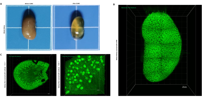
Figure 1: Kidney tissue transparency and FITC-dextran-labeled glomeruli. (A) Kidney before and after transparency treatment. (B) The image of a whole kidney scanned with LSFM. (C) The image of a slice of kidney scanned with the confocal microscope (left: 4x, right: 10x). scale bar = 300 µm (4x, confocal); scale bar = 100 µm (10x, confocal).; scale bar = 700 µm (5x, zoom 0.36, LSFM). Glomeruli were labeled with FITC-Dextran. Please click here to view a larger version of this figure.
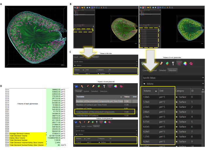
Figure 2: The function Surface applied to the confocal scanned kidney slice. (A) Surface of the slice and all glomeruli are created. Pink = glomeruli, blue = out Surface of the kidney slice, green = original label of vessels (glomeruli and some big vessels). Scale bar = 300 µm. (B) When the Surface of the slice (left) or the glomeruli (right) are selected (selected objects would turn yellow), data could be obtained directly (dotted box highlights where it shows the data). (C) The number, the volume of every single glomerulus, and the volume of total glomeruli could be directly obtained as well as the volume of the slice. (D) Exported data could be further analyzed so the calculation, such as the average volume of the glomeruli and volume ratio of glomeruli and selected area, could be worked out. Please click here to view a larger version of this figure.
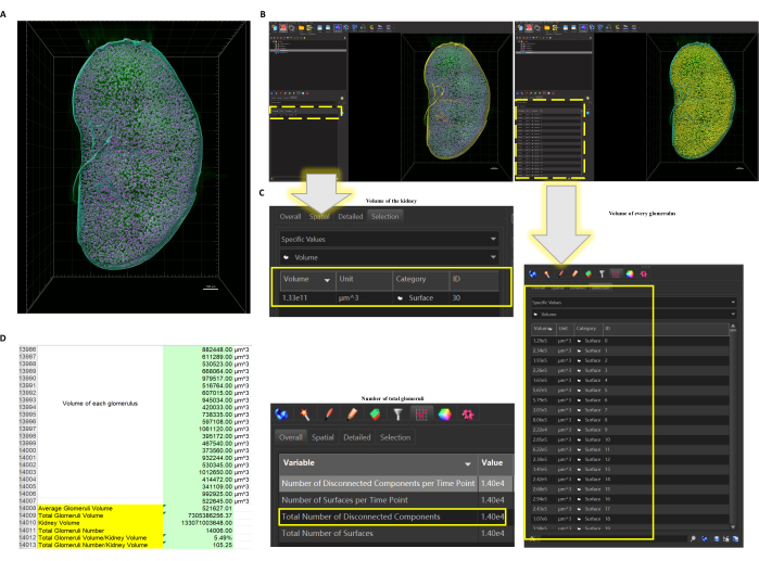
Figure 3: The function Surface applied to the LSFM scanned whole kidney. (A) Surface of the whole kidney and all glomeruli are created. Pink = glomeruli, blue = out Surface of the whole kidney, green = original label of vessels (glomeruli and some big vessels). Scale bar =1000 µm. (B) When the Surface of the kidney (left) or the glomeruli (right) are selected (selected objects would turn yellow), data could be obtained directly (dotted box highlights where it shows the data). (C) The number, the volume of every single glomerulus, and the number of the glomeruli could be directly obtained, as well as the volume of the kidney. (D) Exported data could be further analyzed so the calculation, such as the average volume of glomeruli and volume ratio of glomeruli and the whole kidney, could be worked out. Please click here to view a larger version of this figure.
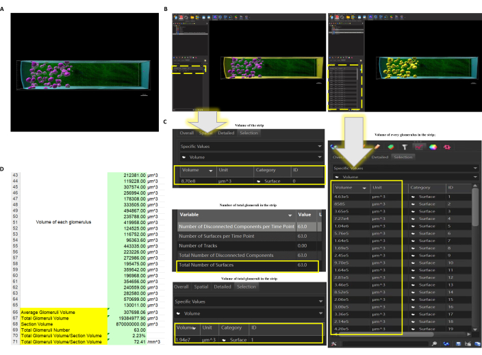
Figure 4: The function Surface applied to the selected kidney strip. (A) Surface of the strip and all glomeruli are created. Pink = glomeruli, blue = out Surface of the kidney strip, green = original label of vessels (glomeruli and some big vessels). Scale bar =150 µm. (B) When the Surface of the strip (left) or the glomeruli (right) are selected (selected objects would turn yellow), data could be obtained directly (dotted box highlights where it displays the data). (C) The number, the volume of every single glomerulus, and the volume of total glomeruli could be directly obtained, as well as the volume of the strip. (D) Exported data could be further analyzed so the calculation, such as the average volume of the glomeruli and volume ratio of glomeruli and selected area, could be worked out. Please click here to view a larger version of this figure.
Tissue-clearing technologies can be classified into 3 or 4 groups29,30,31. Organic solvent-based tissue clearing (e.g., DISCO and PEGASOS), aqueous-based tissue clearing (e.g., CUBIC), and hydrogel embedding tissue clearing (e.g., CLARITY) have all been applied in kidney clearing 25,26,28,32. CUBIC, as we have demonstrated, works best when the glomeruli (vessels) are labeled with FITC-Dextran. In addition, CUBIC has a relatively more convenient operation process and requires no more than ordinary microscopic lenses27, as the processed samples do not need to be soaked in organic reagents33. However, different clearing methods and vessel label methods have their own advantages and disadvantages; researchers may need to experiment with different approaches to meet their specific needs.
Previous studies noticed that the glomeruli could be labeled in cleared kidneys when blood vessels in the kidneys were labeled13,25,26,28. A thorough explanation for this phenomenon still needs to be further investigated, but it can be deduced that a good labeling method for the vascular system also effectively labels the glomeruli. Based on the experimental results, we would recommend the intravascular injection of FITC-Dextran with a medium molecular weight (such as 150 kDa). However, since labeling relies on blood circulation, this method may fail to label the glomeruli when they lack circulation.
LSFM permits large-scale tissue observation, making it more suitable for whole kidney glomeruli detection. However, light-sheet microscopes may not be available in every laboratory. So, we are providing an alternative set of protocols using ordinary confocal microscopes. Although the kidney needs to be sliced into multiple sections, obtaining data such as the counts and volume of glomeruli in the whole kidney is feasible. Besides, confocal microscopy provides a smaller volume of data, which is more data processing friendly.
Imaris provides direct data output. However, care should be taken to match the analog signal to the original closely. Additionally, the software may take a long time to process large files, which can be frustrating. As long as the glomeruli can be clearly labeled and visualized, there is more than one unique solution for data analysis. Researchers from different laboratories could work with their own familiar software and programming languages.
The authors have nothing to disclose.
This study was supported by grants from the National Natural Science Foundation of China (82204951) and Sichuan Science and Technology Program (2020JDRC0102).
4% PFA | Biosharp | 7007171800 | Fixation reaagen |
502 Glue | Deli | 7146 | For fixing the kidney to the sample fixing adapter |
Antipyrine | Aladdin | A110660 | Clearing reagent |
Brain Matrix | RWD Life Science | 1mm 40-75 | Tissue slicing |
Confocal microscopy | Nikon | A1plus | Image acquisition |
FITC-Dextran | Sigma-Aldrich | FD150S | Labeling reagent |
Light sheet fluorescence microscopy | Zeiss | Light sheet 7 | Image acquisition |
Mice | Ensiweier | Adult C57BL/6 mice (6 weeks of age, 25–30 g) | |
N-Butyldiethanolamine | Aladdin | B299095 | Clearing reagent |
Nicotinamide | Aladdin | N105042 | Clearing reagent |
Pentobarbital Natriumsalz | Sigma-Aldrich | P3761 | |
Tail vein fixator | JINUOTAI | JNT-FS35 | Fix the mouse for vail injection |
Triton X-100 | Sigma-Aldrich | T8787 | Clearing reagent |