Summary

바이오 메디컬 응용 프로그램에 대한 실크 실크 단백질 합금 재료 설계

Published: August 13, 2014
doi:

Summary

혼합 속성과 결합하여 광범위한 기능과 생체 물질을 생성 할 수있는 효율적인 방법입니다. 다른 천연 실크 단백질 분자 사이의 상호 작용을 예측함으로써, 가변 기계적 복원력 전기적 응답, 광학 투명성, 화학적 가공성, 생분해 성, 또는 열적 안정성을 가진 새로운 실크 실크 단백질 합금 플랫폼을 설계 할 수있다.

Abstract

섬유 단백질은 바이오 센서, 나노 의학, 조직 재생 및 약물 전달과 같은 생의학 분야에서 다양한 용도로 사용되어왔다 서로 다른 서열과 구조를 표시합니다. 이 단백질 사이의 분자 규모의 상호 작용을 기반으로 자료를 설계하는 것은 조정 가능한 특성을 가진 새로운 다기능 단백질 합금 생체 물질을 생성하는 데 도움이됩니다. 이러한 합금 재료 시스템은 또한 때문에 체내에서 생분해 성 재료, 생체 적합성 및 tenability 전통적인 합성 중합체에 비해 이점을 제공한다. 이 문서에서는 단백질 합금을 생산하는 방법 계산 방법에 의해 단백질 – 단백질 상호 작용을 예측하는 방법을 포함하여이 주제에 관한 유용한 프로토콜을 제공하기 위해 예를 들어 야생 tussah 실크 (Antheraea의 pernyi) 및 국내 뽕나무 실크 (Bombyx 모리)의 단백질 혼합 사용 솔루션은, 어떻게 열 분석에 의해 합금 시스템을 확인하는 방법과 변수 합금 재료를 제조회절 격자와 광학 재료, 회로 코팅과 전기 재료 및 약물 방출 및 전달을위한 제약 재료 포함. 이러한 방법은 다른 단백질 합금에 기초하여 차세대 다기능 생체 재료 설계에 중요한 정보를 제공 할 수있다.

Introduction

활동은 구조 단백질의 제한된 수를 사용하여 가변 다기능 생물학적 행렬을 생성하기위한 전략을 만들었다. 예를 들어, elastins 및 콜라겐은 항상 특정 조직 1,2에 필요한 조정의 장점과 기능을 제공하기 위해 생체 내에서 함께 사용됩니다. 이 전략의 핵심은 블렌딩이다. 블렌딩 비율은 특정 단백질과의 혼합을 포함하고 가변하고 다양한 특성 3-5 간단한 재료 시스템을 생성하는 기술 접근 방법이다. 합성 공학 ​​전략 6,7과 비교하여, 혼합 재료는 균일 인한 운전 8-16의 용이성 물질을 처리 할 수있는 능력을 향상시킬 수있다. 따라서, 다기능, 생체 단백질 합금 재료를 설계하는 의학 연구의 새로운 영역이다. 이 기술은 세포 및 조직의 기능 VIT 모두에서 천연 단백질 기질의 영향에 대한 체계적인 지식을 제공한다RO 및 생체 10,17한다. 다른 단백질 사이의 분자 인터페이스를 최적화함으로써, 단백질 계 합금 재료는, 예컨대 열 상이한 온도에서의 안정성, 가변 장기에 다양한 조직, 전기적 감도를 지원하는 탄성 및 각막 조직 재생 3 광학 특성 등의 물리적 기능의 범위를 포괄 할 수있다 18-27. 이러한 연구의 결과는 조정 조직 수리 및 질병 치료와 자신의 새로운 치료 및 진단 기능은 3을 구상 할 수있다 생분해 성 임플란트 장치에 더 우위에 직접 관련성 생명 과학 분야에서 새로운 단백질 물질 플랫폼을 제공 할 것입니다.

많은 자연의 구조 단백질은 생체 재료 행렬 후보로 악용 될 수 있습니다 중요한 물리적, 생리 활성 특성을 갖는다. 다른 웜 종 실크, 다른 조직으로부터 머리와 양모, elastins 및 콜라겐에서 케라틴 및각종 식물성 단백질 (도 1) 18-27 가변 단백질 계 재료를 설계에 사용되는 가장 일반적인 구조 단백질의 일부이다. 일반적으로, 이들 단백질은 그들의 독특한 반복적 차 아미노산 서열 3,28-35에 다른 이차 구조의 분자 (예 : 실크, 베타 시트, 또는 케라틴 코일 용 코일)을 형성 할 수있다. 이러한 기능은 생체 고분자 물질의 소중한 자원으로 자신의 유틸리티를 자극 생물 인터페이스에 고유 한 기능을 가진 자기 조립 거시적 구조의 형성을 촉진한다. 여기서, 구조 단백질의 두 종류가 사용되었다 (야생 tussah 실크 예로 들여진 뽕나무 실크 단백질로부터 단백질 B)는 각종 단백질 합금 생체 재료의 제조 일반적인 프로토콜을 설명한다. 증명 프로토콜은 1 부 포함 : 단백질 상호 작용 예측 및 시뮬레이션, 2 부 : 단백질 합금 솔루션의 생산 및 부품 (3) : 단백질 합금의 제조를시스템, 광학, 전기 및 제약 응용 프로그램.

그림 1
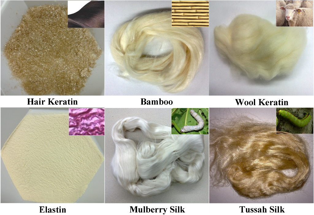
그림은 일반적으로 단백질 기반의 물질을 설계하는 다른 웜 종의 실크를 포함하여 우리의 실험실에서 사용되는 다양한 구조 단백질의 1 원시 자료, 머리와 양모, 다른 조직에서 elastins 및 다양한 식물성 단백질에서 케라틴.

Protocol

단백질 상호 작용의 1 예측 단백질 분자의 분석의 bioinformatics 생명 공학 정보 웹 사이트에 대한 국립 센터 (www.ncbi.nlm.nih.gov/protein/)를 방문하고, 합금 연구에 사용되는 단백질의 이름을 검색 할 수 있습니다. 주 :이 예에서는,이 단백질이 사용되었다 : 야생 tussah 실크 피브로인 인 단백질 A, 및 국내 뽕나무 실크 피브로인 단백질 인 B를,. 단백질의 경우, 아미노산 서열은 "[Antheraea per…

Representative Results

(단백질 및 단백질 B 사이 예) 전형적인 단백질 – 단백질 상호 작용은 전하 – 전하 (정전) 명소, 수소 결합 형성, 소수성 – 친수성 상호 작용뿐만 아니라, 다이폴, 용매, 카운터 이온 및 특정 사이 엔트로피 효과가 포함되어있을 이 단백질의 도메인 (그림 2) 3. 따라서, 근본적으로, 우리가 계산 시뮬레이션에 의한 이러한 상호 작용의 효과를 예측할 수있다. <p class="jove_cont…

Discussion

"합금"단백질 생산 시스템에서 가장 중요한 절차의 하나가 블렌딩 된 단백질의 상용 성을 검증하는 것이다. 그렇지 않으면, 안정된 특성과 동조하지 않고 단지 비혼 화성 혼합물을 단백질 또는 단백질 합성 시스템이다. 실험 열 분석 방법은, 이러한 목적을 위해 사용될 수 있고, 그들의 합금의 특성을 확인하기 위해. 단백질 – 단백질 상호 작용은 플로리 – 허긴의 격자 "용매"(우세한 …

Declarações

The authors have nothing to disclose.

Acknowledgements

저자는이 연구의 지원 로완 대학 감사합니다. 터프 츠 대학의 XH도 덕분에 박사 데이비드 L. 카플란 및 이전 기술 교육에 대한 NIH P41 조직 공학 정보 센터 (TERC).

Materials

Q100 Differential Scanning Calorimeters (DSC) TA Instruments, New Castle, DE, USA
 
N/A You can use any type of DSC with a software to calculate the heat capacity
SS30T Vacuum Sputtering System  T-M Vacuum Products, Inc., Cinnaminson, NJ, USA N/A With custom built parts; You can use any type of sputtering system to coat
VWR 1415M Vacuum Oven  VWR International, Bridgeport, NJ, USA N/A You can use any type of vacuum oven to physically crosslink the samples

Referências

  1. Rosenbloom, J., et al. Extracellular matrix 4: The elastic fiber. FASEB J. 7, 1208-1218 (1993).
  2. Traub, W., et al. On the molecular structure of collagen. Nature. 221, 914-917 (1969).
  3. Hu, X., et al. Protein-Based Composite Materials. Materials Today. 15, 208-215 (2012).
  4. Hardy, J. G., Scheibel, T. R. Composite materials based on silk proteins. Progress in Polymer Science. 35, 1093-1115 (2010).
  5. Kidoaki, S., et al. Mesoscopic spatial designs of nano- and microfiber meshes for tissue-engineering matrix and scaffold based on newly devised multilayering and mixing electrospinning techniques. Biomaterials. 26, 37-46 (2005).
  6. Teng, W. B., et al. Recombinant silk-elastin like protein polymer displays elasticity comparable to elastin. Biomacromolecules. 10, 3028-3036 (2009).
  7. Foo, C. W. P., Kaplan, D. L. Genetic engineering of fibrous proteins, spider dragline, silk and collagen. Adv Drug Delivery Rev. 54, 1131-1143 (2002).
  8. Hu, X., et al. Charge-Tunable Autoclaved Silk-Tropoelastin Protein Alloys That Control Neuron Cell Responses. Adv. Funct. Mater. 23, 3875-3884 (2013).
  9. Hu, X., et al. Biomaterials derived from silk-tropoelastin protein systems. Biomaterials. 31, 8121-8131 (2010).
  10. Hu, X., et al. The influence of elasticity and surface roughness on myogenic and osteogenic-differentiation of cells on silk-elastin biomaterials. Biomaterials. 32, 8979-8989 (2011).
  11. Hu, X., et al. Biomaterials from ultrasonication-induced silk fibroin-hyaluronic acid hydrogels. Biomacromolecules. 11, 3178-3188 (2010).
  12. Gil, E. S., et al. Swelling behavior and morphological evolution of mixed gelatin/silk fibroin hydrogels. Biomacromolecules. 6, 3079-3087 (2005).
  13. Lu, Q., et al. Green process to prepare silk fibroin/gelatin biomaterial scaffolds. Macromol. Biosci. 10, 289-298 (2010).
  14. Lu, S., et al. Insoluble and flexible silk films containing glycerol. Biomacromolecules. 11, 143-150 (2010).
  15. Mandal, B. B., et al. Silk fibroin/polyacrylamide semi-interpenetrating network hydrogels for controlled drug release. Biomaterials. 30, 2826-2836 (2009).
  16. Yeo, I. S., et al. Collagen-based biomimetic nanofibrous scaffolds, preparation and characterization of collagen/silk fibroin bicomponent nanofibrous structures. Biomacromolecules. 9, 1106-1116 (2008).
  17. Holst, J., et al. Substrate elasticity provides mechanical signals for the expansion of hemopoietic stem and progenitor cells. Nat. Biotechnol. 28, 1123-1128 (2010).
  18. Omenetto, F. G., Kaplan, D. L. New Opportunities for an Ancient Material. Science. 329, 528-531 (2010).
  19. Qin, G., et al. Mechanism of resilin elasticity. Nature Communications. 3, 1003 (2012).
  20. Rockwood, D. N., et al. Materials fabrication from Bombyx mori silk fibroin. Nat. Protocols. 6, 1612-1631 (2011).
  21. Wise, S. G., et al. Engineered tropoelastin and elastin-based biomaterials. Adv Protein Chem Struct Biol. 78, 1-24 (2009).
  22. Amsden, J. J., et al. Rapid nanoimprinting of silk fibroin films for biophotonic applications. Adv. Mater. 22, 1746-1749 (2010).
  23. Lawrence, B. D., et al. Silk film biomaterials for cornea tissue engineering. Biomaterials. 30, 1299-1308 (2009).
  24. Kim, D. H., et al. Dissolvable films of silk fibroin for ultrathin conformal bio-integrated electronics. Nat. Mater. 9, 511-517 (2010).
  25. Zhang, J., et al. Stabilization of vaccines and antibiotics in silk and eliminating the cold chain. Proc Natl Acad Sci U S A. 109, 11981-11986 (2012).
  26. Pritchard, E. M., et al. Effect of silk protein processing on drug delivery from silk films. Macromolecular Bioscience. 13, 311-320 (2013).
  27. Lammel, A. S., et al. Controlling silk fibroin particle features for drug delivery. Biomaterials. 31, 4583-4591 (2010).
  28. Urry, D. W. Physical chemistry of biological free energy transduction as demonstrated by elastic protein-based polymers. J Phys Chem B. 101, 11007-11028 (1997).
  29. Shao, Z., Vollrath, F. Materials: Surprising strength of silkworm silk. Nature. 418, 741-741 (2002).
  30. Jin, H. J., Kaplan, D. L. Mechanism of silk processing in insects and spiders. Nature. 424, 1057-1061 (2003).
  31. Hu, X., et al. Determining Beta-Sheet Crystallinity in Fibrous Proteins by Thermal Analysis and Infrared Spectroscopy. Macromolecules. 39, 6161-6170 (2006).
  32. Hu, X., et al. Dynamic Protein-Water Relationships during β-Sheet Formation. Macromolecules. 41, 3939-3948 (2008).
  33. Hu, X., et al. Microphase separation controlled beta-sheet crystallization kinetics in fibrous proteins. Macromolecules. 42, 2079-2087 (2009).
  34. Cebe, P., et al. Beating the Heat – Fast Scanning Melts Beta Sheet Crystals. Scientific Reports. 3, 1130 (2013).
  35. Pyda, M., et al. Heat Capacity of Silk Fibroin Based on the Vibrational Motion of Poly(amino acid)s in the Presence and Absence of Water. Macromolecules. 41, 4786-4793 (2008).
  36. Buxton, G. A., et al. A lattice spring model of heterogeneous materials with plasticity. Model. Simul. Mater. Sci. Eng. 9, 485-497 (2001).
  37. Buxton, G. A., Balazs, A. C. Modeling the dynamic fracture of polymer blends processed under shear. Phys. Rev. B. 69, 054101 (2004).
  38. Kolmakov, G. V., et al. Harnessing labile bonds between nanogel particles to create self-healing materials. ACS Nano. 3, 885-892 (2009).
  39. Duki, S. F., et al. Modeling the nanoscratching of self-healing materials. J. Chem. Phys. 134, 084901 (2011).
  40. Bell, G. I. Models for the specific adhesion of cells to cells. Science. 200, 618-627 (1978).
  41. Bell, G. I., et al. Cell adhesion. Competition between nonspecific repulsion and specific bonding. Biophys. J. 45, 1051-1064 (1984).
  42. Wang, Q., et al. Effect of various dissolution systems on the molecular weight of regenerated silk fibroin. Biomacromolecules. 14, 285-289 (2013).
  43. Wray, L. S., et al. Effect of processing on silk-based biomaterials: reproducibility and biocompatibility. J Biomed Mater Res B Appl Biomater. 99, 89-101 (2011).
  44. Lawrence, B. D., et al. Silk film culture system for in vitro analysis and biomaterial design. J. Vis. Exp. (62), e3646 (2012).
  45. Hu, X., et al. Regulation of Silk Material Structure by Temperature-Controlled Water Vapor Annealing. Biomacromolecules. 12, 1686-1696 (2011).
  46. Yucel, T., et al. Vortex-induced injectable silk fibroin hydrogels. Biophys J. 97, 2044-2050 (2009).
  47. Yucel, T., et al. Non-equilibrium silk fibroin adhesives. J Struct Biol. 170, 406-412 (2010).
  48. Flory, P. J. . Principles of polymer chemistry. , (1953).
  49. Chen, H., et al. Thermal properties and phase transitions in blends of Nylon-6 with silk fibroin. J Therm Anal Calorim. 93, 201-206 (2008).
  50. Scabarozi, T. H., et al. Epitaxial growth and electrical-transport properties of Ti7Si2C5 thin films synthesized by reactive sputter deposition. Scripta Materialia. 65, 811-814 (2011).
  51. Tao, H., et al. Silk materials-a road to sustainable high technology. Adv Mater. 24, 2824-2837 (2012).
  52. Annabi, N., et al. Cross-linked open-pore elastic hydrogels based on tropoelastin, elastin and high pressure CO2. Biomaterials. 31, 1655-1665 (2010).
  53. Moll, R., et al. The human keratins: biology and pathology. Histochem Cell Biol. 129, 705-733 (2008).
check_url/pt/50891?article_type=t

Play Video

Citar este artigo
Hu, X., Duki, S., Forys, J., Hettinger, J., Buchicchio, J., Dobbins, T., Yang, C. Designing Silk-silk Protein Alloy Materials for Biomedical Applications. J. Vis. Exp. (90), e50891, doi:10.3791/50891 (2014).

View Video