设计丝绸蚕丝蛋白合金生物医用材料
Summary
勾兑是一种有效的方法来生成生物材料具有广泛的性能和功能相结合的。通过预测不同的天然丝蛋白之间的分子相互作用,新丝的丝蛋白合金平台具有可调的机械弹性,电响应,光学透明性,化学加工性,生物降解性,或热稳定性可以被设计。
Abstract
纤维蛋白显示已用于在生物医学领域的各种应用,例如生物传感器,纳米医学,组织再生,和药物递送不同的序列和结构。设计基于这些蛋白之间的分子级材料相互作用将有助于产生新的多功能蛋白生物材料的合金具有可调性。这种合金材料系统也由于材料的生物降解性,生物相容性,并在体内可维持提供相比于传统的合成聚合物的优点。本文所用的野生柞蚕丝( 柞蚕 )和国内桑蚕丝( 家蚕 )的蛋白质混合物,例如,提供有关这些主题,包括如何计算方法,如何产生蛋白质合金预测蛋白质-蛋白质相互作用有用的协议解决方案,如何通过热分析验证合金系统,以及如何制造可变合金材料包括光学材料,具有衍射光栅,电子材料用的电路的涂层,以及用于药物释放和递送的药物的材料。这些方法可以提供重要信息,根据不同的蛋白质的合金设计新一代多功能生物材料。
Introduction
大自然创造战略,以利用产生结构蛋白数量有限可调,多功能的生物基质。例如,弹性蛋白和胶原蛋白总是一起使用的体内 ,以提供所需的特定组织1,2的可调节的优点和功能。这里的关键策略是混合。共混包括混合蛋白质与特定的比例,是一个技术的方法来生成具有可调和不同性质3-5简单的材料系统。用合成的工程化策略6,7相比,共混还可以提高材料的均匀性和对材料的过程中,由于操作方便8-16的能力。因此,多功能的设计,生物相容性蛋白质合金材料是医学研究的一个新兴领域。此技术也将提供的天然蛋白基质的影响系统的知识上都在维生素细胞和组织的功能RO和体内 10,17。通过优化不同蛋白质之间的分子的接口,以蛋白质为基础的合金材料可以包括一系列的生理功能,例如在不同温度下的热稳定性,弹性,以支持不同的组织中,电灵敏度的可变机构,和光学性能用于角膜组织再生3, 18-27。这些研究成果将提供一个新的蛋白质材料平台在生物医学科学领域有直接关系的调谐组织修复和疾病治疗,并进一步导致他们在那里新的治疗和诊断功能,可以预见3可生物降解的植入装置。
许多天然结构蛋白具有关键物理和生物活性性质,可以利用作为候选生物材料矩阵。不同虫种丝绸,角蛋白来自不同组织的毛发和羊毛,弹性蛋白和胶原蛋白,并各种植物蛋白是一些用于设计变量蛋白基材料( 图1)18-27中最常见的结构蛋白。在一般情况下,这些蛋白可形成不同的分子的二级结构( 例如 ,β片层为丝绸,或者用于角蛋白螺旋线圈)由于其独特的重复性的一级氨基酸序列,3,28-35。这些功能推动形成自组装的宏观结构与独特的功能,在生物界面提示它们作为生物高分子材料的珍贵资源。在这里,两种结构蛋白被用于(野生柞蚕丝和家养桑蚕丝,例如,蛋白B蛋白A),以证明生产各种蛋白质合金生物材料的通用协议。演示的协议包括第1部分:蛋白质相互作用的预测和模拟,第2部分:生产蛋白质合金的解决方案,第3部分:蛋白质合金制造系统以及用于光,电,和制药应用。
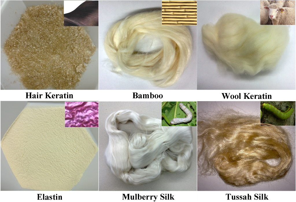
图是在我们的实验室中设计的基于蛋白质的材料,包括来自不同虫物种的丝绸常用的各种结构蛋白1,原料,角蛋白从头发和羊毛,从不同组织中弹性蛋白和各种植物的蛋白质。
Protocol
Representative Results
Discussion
一种生产“合金”蛋白系统中最关键的步骤是,以验证混合蛋白质的混溶性。否则,它是唯一的一个不混溶的蛋白混合物或蛋白质没有稳定和调谐性能的复合系统。实验热分析法可用于该目的,并确认其合金的特性。蛋白质-蛋白质相互作用可以根据弗洛里-哈金斯的晶格模型48为“溶剂”(主要的蛋白成分)之间的相互作用和“溶质”(次要蛋白成分)来查看。基于该模型中,“溶剂”和?…
Declarações
The authors have nothing to disclose.
Acknowledgements
作者感谢罗文大学支持本研究。 XH也感谢大卫L卡普兰博士塔夫茨大学和美国国立卫生研究院P41组织工程资源中心(TERC)在以前的技 术培训。
Materials
| Q100 Differential Scanning Calorimeters (DSC) | TA Instruments, New Castle, DE, USA |
N/A | You can use any type of DSC with a software to calculate the heat capacity |
| SS30T Vacuum Sputtering System | T-M Vacuum Products, Inc., Cinnaminson, NJ, USA | N/A | With custom built parts; You can use any type of sputtering system to coat |
| VWR 1415M Vacuum Oven | VWR International, Bridgeport, NJ, USA | N/A | You can use any type of vacuum oven to physically crosslink the samples |
Referências
- Rosenbloom, J., et al. Extracellular matrix 4: The elastic fiber. FASEB J. 7, 1208-1218 (1993).
- Traub, W., et al. On the molecular structure of collagen. Nature. 221, 914-917 (1969).
- Hu, X., et al. Protein-Based Composite Materials. Materials Today. 15, 208-215 (2012).
- Hardy, J. G., Scheibel, T. R. Composite materials based on silk proteins. Progress in Polymer Science. 35, 1093-1115 (2010).
- Kidoaki, S., et al. Mesoscopic spatial designs of nano- and microfiber meshes for tissue-engineering matrix and scaffold based on newly devised multilayering and mixing electrospinning techniques. Biomaterials. 26, 37-46 (2005).
- Teng, W. B., et al. Recombinant silk-elastin like protein polymer displays elasticity comparable to elastin. Biomacromolecules. 10, 3028-3036 (2009).
- Foo, C. W. P., Kaplan, D. L. Genetic engineering of fibrous proteins, spider dragline, silk and collagen. Adv Drug Delivery Rev. 54, 1131-1143 (2002).
- Hu, X., et al. Charge-Tunable Autoclaved Silk-Tropoelastin Protein Alloys That Control Neuron Cell Responses. Adv. Funct. Mater. 23, 3875-3884 (2013).
- Hu, X., et al. Biomaterials derived from silk-tropoelastin protein systems. Biomaterials. 31, 8121-8131 (2010).
- Hu, X., et al. The influence of elasticity and surface roughness on myogenic and osteogenic-differentiation of cells on silk-elastin biomaterials. Biomaterials. 32, 8979-8989 (2011).
- Hu, X., et al. Biomaterials from ultrasonication-induced silk fibroin-hyaluronic acid hydrogels. Biomacromolecules. 11, 3178-3188 (2010).
- Gil, E. S., et al. Swelling behavior and morphological evolution of mixed gelatin/silk fibroin hydrogels. Biomacromolecules. 6, 3079-3087 (2005).
- Lu, Q., et al. Green process to prepare silk fibroin/gelatin biomaterial scaffolds. Macromol. Biosci. 10, 289-298 (2010).
- Lu, S., et al. Insoluble and flexible silk films containing glycerol. Biomacromolecules. 11, 143-150 (2010).
- Mandal, B. B., et al. Silk fibroin/polyacrylamide semi-interpenetrating network hydrogels for controlled drug release. Biomaterials. 30, 2826-2836 (2009).
- Yeo, I. S., et al. Collagen-based biomimetic nanofibrous scaffolds, preparation and characterization of collagen/silk fibroin bicomponent nanofibrous structures. Biomacromolecules. 9, 1106-1116 (2008).
- Holst, J., et al. Substrate elasticity provides mechanical signals for the expansion of hemopoietic stem and progenitor cells. Nat. Biotechnol. 28, 1123-1128 (2010).
- Omenetto, F. G., Kaplan, D. L. New Opportunities for an Ancient Material. Science. 329, 528-531 (2010).
- Qin, G., et al. Mechanism of resilin elasticity. Nature Communications. 3, 1003 (2012).
- Rockwood, D. N., et al. Materials fabrication from Bombyx mori silk fibroin. Nat. Protocols. 6, 1612-1631 (2011).
- Wise, S. G., et al. Engineered tropoelastin and elastin-based biomaterials. Adv Protein Chem Struct Biol. 78, 1-24 (2009).
- Amsden, J. J., et al. Rapid nanoimprinting of silk fibroin films for biophotonic applications. Adv. Mater. 22, 1746-1749 (2010).
- Lawrence, B. D., et al. Silk film biomaterials for cornea tissue engineering. Biomaterials. 30, 1299-1308 (2009).
- Kim, D. H., et al. Dissolvable films of silk fibroin for ultrathin conformal bio-integrated electronics. Nat. Mater. 9, 511-517 (2010).
- Zhang, J., et al. Stabilization of vaccines and antibiotics in silk and eliminating the cold chain. Proc Natl Acad Sci U S A. 109, 11981-11986 (2012).
- Pritchard, E. M., et al. Effect of silk protein processing on drug delivery from silk films. Macromolecular Bioscience. 13, 311-320 (2013).
- Lammel, A. S., et al. Controlling silk fibroin particle features for drug delivery. Biomaterials. 31, 4583-4591 (2010).
- Urry, D. W. Physical chemistry of biological free energy transduction as demonstrated by elastic protein-based polymers. J Phys Chem B. 101, 11007-11028 (1997).
- Shao, Z., Vollrath, F. Materials: Surprising strength of silkworm silk. Nature. 418, 741-741 (2002).
- Jin, H. J., Kaplan, D. L. Mechanism of silk processing in insects and spiders. Nature. 424, 1057-1061 (2003).
- Hu, X., et al. Determining Beta-Sheet Crystallinity in Fibrous Proteins by Thermal Analysis and Infrared Spectroscopy. Macromolecules. 39, 6161-6170 (2006).
- Hu, X., et al. Dynamic Protein-Water Relationships during β-Sheet Formation. Macromolecules. 41, 3939-3948 (2008).
- Hu, X., et al. Microphase separation controlled beta-sheet crystallization kinetics in fibrous proteins. Macromolecules. 42, 2079-2087 (2009).
- Cebe, P., et al. Beating the Heat – Fast Scanning Melts Beta Sheet Crystals. Scientific Reports. 3, 1130 (2013).
- Pyda, M., et al. Heat Capacity of Silk Fibroin Based on the Vibrational Motion of Poly(amino acid)s in the Presence and Absence of Water. Macromolecules. 41, 4786-4793 (2008).
- Buxton, G. A., et al. A lattice spring model of heterogeneous materials with plasticity. Model. Simul. Mater. Sci. Eng. 9, 485-497 (2001).
- Buxton, G. A., Balazs, A. C. Modeling the dynamic fracture of polymer blends processed under shear. Phys. Rev. B. 69, 054101 (2004).
- Kolmakov, G. V., et al. Harnessing labile bonds between nanogel particles to create self-healing materials. ACS Nano. 3, 885-892 (2009).
- Duki, S. F., et al. Modeling the nanoscratching of self-healing materials. J. Chem. Phys. 134, 084901 (2011).
- Bell, G. I. Models for the specific adhesion of cells to cells. Science. 200, 618-627 (1978).
- Bell, G. I., et al. Cell adhesion. Competition between nonspecific repulsion and specific bonding. Biophys. J. 45, 1051-1064 (1984).
- Wang, Q., et al. Effect of various dissolution systems on the molecular weight of regenerated silk fibroin. Biomacromolecules. 14, 285-289 (2013).
- Wray, L. S., et al. Effect of processing on silk-based biomaterials: reproducibility and biocompatibility. J Biomed Mater Res B Appl Biomater. 99, 89-101 (2011).
- Lawrence, B. D., et al. Silk film culture system for in vitro analysis and biomaterial design. J. Vis. Exp. (62), e3646 (2012).
- Hu, X., et al. Regulation of Silk Material Structure by Temperature-Controlled Water Vapor Annealing. Biomacromolecules. 12, 1686-1696 (2011).
- Yucel, T., et al. Vortex-induced injectable silk fibroin hydrogels. Biophys J. 97, 2044-2050 (2009).
- Yucel, T., et al. Non-equilibrium silk fibroin adhesives. J Struct Biol. 170, 406-412 (2010).
- Flory, P. J. . Principles of polymer chemistry. , (1953).
- Chen, H., et al. Thermal properties and phase transitions in blends of Nylon-6 with silk fibroin. J Therm Anal Calorim. 93, 201-206 (2008).
- Scabarozi, T. H., et al. Epitaxial growth and electrical-transport properties of Ti7Si2C5 thin films synthesized by reactive sputter deposition. Scripta Materialia. 65, 811-814 (2011).
- Tao, H., et al. Silk materials-a road to sustainable high technology. Adv Mater. 24, 2824-2837 (2012).
- Annabi, N., et al. Cross-linked open-pore elastic hydrogels based on tropoelastin, elastin and high pressure CO2. Biomaterials. 31, 1655-1665 (2010).
- Moll, R., et al. The human keratins: biology and pathology. Histochem Cell Biol. 129, 705-733 (2008).
