This is a cadaveric study investigating the landmarks for the posterior approach for irrigation and debridement of the psoas abscess. The interval between the transverse processes (TP) was used to access the substance of the psoas muscle.
This method focuses on outlining a safe zone for irrigation and debridement of a psoas abscess through a posterior approach. Initially, an anterior approach to the spine was performed to ensure that the anterior longitudinal ligament and the psoas muscle could be visualized. All the abdominal organs were removed. Subsequently, a posterior approach was performed to remove the paraspinal muscles from L1–L5. The transverse processes, pars interarticularis and lamina of L1–L5 were identified. The exiting nerve root was identified between the transverse processes and followed into the substance of the psoas muscle. Using the anterior and posterior approach, the lumbar plexus was isolated from the substance of the psoas muscle. Before and after various steps of dissection, digital photographs were obtained. These images were uploaded into ImageJ and multiple measurements, including the distance between the lateral superior and inferior tip of each TP to the most lateral region of the plexus, the distance between the lateral superior and inferior tip of the TP to the lateral edge of the psoas, and the width of the lumbar plexus were recorded. The safe zone for entering the substance of the psoas muscle was defined between the lateral edge of the psoas muscle and the lateral edge of the lumbar plexus. The relationship of this interval to the tip of the transverse process at each level was measured and reported.
A psoas abscess (PA) is a rare diagnosis reported in 0.4–1 per 10,000 admissions to the hospital1,2. A PA can be caused by hematogenous spread, lymphatic spread from a distant infectious site, or by contiguous spread from adjacent organs3. Currently, percutaneous drainage under real-time computed tomography is a safe and first-line treatment for a PA4. Dietrich et al. found that it has a higher success rate compared to open surgery, offering a lower mortality rate and shorter hospital length of stay2. However, Tabrizian et al. found that 44% of the patients treated with percutaneous drainage underwent open surgery to eradicate the infection5.
Given the rate of open surgeries performed for treatment of psoas abscesses, the aim of the method developed in this study is to find a "safe zone" for irrigation and debridement (I&D) of a psoas abscess via a dorsal approach. An initial ventral approach is taken to identify and isolate key anatomical structures, including the transverse processes (TP), psoas muscle, and lumbar plexus (LP). A dorsal approach is then taken to further isolate the TP and LP from the surrounding structures. Throughout the various steps of dissection, photographs are obtained. Once the dissection is complete, the images are uploaded to ImageJ (1.48v, National Institute of Health) and several measurements are taken to identify the relationship between the TP, LP, and psoas muscle. Finally, using the measurements obtained, a "safe zone" for entering the substance of the psoas and minimizing the risk of lumbar plexus injury is calculated. To our knowledge, this is the first work that uses the transverse processes as reference points for the purpose of finding a safe zone for I&D of a psoas abscess.
The use of deceased human specimens, for the purposes of developing this method, has been approved by the WellStar Research Institute's institutional review board and conforms with the ethical guidelines established by the institution's ethics committee. The specimens used in this method were all formalin-fixed, transected at the T12 vertebral body, and had their abdominal organs and viscera removed.
1. Preparation of the cadaveric specimen and surgical field
2. Isolation and identification of key anatomical structures using a ventral approach
3. Further isolation of the transverse processes and obtaining digital photographs for analysis
4. Calibrating and taking measurements using ImageJ software
5. Calculating the "safe zone" for maximal I&D
6. Calculating the safe zone for maximal irrigation and debridement using the measurements obtained in Section 5
Eleven formalin-fixed cadavers were included, with an average age of 80.5 years6. Two cadavers had damaged lumbar vertebrae on the right side and were removed. Thus, a total of 20 measurements were included, 13 female and 7 male. The medial border of the safe zone, defined by the most lateral aspect of the lumbar plexus, was found to be approximately 1 cm medial to the tip of the TP at L1–L3, but narrowed down to approximately 6 and 1 mm at L4 and L5, respectively. The medial border of the safe zone was then subtracted by the distances of the cranial and caudal tip of each TP to the lateral edge of the psoas muscle at each level. This calculation gave us the medial to lateral area of safe debridement. The average area of the safe zone for debridement, at all levels of the lumbar spine, was found to be between 4 and 8 mm.
The area for maximum medial to lateral irrigation and debridement in the coronal plane was determined by measuring the distance from the superior and inferior tips of the TP to the lateral edge of the psoas muscle at the corresponding vertebral level. On average, the maximal extent of lateral debridement one can perform is as follows: 5.5 and 5.7 mm medial to the inferior and superior tips of the TP at L1, 4.7 and 5.1 mm medial at L2, 1.8 and 2.5 mm medial at L3, 0 and 0.4 mm medial at L4, and 3.8 and 3.7 mm lateral at L5. In the sagittal plane, the lumbar plexus was approximately 15–20 mm anterior to the TP. (Table 1).
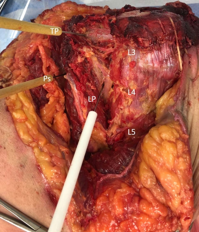
Figure 1: Cadaveric image of an anterior approach dissection. The TP, LP and psoas are clearly defined and used as the reference points for measurements. TP = transverse process; LP = lumbar plexus; Ps = lateral edge of psoas; L3, L4, L5 = lumbar vertebrae. Please click here to view a larger version of this figure.
Lumbar Vertebra | ||||||
Measurement | L1 | L2 | L3 | L4 | L5 | |
Lateral superior tip of TP to lateral edge of psoas | -5.7 ± 1.92 | -5.11 ± 1.84 | -2.52 ± 2.63 | -0.42 ± 2.39 | 3.70 ± 1.60 | |
Lateral inferior tip of TP to lateral edge of psoas | -5.49 ± 2.02 | -4.70 ± 1.71 | -1.84 ± 2.56 | -0.03 ± 2.31 | 3.77 ± 1.57 | |
Lateral superior tip of TP to lateral edge of LP | -9.3 ± 1.94 | -11.75 ± 1.59 | -10.47 ± 2.90 | -6.64 ± 2.53 | -1.01 ± 2.31 | |
Lateral inperior tip of TP to lateral edge of LP | -9.2 ± 2.06 | -11.71 ± 1.54 | -9.8 ± 2.47 | 6.24 ± 2.43 | -0.89 ± 2.40 | |
Lateral edge of LP to lateral edge of psoas calculated using the lateral superior tip of the TP | 3.96 ± 0.98 | 6.64 ± 1.17 | 7.94 ± 2.00 | 6.22 ±1.28 | 4.71 ± 1.38 | |
Lateral edge of LP to lateral edge of psoas calculated using the lateral inferior tip of the TP | 4.03 ± 1.05 | 7.0 ± 1.15 | 7.96 ± 2.00 | 6.21 ± 1.77 | 4.66 ± 1.59 | |
Lateral superior tip of the TP to the LP in the saggital plane | 17.89 ± 2.72 | 19.08 ± 1.93 | 19.50 ± 3.12 | 16.26 ± 2.69 | 14.97 ± 1.90 | |
Lateral inferior tip of the TP to the LP in the saggital plane | 17.84 ± 3.03 | 18.65 ± 1.60 | 19.19 ± 3.06 | 16.27 ± 2.93 | 14.92 ± 1.85 |
Table 1: Mean value in millimeters of 20 sets of measurements, 13 female and 7 male. Negative values indicate that the measurement is medial to the tip of the TP. The distance of the lateral edge of the psoas to the most lateral region of the LP defines the safe zone for medial to lateral debridement. The distance from the tip of the TP to the LP in the sagittal plane defines the safe zone for debridement in the sagittal plane.
The most critical steps for finding the safe zone for irrigation and debridement of a psoas muscle abscess through a dorsal approach are 1) careful blunt dissection of the lumbar plexus during the ventral and dorsal approach; 2) preservation of the transverse processes during their isolation, as they can be fragile, for better visualization; and 3) calibrating the ImageJ software with the surgical ruler and taking careful measurements to elucidate the relationship between the TP, LP, and psoas muscle.
Surgical instruments used for dissection can be changed according to personal preference; it is crucial however to perform a careful and thorough dissection of the lumbar plexus and to ensure no damage is done to the transverse processes. When taking the photographs, make sure that the ruler is parallel to the field of dissection and that the pictures taken are of high quality. Use of both the cranial and caudal tip of the TP should be included for precise measurements.
A significant limitation of this study is a high probability of psoas muscle atrophy in the cadaveric specimens used. The average age of the specimens used was 80.5 years and cross-sectional area of skeletal muscle has been shown to be negatively correlated with age7. In addition, the cadavers used were formalin-fixed, leading to less tissue pliability as compared to the natural tissue that would be found in a living human being needing an operation. Another limitation is that, because their course can be highly variable after exiting the neuroforamina, the sensory nerves were not included when measuring the lumbar plexus8. Care should still be taken to avoid these nerves when performing I&D of a psoas abscess.
This method focuses on different measurements than previous studies, which may account for the homogenous distance found from the tip of each TP to the lateral edge of the psoas muscle. Previous methods studying psoas anatomy, such as in the studies by Spivak et al., Reid et al., Hanson et al., and Ilayperuma et al., directly measure the width of the psoas muscle9,10,11,12. Compared to the study by Spivak et al., for example, the range of measurements found by measuring from the TP to the psoas using this method was less than the range found when measuring the psoas muscle directly. An increase of 5 mm from L1–L4 and 8 mm from L1–L5 was found when measuring from the TP to the psoas; with a direct measurement, Spivak et. al. found an increase from 15.9 mm at L2–L3 to 23.3 mm at L3–L4 and 28.7 mm at L4–L59. Analysis of the data obtained indicates that the psoas muscle is generally medial to the TP and that aiming laterally would not allow access unless you are at the level of L5. Irrigation and debridement less than 1.5 cm past the transverse process in the sagittal plane should allow for avoidance of the lumbar plexus. However, pathologically, a psoas abscess may expand beyond the normal anatomical margins and a more lateral approach may be possible. MRI is recommended to clearly define the depth and medial to lateral extent of the psoas abscess and a future radiological study would be beneficial to further assess the findings of this method.
Using the measurements found in this study as a guide, one can determine the ability to safely perform irrigation and debridement of a psoas abscess, through a dorsal spinal approach, by staying within the confines of the safe zone at each level and reduce the risk lumbar plexus injury.
The authors have nothing to disclose.
The authors would like to acknowledge all those who donate their bodies for research and keep impacting others after they have gone.
Blunt ended dissecting scissors | To cover the specimens | ||
Camera | Sony a7 III | ILCE-7 | |
Cobb elevator | Sklar | 40-6950 | For isolation of TP and general dissection |
Dissection table | |||
Formalin fixed cadavers | Restore Life USA | N/A | Transected at T12 with abdominal organs removed |
Hemostats | |||
Retractors | For blunt dissection | ||
Rongeur | Sklar | 40-4085 | |
Scissors | Sklar | 15-2555 | |
Surgical absorbent pads | Placed under the cadaver to absorb fluids | ||
Surgical blades/scalpels | Dynarex | 4110 | |
Surgical drape/blanket | For blunt dissection | ||
Surgical gauze sponges | |||
Surgical lights | |||
Surgical Ruler | Aspen Surgical Products | 42182702 | |
Tissue forceps | |||
Tool tray |