“发光法”的分类检测细菌病原体的记者噬菌体
Summary
确定优先的细菌病原体的一个简单的方法是使用基因工程的记者噬菌体。这些记者的噬菌体,这是具体到特定主机物种,的宿主细胞迅速传导一个发光的信号响应能力。在这里,我们描述了记者的噬菌体检测<em>鼠疫耶尔森氏菌</em>。
Abstract
鼠疫耶尔森氏菌和炭疽芽孢杆菌 ,鼠疫,炭疽病原体,分别为1 A类的细菌病原体。虽然这两种疾病的“自然的发生,是目前比较少见的,使用这些病原体作为一个生物武器的恐怖团体的可能性是真实的。由于固有的传染性疾病的,快速的临床过程,和死亡率高,关键是快速检测爆发。因此,提供快速检测和诊断的方法,是必须立即执行,以确保公众健康的措施,并启动危机管理。
重组记者噬菌体可能会提供一个快速检测Y 的具体做法鼠疫杆菌和B 。 炭疽。疾病控制和预防中心目前使用的古典噬菌体裂解试验证实这些2-4细菌病原体识别。这些检测利用自然产生的噬菌体,这是具体和裂解细菌主机优势。隔夜后在特定的噬菌体的存在耕地细菌生长,形成斑块(细菌裂解)提供了一个积极识别细菌的目标。虽然这些实验是强大的,他们遭受三个缺点:1)他们是实验室的基础; 2)他们需要从怀疑样品的细菌分离和种植,和3),他们采取24-36 h内完成。为了解决这些问题,基因工程重组“轻标签”记者噬菌体哈维氏弧菌luxAB基因融入Y 的基因组鼠疫杆菌和B 。 炭疽特定的噬菌体5-8。由此产生的luxAB记者噬菌体是能迅速检测其具体目标(在几分钟之内),并巧妙地赋予一个发光的受体细胞的表型。更重要的是,检测,获得与耕地的受体细胞或模拟感染的临床标本7。
出于演示的目的,我们在这里描述为已知Y。噬菌体介导的检测方法鼠疫隔离使用噬菌体诊断的疾病预防控制中心鼠疫噬菌体ΦA11226,7(图1)建造一个luxAB记者。一个细微的修改(例如改变生长温度和媒体),类似的方法,可用于检测B 的炭疽杆菌菌株使用的B.炭疽记者噬菌体Wβ:luxAB 8。该方法描述了一个biolumescent表型的噬菌体介导的转耕地 Y。 鼠疫随后使用微孔板光度计测量细胞。传统的噬菌体裂解试验这种方法的主要优点是易于使用,快速的结果,并且能够同时测试多个样本,在96孔微孔板格式。

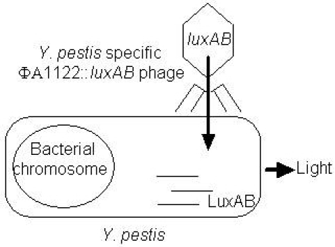
图1。检测原理图。噬菌体混合样品,噬菌体感染的细胞,luxAB表示,和细胞bioluminesces。样品的处理是没有必要的;噬菌体和细胞混合在一起,其后轻。
Protocol
Discussion
这种方法证明了记者的噬菌体的能力,以快速检测Y 。因为记者噬菌体的菌可以发光信号转导的一个培养Y 。 鼠疫细胞在20分钟后,噬菌体此外。记者噬菌体也能够直接检测Y。鼠疫在临床上的矩阵,没有隔离的前提条件和后续培养 7 。相比标准的噬菌体裂解试验,一般需要48小时完成,这大大降低了检测时间。
以往的研究表明,ΦA1122?…
Disclosures
The authors have nothing to disclose.
Acknowledgements
这项研究是支持小企业创新研究计划,美国国立卫生研究院(NIAID的,1R43AI082698 – 01)和美国农业部国家食品和农业研究所(NifA蛋白,2009-33610-20028)。
Materials
| Name of the reagent | Company | Catalogue number |
|---|---|---|
| Difco LB agar, Miller | VWR | 90003-346 |
| Difco LB broth, Miller | VWR | 90003-350 |
| 17 x 100 mm culture tubes | USA Scientific | 1485-0810 |
| n-Decanal | Sigma | D7384 |
| Veritas microplate luminometer | Turner Biosystems | 9100-001 |
| Microlite microtiter 96-well plate | VWR | 62402-984 |
References
- Darling, R. G., Catlett, C. L., Huebner, K. D., Jarrett, D. G. Threats in bioterrorism. I: CDC category A agents. Emerg Med Clin North Am. 20, 273-309 (2002).
- Chu, M. C. . Laboratory manual of plague diagnostic tests.. , (2000).
- . . , (1999).
- Inglesby, T. V. Anthrax as a biological weapon, 2002: updated recommendations for management. JAMA. 287, 2236-2252 (2002).
- Schuch, R., Fischetti, V. A. Detailed genomic analysis of the Wbeta and gamma phages infecting Bacillus anthracis: implications for evolution of environmental fitness and antibiotic resistance. J Bacteriol. 188, 3037-3051 (2006).
- Garcia, E. The genome sequence of Yersinia pestis bacteriophage phiA1122 reveals an intimate history with the coliphage T3 and T7 genomes. J Bacteriol. 185, 5248-5262 (2003).
- Schofield, D. A., Molineux, I. J., Westwater, C. Diagnostic bioluminescent phage for detection of Yersinia pestis. Journal of Clinical Microbiology. 47, 3887-3894 (2009).
- Schofield, D. A., Westwater, C. Phage-mediated bioluminescent detection of Bacillus anthracis. Journal of Applied Microbiology. 107, 468-478 (2009).
- Chu, M. C. . CDC: Basic laboratory protocols for the presumptive identification of Yersinia pestis. , 1-19 (2001).
- Gunnison, J. B., Larson, A., Lazarus, A. S. Rapid differentiation between Pasteurella pestis and Pasteurella pseudotuberculosis by action of bacteriophage. J Infect Dis. 88, 254-255 (1951).
- Lazarus, A. S., Gunnison, J. B. The Action of Pasteurella pestis Bacteriophage on Strains of Pasteurella, Salmonella, and Shigella. J Bacteriol. 53, 705-714 (1947).
- Sergueev, K. V., He, Y., Borschel, R. H., Nikolich, M. P., Filippov, A. A. Rapid and sensitive detection of Yersinia pestis using amplification of plague diagnostic bacteriophages monitored by real-time PCR. PLoS One. 5, e11337-e11337 (2010).

