Small animal imaging techniques allow serial diagnostic examinations and therapeutic interventions in vivo. Recently, the scope of applications has significantly widened and currently includes assessment of colonic tumor development, wound healing and monitoring of inflammation. This protocol illustrates these diverse potential applications of murine endoscopy.
Mouse models are widely used to study pathogenesis of human diseases and to evaluate diagnostic procedures as well as therapeutic interventions preclinically. However, valid assessment of pathological alterations often requires histological analysis, and when performed ex vivo, necessitates death of the animal. Therefore in conventional experimental settings, intra-individual follow-up examinations are rarely possible. Thus, development of murine endoscopy in live mice enables investigators for the first time to both directly visualize the gastrointestinal mucosa and also repeat the procedure to monitor for alterations. Numerous applications for in vivo murine endoscopy exist, including studying intestinal inflammation or wound healing, obtaining mucosal biopsies repeatedly, and to locally administer diagnostic or therapeutic agents using miniature injection catheters. Most recently, molecular imaging has extended diagnostic imaging modalities allowing specific detection of distinct target molecules using specific photoprobes. In conclusion, murine endoscopy has emerged as a novel cutting-edge technology for diagnostic experimental in vivo imaging and may significantly impact on preclinical research in various fields.
Animal models have greatly enriched our understanding of numerous intestinal pathologies. The laboratory mouse (Mus musculus) has emerged as a prime animal model in biomedical research due to its abundant genetic and genomic information and is readily available in transgenic and knockout strains. In addition to enhancing understanding disease pathogenesis, animal models are also importantly used for testing drug candidates as well as preclinical diagnostic or therapeutic interventions. However, despite the variety of mouse models mimicking human disease, many diagnostic and interventional options that are routinely used in patient care are not available for mice. Accordingly, surveillance strategies to monitor the course of murine disease or the effect of therapeutic interventions are often limited to indirect observations or post mortem analyses. While non-invasive procedures exist for monitoring mice vitality like disease activity indices, quantification of weight loss or gain, blood, urine and feces analyses, these are only indirect indicators and are biased by inter-individual variability. Additionally, post mortem analyses prevent longitudinal observations at repetitive time points. Sophisticated imaging techniques to monitor disease activity in mice have only recently been introduced 1,2. Although these imaging techniques allow for repetitive analyses, they only provide a descriptive and often imprecise view on the gut, do not enable direct mucosal visualization or allow diagnostic or therapeutic interventions such as biopsy acquisition or topical and intramucosal application of drug candidates.
Recently, high-resolution endoscopic systems for use in live mice have been developed 3,4. For the first time these endoscopic techniques allow direct visualization of endoluminal colonic disease pathologies such as wound healing or intestinal inflammation providing objective, real-time status allowing longitudinal studies in the same animal at repetitive time points. Aside from allowing repeated biopsies in an individual mouse, endoscopic systems can also be used to therapeutically influence a distinct tumor or localized inflammation by allowing direct application of a substance to the area of interest. Furthermore, as therapeutic and control substances can be delivered directly to the area of interest, this can be performed in the same mouse, excluding inter-individual variability. These systems have now been employed for the assessment of colonic inflammation, wound healing, laparoscopic liver biopsies and orthotopic induction of liver tumors 8 and tumor development using various scoring systems such as the murine endoscopic index of colitis severity (MEICS) 5-7. MEICS consists of five parameters to assess inflammation: thickening of the colon wall, changes of the vascular pattern, presence of fibrin, granularity of the mucosal surface, and stool consistence.
In this protocol we describe the use of rigid endoscopy in murine models of intestinal wound healing, inflammation and colon cancer. First, we demonstrate the endoscopic evaluation of wound healing and colonic inflammation as well as longitudinal assessment of colitis activity and the study of cancerogenesis in the murine colon. Beyond the descriptive use of murine endoscopy, we provide detailed instructions on the use of endoscopic instrumentation to obtain biopsies, and the topical and intramucosal application of different components of interest (e.g., drug candidates or tumor cells). Finally, we demonstrate the use of murine fluorescence endoscopy, which employs sophisticated molecular imaging techniques, in the setting of colorectal tumors.
All animal experiments were approved by the Landesamt für Natur, Umwelt und Verbraucherschutz (LANUV) according to German Animal Protection Law.
1. Materials and Experimental Setup
2. Technical Equipment
3. Anesthesia of Animals
4. Colonoscopy
5. Post-colonoscopy
In vivo monitoring of intestinal wound healing
During routine endoscopy, mucosal wounds were induced mechanically by miniature biopsy forceps with a diameter of 3 French (equals 1 mm; Figure 1A). Subsequently, wound healing was monitored by daily endoscopic examinations and quantified by measurement of the residual wound area using image editing software, e.g., ImageJ (Figure 1B). The individual wound closure over time is expressed by the quotient of actual wound area / initial wound area. For example, at day 3 after wound generation, 41% ± 4.1% of the wound area was recovered, whereas at day 7 the wound was usually completely healed (Figure 1C). Additionally, at the end of the experiment, wounds can be resected for histological evaluation ex vivo. Depicted are representative images of hematoxylin and eosin (H&E)-stained wound beds at day 0 and day 5 (Figure 1D).
Endoscopy-guided intramucosal injection therapy
For intramucosal application of pharmacological agents, a flexible tube (diameter of 0.96 mm) with a cannula fixed to the end (30 G) was introduced to the working channel of the endoscope (Figure 2A). After intramucosal placement of the needle, a maximum of 50 µl was carefully injected. Indicative of successful intramucosal application, lifting of the colonic mucosa can be easily observed macroscopically (Figure 2B,C).
In vivo assessment of experimental colitis
After induction of colitis, mice showed weight loss from day 3 with maximum loss of body weight of 19% occurring at day 7 (Figure 3A). In addition to daily measurement of body weight, disease activity was monitored by repetitive endoscopies and macroscopic quantification of inflammation by the murine endoscopic index of colitis severity (MEICS). In accordance with the loss of body weight, MEICS score was increased at day 7 after DSS start indicating a massive inflammatory damage of the colonic mucosa, which was ameliorated at day 13 (Figure 3B). For ex vivo correlation of histological damage, inflammatory alterations of colonic H&E-stained sections were quantified according to the Dieleman Score 11. At day 7 after DSS start, histological damage was significantly higher in DSS-treated mice compared to controls as reflected by epithelial denudation, mucosal ulcerations as well as increased neutrophil infiltration and was significantly improved at day 13 (Figure 3C, E). In addition, histological evaluation of mucosal biopsies, routinely obtained during endoscopic examinations, corroborated the advanced stage of colitis at day 7 (Figure 3F-H).
Fluorescence endoscopy of colorectal tumors
Approximately 80 days after tumor induction by AOM and three cycles of DSS (Figure 4A), multiple colonic tumors (Figure 4C) as well as macroscopic signs of chronic inflammation such as granulated mucosa (Figure 4B) 10 were observed endoscopically. Histological evaluation of colorectal tumors by H&E staining revealed adenomas with and without high-grade intraepithelial neoplasia. Therefore, the AOM-DSS-model resembles a perfect model to study molecular processes of carcinogenesis 12, as well as to evaluate new diagnostic devices13. Fluorescence imaging targeting specific molecules allows in vivo molecular imaging with ‘photographic methods’ 14,15. To demonstrate feasibility of FE, we used FITC, a widely used fluorochrome. For specific FITC detection, a bandpass filter system combined with the light source provided specific excitation wavelength needed (490 nm; Figure 4D). For accurate detection of FITC-specific emission wavelength (525 nm), a second bandpass filter was interposed between the camera head and the endoscope (Figure 4E). FE without tracer application did not detect any specific signal and no interaction with colonic tissue or fecal autofluorescence (Figure 4F, G). In contrast, immediately after intravenous application of FITC-dextran, the fluorochrome could be observed at the colonic mucosa and may be used for assessment of increased vascularity in regions of chronic inflammation (Figure 4H) as well as malignant mucosa (Figure 4I). Accordingly, quantification of fluorescence intensity by an image editing software showed significantly increased uptake of the fluorochrome within malignant tissue as compared to non-affected colonic mucosa (Figure 4K).
Figure 1. Endoscopic monitoring of epithelial wound healing in vivo as well as quantitative and histological assessment of wound healing. After generation of colonic wounds, wound border and wound closure can be easily detected. The wound area (white arrows) is assessed during daily follow-up endoscopies to quantitatively follow epithelial wound healing (A–C). Ex vivo, wounds were resected and H&E-stained for histological analysis of wound healing (D). Scales are defined by depicted scale bar. Please click here to view a larger version of this figure.
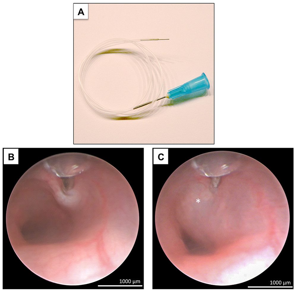
Figure 2. Endoscopy-guided intramucosal injection therapy. Under visual control, the tip of the needle (A) is softly placed into the colonic mucosa and 50 µl of dissolved substances is injected (B). Subsequently, marked mucosal lifting can be recognized (asterisk) without any signs of acute bleeding (C). Scales are defined by depicted scale bar. Please click here to view a larger version of this figure.
Figure 3. Endoscopic evaluation of the course of experimental DSS colitis. The course of colitis was evaluated by changes in body weight, endoscopic examinations as well as histological analysis of inflamed colonic sections and endoscopic biopsies. In line with massive loss of body weight and advanced histological damage at day 7 (A, C, E; magnification 10X), endoscopic examinations and histological evaluation of obtained biopsies depicted signs of severe inflammation (B, D, G, H; magnification 5X and 10X) whereas at day 13 after DSS start inflammatory alterations were significantly ameliorated. Scales are defined by depicted scale bar. Please click here to view a larger version of this figure.
Figure 4. FE of colorectal tumors. After induction of colorectal cancerogenesis by AOM and cyclic DSS administration for 11 weeks (A), white light endoscopy detected granulated mucosa indicative of chronic colitis (B) and numerous endoluminal lesions (C) which were diagnosed as adenomas with high-grade intraepithelial neoplasia by H&E staining ex vivo (G). While visualization of chronic inflammation (H) and tumors (I) was easily possible using FE targeted FITC, FE without tracer application did not allow definitive tumor detection (F, G). Accordingly, quantification of fluorescence intensity was significantly increased within malignant tissue as compared to non-affected colonic mucosa, shown by gray scale profiles (E, F). To switch to the fluorescence mode during white light colonoscopy, a specific bandpass filter is additionally plugged into the cold-light source to provide the specific excitation wavelength (e.g., 490 nm for FITC; D). This filter facilitates switching (white arrow) between white light and fluorescence modes (D). To capture the specific emission wavelength (e.g., 525 nm for FITC), a second bandpass filter is interposed between endoscope and camera head using a bayonet joint (E). Scales are defined by depicted scale bar. Please click here to view a larger version of this figure.
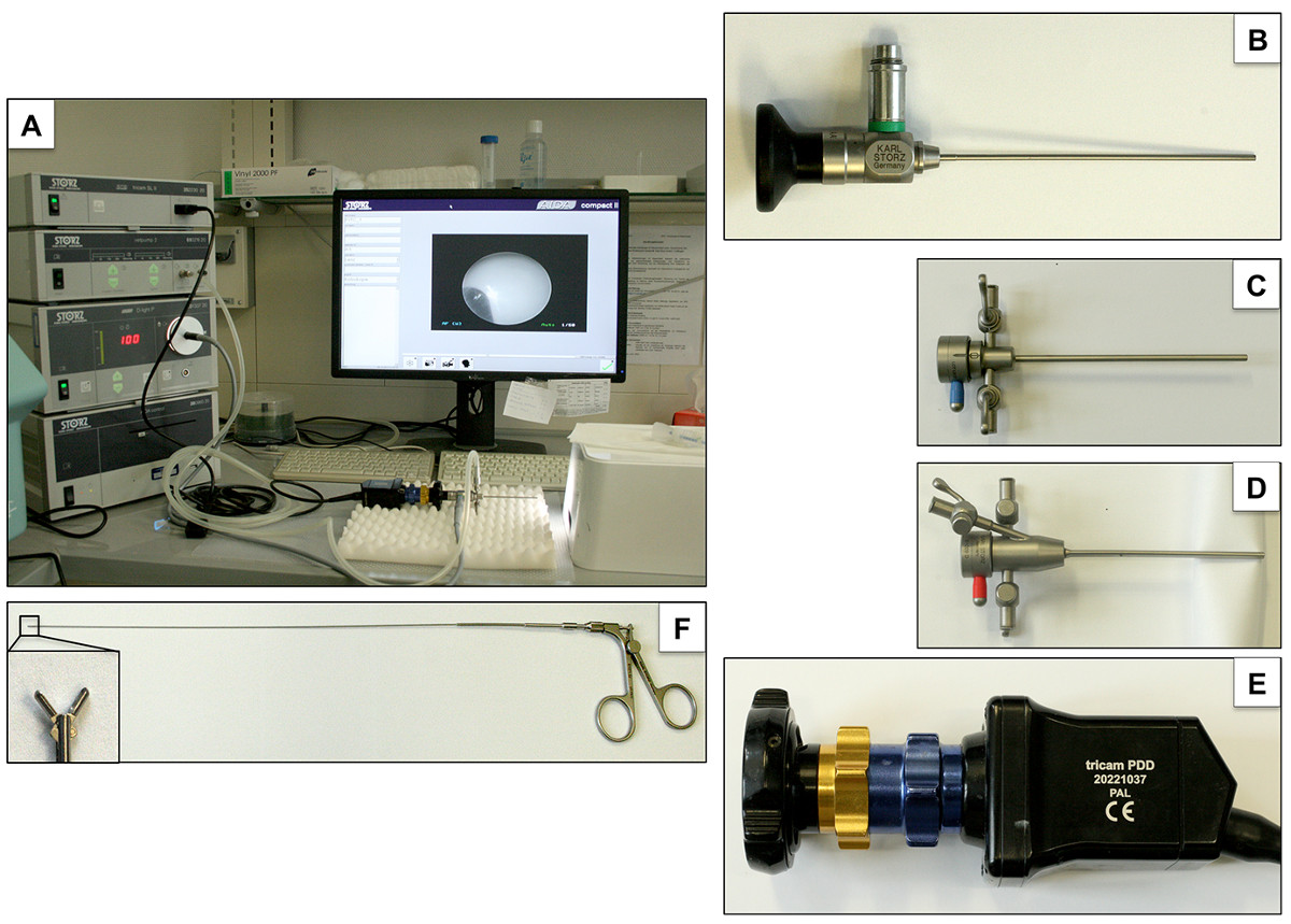
Figure 5. Experimental set-up of endoscopic working station. The endoscopic working station (A) consists of the following components: straightforward telescope (0°, diameter: 1.9 mm, length: 10 cm; B), endoscope sheath (9 Charr.) without (C) and with working channel (D), camera (E) and biopsy forceps (F). Please click here to view a larger version of this figure.
Epithelial wound healing is an ongoing process. Continuous physiological exfoliation of surface cells within the gastrointestinal mucosa occurs requiring frequent regeneration of epithelial cells 16. Consequently, impaired wound healing has an immense impact on several diseases including gastrointestinal ulcers and 17 anastomotic leakage 18. Evaluation of molecular background as well as potential drug candidates to stimulate epithelial healing may only be incompletely performed in cell culture systems in vitro19,20. Thus, more sophisticated experimental setups such as murine colonoscopy with generation of defined mucosal wounds by a biopsy forceps are needed to enable reliable in vivo evaluation of gastrointestinal wound healing and to assess possible interactions between intestinal inflammation and wound healing processes.
In addition, an injection needle may be used for local intramucosal administration of diagnostic dyes or potential drug candidates. This can be achieved using a flexible tube (diameter of 0.96 mm) with a needle fixed to the end can be introduced through the working channel. Given that a test agent as well as the placebo control may be delivered in separate inflammatory or neoplastic lesions within the same animal, this approach provides an advantage in reliability compared to traditional experimental settings. A further application of local injections is the implantation of human or murine tumor cells to generate orthotopic tumors in the murine colon 21.
Murine models of colitis are needed to elucidate pathophysiology as well as to evaluate potential therapeutic agents preclinically. Therefore, accurate monitoring of the disease course is of utmost importance. Conventionally, severity of disease is usually assessed by indirect parameters such as body weight, haemoccult testing as well as analysis of blood and feces. In contrast, direct determination of colitis severity is often limited to histological analysis post mortem, which requires the death of the animal. However, murine colonoscopy offers a direct visualization of the colonic mucosa of live mice. Furthermore, direct and repetitive monitoring for features of colitis is possible, which is a requisite in experimental models with inhomogeneous disease onset, e.g., IL-10 deficient mice or in the model of transfercolitis in RAG-deficient mice. Consequently, a murine endoscopic index of colitis has been established 6 which allows objective quantification of mucosal inflammation and serial follow-up examinations of the same animal.
In the context of colorectal carcinogenesis, colonoscopy offers various beneficial opportunities. For example, in contrast to non-invasive methods, endoscopy is the first approach to allow in vivo determination of tumor size and tumor numbers. Furthermore, the use of fluorescent photoprobes targeting specific molecules enables visualization and quantification of molecular processes. In a translational study performed by Foersch et al. specific targeting of VEGF expression within malignant colonic mucosa was shown to be feasible and may be used for lesion characterization and prediction of treatment response in human patients with colorectal cancer 22. Furthermore, information resulting from this molecular imaging approach may be able to be translated for use in human patients. This would allow live characterization of suspect lesions during endoscopy. Finally, so called ‘smart probes’ increase specificity of these tracers by activation of the fluorophores by enzymatic processes at the side of the targeted lesion 23.
When performing murine endoscopy, certain steps of the given protocol are particularly critical. For example, different mouse strains differ in their susceptibility to anesthesia and DSS concentrations. Therefore this protocol may be required to be adapted to local settings. Furthermore, experience in performing endoscopic examinations and exact knowledge of murine anatomy is required to perform optimal murine endoscopy that is safe and target-oriented. With regard to possible limitations of this technique, we highlight that the endoscope system used is rigid, therefore limiting the procedure to the colon as far as the right flexure. Furthermore, most fluorochromes applicable for FE are currently being evaluated regarding their safety profiles and, therefore while available for murine studies, are not yet approved for use in human patients.
The following are critical steps concerning the practicalities of the procedure: (1) since susceptibility to DSS induced colitis may vary between different strains, the induction of acute DSS colitis may risk death of animals if there is advanced severity of colitis. Therefore, consider evaluating several DSS concentrations to identify the most suitable for an individual strain and the specific DSS batch used. Endoscopic examination might be difficult in the presence of large intracolonic stool masses. Inducing bowel movement prior to the procedure using rectal application of 2 ml of enema fluid via buttoned cannula will improve visibility if significant fecal loading is suspected that may obscure the view. If high perforation rates during biopsy occur, decrease air supply into the colon before obtaining mucosal biopsies and diminish pressure of the biopsy forceps on the mucosal surface before closing the branches.
Taken together, in contrast to conventional methods for assessing disease activity of experimental colitis or cancerogenesis by indirect parameters such as body weight, occurrence of fecal blood, analysis of peripheral blood or post mortem histological analysis, endoscopy-based techniques enable live monitoring of disease course with the opportunity to perform biopsies under visual control. In addition, wound healing and therapeutic impact of topically applied drug candidates may be evaluated in vivo.
The authors have nothing to disclose.
We thank Sonja Dufentester and Elke Weber for expert technical assistance. We thank Faekah Gohar for proofreading the manuscript and Stefan Brückner for medical informatics support. This work was supported by an interdisciplinary grant from the Else-Kröner-Fresenius-Stiftung (2012_A94). D. Bettenworth was supported by a research fellowship from the Faculty of Medicine, Westfälische Wilhelms-Universität Münster. M. Brückner was supported by a “Gerok” rotational position of the Deutsche Forschungsgemeinschaft (DFG SFB1009B8). We thank Heike Blum for illustration of the mouse cartoon.
Name | Company | Catalogue Number | Comment |
Reagents | |||
Alfalfa-free diet | Harlan Laboritories, Madison, USA | 2014 | |
Azoxymethane (AOM) | Sigma – Aldrich, Deisenhofen, Germany | A5486 | |
Bepanthen eye ointment | Bayer, Leverkusen, Germany | 80469764 | |
Dextran sulphate sodium (DSS) | TdB Consulatancy, Uppsala, Sweden | DB001 | |
Eosin | Sigma – Aldrich, Deisenhofen, Germany | E 4382 | |
Ethylenediaminetetraacetic acid (EDTA) | Sigma – Aldrich, Deisenhofen, Germany | E 9884 | |
Falcon Tube 50ml | BD Biosciences, Erembodegem, Belgium | 352070 | |
Florene 100V/V | Abbott, Wiesbaden, Germany | B506 | |
Fluorescein-Isothiocyanat (FITC)–dextrane | Sigma – Aldrich, Deisenhofen, Germany | FD4-250MG | |
Haematoxylin | Sigma – Aldrich, Deisenhofen, Germany | HHS32-1L | |
Isopentane (2- Methylbutane) | Sigma – Aldrich, Deisenhofen, Germany | M32631-1L | |
Methylene blue | Merck, Darmstadt, Germany | 1159430025 | |
O.C.T. Tissue Tek compound | Sakura, Zoeterwonde, Netherlands | 4583 | |
Omnican F – canula | Braun, Melsungen, Germany | 9161502 | |
Phosphate buffered saline, PBS | Lonza, Verviers, Belgium | 4629 | |
Sodium Chloride 0,9% | Braun, Melsungen, Germany | 5/12211095/0411 | |
Standard diet | Altromin, Lage, Germany | 1320 | |
Tissue-Tek Cryomold | Sakura, Leiden, Netherlands | 4566 | |
Vitro – Clud | R. Langenbrinck, Teningen, Germany | 04-0002 | |
Equipment | |||
AIDA Control | Karl Storz – Endoskope, Tuttlingen, Germany | 20 096020 | |
Bandpass filter | Semrock, Rochester, USA | HC 716/40 | |
Bandpass filter | Semrock, Rochester, USA | HC 809/81 | |
Biopsy Forceps, 3 Fr., 28cm | Karl Storz – Endoskope, Tuttlingen, Germany | 61071ZJ | |
Dell Monitor | Dell, Frankfurt am Main, Germany | U2412Mb | |
Examination Sheath, 9 Fr. | Karl Storz – Endoskope, Tuttlingen, Germany | 61029D | |
Examination Sheath, 9 Fr. | Karl Storz – Endoskope, Tuttlingen, Germany | 61029C | |
Fiber Optic Light Cable, 3.5mm | Karl Storz – Endoskope, Tuttlingen, Germany | 69495NL | |
Fluorescein Blue Filter System | Karl Storz – Endoskope, Tuttlingen, Germany | 20100032 | |
Fluorescein Barrier Filter | Karl Storz – Endoskope, Tuttlingen, Germany | 20100033 | |
Foot switch | Karl Storz – Endoskope, Tuttlingen, Germany | 20010430 | |
HOPKINS Telescope, 1.9mm, Length 10cm | Karl Storz – Endoskope, Tuttlingen, Germany | 1830231 | |
SCB D-light P | Karl Storz – Endoskope, Tuttlingen, Germany | 20 133720 | |
SCB tricam SL II | Karl Storz – Endoskope, Tuttlingen, Germany | 20 2230 20 | |
Tubing set instruments VETPUMP II | Karl Storz – Endoskope, Tuttlingen, Germany | 69811 | |
Tricam PDD PAL | Karl Storz – Endoskope, Tuttlingen, Germany | 20221037 | |
UniVet Porta | Groppler Medizintechnik, Deggendorf, Germany | BKGM 0451 | |
Vetpump 2 | Karl Storz – Endoskope, Tuttlingen, Germany | 69321620 |