The alkaline comet assay measures DNA strand breaks in eukaryotic cells. By adding an Endonuclease III or human 8-oxoguanine-DNA-N-glycosylase digestion step, the assay can efficiently detect oxidative DNA damage. We describe methods for using these assays to detect DNA damage in rat liver.
Unrepaired DNA damage can lead to genetic instability, which in turn may enhance cancer development. Therefore, identifying potential DNA damaging agents is important for protecting public health. The in vivo alkaline comet assay, which detects DNA damage as strand breaks, is especially relevant for assessing the genotoxic hazards of xenobiotics, as its responses reflect the in vivo absorption, tissue distribution, metabolism and excretion (ADME) of chemicals, as well as DNA repair process. Compared to other in vivo DNA damage assays, the assay is rapid, sensitive, visual and inexpensive, and, by converting oxidative DNA damage into strand breaks using specific repair enzymes, the assay can measure oxidative DNA damage in an efficient and relatively artifact-free manner. Measurement of DNA damage with the comet assay can be performed using both acute and subchronic toxicology study designs, and by integrating the comet assay with other toxicological assessments, the assay addresses animal welfare requirements by making maximum use of animal resources. Another major advantage of the assays is that they only require a small amount of cells, and the cells do not have to be derived from proliferating cell populations. The assays also can be performed with a variety of human samples obtained from clinically or occupationally exposed individuals.
The alkaline comet assay measures DNA strand breaks at the single-cell level. Suspensions of single cells are embedded in agarose on a microscope slide and the cells lysed to form nucleoids, which contain supercoiled loops of DNA. Electrophoresis at pH>13 results in the loss of supercoiling in DNA loops containing strand breaks, with the freed strands of DNA migrating toward the anode, creating comet-like structures that can be observed by fluorescence microscopy. Fragmented DNA migrates from the "head" of the comet into the "tail" based on the size of the fragment, and the relative fluorescence of the comet tail compared to the total intensity (head and tail) can be used to quantify DNA breakage1,2. The assay is simple, sensitive, versatile, rapid, and relatively inexpensive1. The detection of fragmented DNA caused by DNA-damaging agents is used an assay for quantifying DNA damage in cells or isolated nuclei from individual tissues of animals treated with potentially genotoxic material(s). Due to its advantages, the in vivo comet assay is recommended as a second in vivo genotoxicity assay (paired with the in vivo micronucleus assay) for conducting product safety evaluations in current International Conference on Harmonisation (ICH)3 and European Food Safety Authority (EFSA)4 regulatory guidelines. In our lab, we have employed the assay for evaluating in vivo DNA damage induced by food ingredients, pharmaceuticals, and nanomaterials5-10. Rat liver will be used as an example in this protocol, but the comet assay can be performed with other tissues/organs of experimental animals, as long as intact single cells can be isolated from the tissue.
Certain types of DNA damage are difficult to detect as DNA strand breaks without modifying the basic alkaline comet assay. In the case of oxidative DNA damage, strand breaks can be created at oxidative lesions in DNA by digesting with repair enzymes such as human 8-oxoguanine-DNA-N-glycosylase 1 (hOGG1, which creates breaks at 8-oxoguanine (8-oxoGua) and methyl-fapy-guanine11. Also, Endonuclease III (Endo III) creates breaks mainly at oxidized pyrimidines1. Thus, the addition of an enzyme-digestion step makes the assay a specific and sensitive method for measuring oxidative DNA damage in vivo12. Utilizing these assays, we have demonstrated toxicant-induced oxidative DNA damage in the liver of rats and mice6-8 and in the heart of rats10.
The alkaline comet assay has many applications in genetic toxicology and human biomonitoring: 1) as a follow-up in vivo assay for genotoxins identified by sensitive in vitro tests3,13, 2) to evaluate mechanisms of xenobiotic-induced DNA damage in multiple tissues14, 3) to investigate if a carcinogen operates using a genotoxic or a non-genotoxic mode of action (MOA)7, 4) to evaluate DNA damage repair15, 5) to investigate human diseases and occupational exposures 16, and 6) as a potential high-throughput screening assay for organ-specific genotoxicity17.
Ethics statement: Procedures involving animals have been approved by the Institutional Animal Care and Use Committee (IACUC) at the US FDA/National Center for Toxicological Research.
NOTE: The study design described here is based on the protocol developed by the Japanese Center for the Validation of Alternative Methods (JaCVAM) for their validation of the in vivo rodent alkaline comet assay18, and further modified based on recommendations in OECD guideline TG48919.
1. Preparation
2. Preparation of Single Cell Suspensions from Rat Liver
3. Preparation of Comet Slides
4. Enzyme Treatment of Comet Slides
5. Unwinding and Electrophoresis
6. DNA Staining, Comet Visualization and Analysis
7. Histopathology Analysis
8. Data analysis
The in vivo alkaline comet assay was performed in conjunction with the enzyme-modified comet assay to measure both direct and oxidative DNA damage in the liver of rats treated with cyproterone acetate (CPA)5. CPA is a synthetic hormonal drug that induces rat liver tumors in a sex-specific manner, with five-fold higher doses needed to induce liver tumors in male rats compared to females24. We found that the direct DNA damage produced by CPA in the liver of male and female rats has the same sex-specific pattern as its hepatotumorigenicity: a five-fold-higher dose of CPA is needed to induce a significant increase in DNA damage in the livers of males compared to females (Figure 1).
We hypothesize that the sex-specific comet assay result is due to the activity of hydroxysteroid sulfotransferase(s) (HST), which is 15-fold higher in adult female rats than in adult males. HST metabolism is a rate-limiting step in the activation of CPA to DNA-binding metabolite(s)25. In contrast, CPA-induced oxidative DNA damage was generally greater in male than female rat livers, and thus less likely to be a rate-limiting step in tumor formation (Figure 2). Histopathology evaluation of livers from CPA-treated rats showed no evidence of agent-induced apoptosis or necrosis (Tables 1 and 2), indicating that the positive comet assay results were not a secondary effect of cytotoxicity5. Figures 1 & 2 and Tables 1 & 2 are reprinted with permission of Elsevier B.V. (Reference 5).
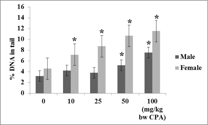
Figure 1. DNA damage in livers of CPA-treated male and female rats measured with the in vivo alkaline comet assay. Groups of five seven-week-old male and female F344 rats were treated with olive oil or with 10, 25, 50, or 100 mg/kg/day CPA in olive oil. Treatments were conducted at 0, 24, and 45 hr, the rats were sacrificed at 48 hr, and DNA damage was measured in liver as % tail DNA using the alkaline comet assay. CPA treatment induced an increase in % tail DNA in the livers of male rats in a threshold-like manner, with significant increases being detected only with the 50 and 100 mg/kg/day doses. CPA treatment induced DNA strand breaks in the livers of female rats in a near-linear dose-dependent manner, with significant increases in % tail DNA detected in all CPA groups. The Lowest Observed Genotoxicity Effect Levels (LOGELs) for CPA-induced DNA damage in the livers of female and male rats were estimated to be 10 and 50 mg/kg/day, respectively. *Significant at p≤0.05 relative to the vehicle control; error bars represent standard deviation. Please click here to view a larger version of this figure.
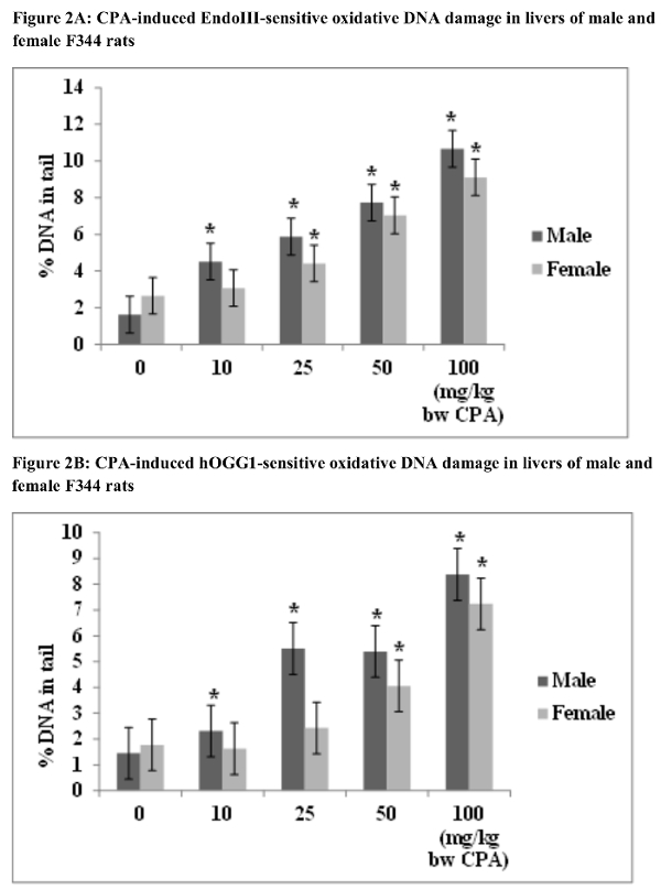
Figure 2. Oxidative DNA damage in livers of CPA-treated male and female rats measured with the enzyme-modified in vivo alkaline comet assay. Groups of five seven-week-old male and female F344 rats were treated with olive oil or with 10, 25, 50, or 100 mg/kg/day CPA in olive oil. Treatments were conducted at 0, 24, and 45 hr, the animals were sacrificed at 48 hr, and the enzyme-modified comet assay was conducted using % tail DNA as a metric of DNA damage. All CPA doses produced significant increases in Endo III-sensitive DNA damage in the livers of CPA-treated male rats; while Endo III-sensitive DNA damage was detected only in female rats treated with 25, 50, and 100 mg/kg/day CPA (A). All CPA doses resulted in significant increases in hOGG1-sensitive oxidative DNA damage in the livers of male rats; increases in hOGG1-sensitive DNA damage were detected only in female rats treated with 50 and 100 mg/kg/day CPA (B). *Significant at p≤0.05 relative to the vehicle control; error bars represent standard deviation. Please click here to view a larger version of this figure.
Tables 1&2. Histopathology analysis results. Since cytotoxicity can generate false-positive comet assay results, histopathological assessments for apoptotic and/or necrotic cells should be conducted for comet-positive tissues. In the current study, no CPA-induced hepatocyte apoptosis or necrosis was observed in livers of both male and female rats, which excluded the possibility of a false positive comet assay response.
Lesion | Data | Dose CPA | ||||
0 mg/kg | 10 mg/kg | 25 mg/kg | 50 mg/kg | 100 mg/kg | ||
Hepatocyte cytoplasmic vacuolization | Lesion Count | 1 | 1 | 5 | 5 | 6 |
# Examined | 6 | 5 | 5 | 5 | 6 | |
Lesion% | 17% | 20% | 100% | 100% | 100% | |
Avg Severity | 1.0 | 1.0 | 1.2 | 1.8 | 2.0 | |
Hepatocyte mitosis | Lesion Count | 0 | 0 | 5 | 5 | 6 |
# Examined | 6 | 5 | 5 | 5 | 6 | |
Lesion% | 0 | 0 | 100% | 100% | 100% | |
Avg Severity | 0 | 0 | 1.0 | 1.4 | 1.8 |
Table 1. Incidence of non-neoplastic lesions in livers of vehicle- and CPA-treated male F344 rats.
Lesion | Data | Dose CPA | ||||
0 mg/kg | 10 mg/kg | 25 mg/kg | 50 mg/kg | 100 mg/kg | ||
Hepatocyte cytoplasmic vacuolization | Lesion Count | 1 | 3 | 5 | 5 | 5 |
# Examined | 5 | 5 | 5 | 5 | 5 | |
Lesion% | 20% | 60% | 100% | 100% | 100% | |
Avg Severity | 1.0 | 1.3 | 1.8 | 1.8 | 2.0 | |
Hepatocyte mitosis | Lesion Count | 0 | 5 | 5 | 5 | 5 |
# Examined | 5 | 5 | 5 | 5 | 5 | |
Lesion% | 0 | 100% | 100% | 100% | 100% | |
Avg Severity | 0 | 1.0 | 1.2 | 1.8 | 2.0 | |
Hepatocyte Karyomegaly | Lesion Count | 0 | 0 | 5 | 5 | 5 |
# Examined | 5 | 5 | 5 | 5 | 5 | |
Lesion% | 0 | 0 | 100% | 100% | 100% | |
Avg Severity | 0 | 1.0 | 1.2 | 1.8 | 2.0 |
Table 2. Incidence of non-neoplastic lesions in livers of vehicle-and CPA-treated female F344 rats.
This protocol describes the concurrent measurement of both direct and oxidative DNA damage in rat liver at the single cell level. The general protocol is applicable to any tissue from which single cells or nuclei can be isolated with minimal processing-induced DNA damage (i.e., DNA damage induced not by the test agent, but by the handling and processing of the animal tissues). In our research, we have conducted alkaline comet assays on cells from bone marrow7,9, stomach6, kidney9, bladder9, lung9, heart10, mammary gland5, uterus5, testis5, and blood5. These assays can provide a quick, simple and inexpensive method for studying xenobiotic-induced DNA damage in multiple organs and tissues of experimental animals. In addition, this method can be used for human biomonitoring studies by conducting assays, for instance, on peripheral blood, exfoliated cells of the urinary tract, or the buccal or nasal epithelia obtained from clinically or occupationally exposed individuals. The basic approach also can be used for evaluating DNA damage in cultured cells in vitro.
When the comet assay is integrated into acute or subchronic toxicology studies, the sampling time, i.e., the time between the last treatment and the tissue collection, is a critical variable that sometimes must be reconciled with the collection of other toxicological data. If possible, sampling times should be based on time course comet data, the time at which the peak plasma or tissue concentration (Cmax) is achieved, or after steady state is achieved following multiple administrations of the test article19. In cases were kinetic data or tissue concentrations are not available, OECD TG489 recommends conducting two or more treatments and collecting tissues 2-6 hr after the last treatment, or, if a single treatment is conducted, sampling tissues at both 2-6 and 16-26 hr after the treatment19.
It is crucial that the length of time from euthanasia to comet slide preparation be as short as possible. It is advisable that experimental competency be established by demonstrating proficiency in obtaining high-quality single cell suspensions quickly from the tissues that are assayed. Generally, comet slides should be prepared as soon as possible after animal sacrifice, with single cell preparation taking a maximum of one hr. Establishment of an historical database to establish ranges and distributions of negative (vehicle) controls is highly recommended to ensure that the assay is under proper control. Excessive levels of DNA damage may be observed in vehicle/negative control animals when the following occur: 1) improper handling of the tissue; 2) too long a time between euthanasia and comet slide preparation; or 3) exposure of the single cell suspension to UV-containing light. Establishment of an historical database for the ranges and distributions of positive controls is also highly recommended to assure that the assay displays appropriate sensitivity to known genotoxins. Methyl methanesulfonate (MMS) was used as positive control in the current study; ethyl methane sulfonate (EMS) is recommended by JaCVAM as a positive control for the in vivo comet assay18.
Incorporating digestions with lesion-specific endonucleases into the comet assay protocol increases the sensitivity and specificity of the assay through the recognition of particular types of DNA lesions-in the example presented here, oxidized DNA bases. However, it should be recognized that some endonucleases may be capable of recognizing and cleaving at various classes of DNA lesions; in the case of Endo III, this includes lesions not associated with oxidative DNA damage. A positive Endo III-modified comet assay result may indicate a mixed MOA. By comparison, hOGG1 is more specific for oxidative lesions and data from an hOOG1-modified assay may be more useful for establishing a MOA that involves oxidative DNA damage11.
Cytotoxicity (cell apoptosis and necrosis) may result in DNA strand breaks and a false positive comet assay result: positive comet assay results by themselves cannot be interpreted as genotoxicity. Information on cytotoxicity is required to establish the biological relevance of a positive comet assay result. Histopathological analysis of comet-positive tissues is recommended by OECD TG489 as a relevant measure of tissue toxicity19. Positive comet assay data with clear evidence of tissue toxicity (cell apoptosis or necrosis) should be interpreted carefully.
The in vivo alkaline comet assay and the Endo III- and hOGG1-modified alkaline comet assays can be used in a quantitative manner to make decisions about the genotoxicity of xenobiotic exposures. They also can be used to conduct fundamental research in DNA damage and repair in multiple animal tissues.
The authors have nothing to disclose.
This work was supported by the US Food and Drug Administration. We acknowledge the original publication of the CPA study by Elsevier B.V.: Ding W, Bishop ME, Peace MG, Davis KJ, White GA, Lyn-Cook LE, Manjanatha MG. Sex-specific dose-response analysis of genotoxicity in cyproterone acetate-treated F344 rats. Mutation Research 774: 1-7, 2014 (PMID: 25440904)
Coverslips (No. 1, 24 x 50 mm) | Fisher | 12-544-14 | |
Microscope Slides | Fisher | 12-550-123 | |
Dimethylsulfoxide (DMSO) | Fisher | 67-68-5 | |
EDTA, Disodium | Fisher | BP120-1 | |
Phosphate buffered saline | Fisher | ICN1860454 | |
1X Hanks Balanced Salt Solution (HBSS) (Ca++, Mg++ free) | HyClone | SH30588.02 | |
HEPES | Fisher | BP310-1 | |
Low Melting Point Agarose (LMP) | Lonza | 50081 | NuSieve GTG Agarose |
Normal Melting Agarose (NMA) | Fisher | BP1356-100 | |
pH testing paper strips (pH 7.5-14) | Fisher | M95873 | |
Potassium Cloride | Fisher | 7447-40-7 | |
Potassium Hydroxide | Fisher | 1310-58-3 | |
slide labels, (0.94 x 0.5 in.) | Fisher | NC9822036 | |
Sodium Chloride (NaCl) | Fisher | 7647-14-5 | |
Sodium Hydroxide (NaOH) | Fisher | 1310-73-2 | |
SYBR™ Gold | Invitrogen | S11494 | |
Triton X-100 | Fisher | 9002-93-1 | |
Trizma Base | Fisher | 77-86-1 | |
2.0 mL microcentrifuge tubes | Fisher | 05-402-6 | |
Cell strainer (40 µm) | Fisher | 22363547 | |
Endonuclease III (Nth) | New England Biolabs | M0268S | Dilution 1:1000 |
hOGG1 | New England Biolabs | M0241S | Dilution 1:1000 |