Summary

Endoplazmik retikulum ve mitokondri Etkileşimleri Çalışma ile<em> In Situ</em> Proximity ligasyon Testi Sabit Hücrelerde

Published: December 10, 2016
doi:

Summary

Burada, sabit hücrelerde endoplazmik retikulum ve mitokondri arasındaki endojen etkileşimleri görselleştirmek ve yüksek hassasiyet ile ölçmek için bir prosedür açıklanmaktadır. Protokol, bir mitokondri ilişkili zar arayüzünde inositol 1,4,5-trifosfat reseptörü / glukoz-regüle proteini 75 / voltaj bağımlı anyon kanalı / siklofilin D kompleksi hedefleyen in situ yakınlık ligasyonu deneyinde optimize özellikleri.

Abstract

Structural interactions between the endoplasmic reticular (ER) and mitochondrial membranes, in domains known as mitochondria-associated membranes (MAM), are crucial hubs for cellular signaling and cell fate. Particularly, these inter-organelle contact sites allow the transfer of calcium from the ER to mitochondria through the voltage-dependent anion channel (VDAC)/glucose-regulated protein 75 (GRP75)/inositol 1,4,5-triphosphate receptor (IP3R) calcium channeling complex. While this subcellular compartment is under intense investigation in both physiological and pathological conditions, no simple and sensitive method exists to quantify the endogenous amount of ER-mitochondria contact in cells. Similarly, MAMs are highly dynamic structures, and there is no suitable approach to follow modifications of ER-mitochondria interactions without protein overexpression. Here, we report an optimized protocol based on the use of an in situ proximity ligation assay to visualize and quantify endogenous ER-mitochondria interactions in fixed cells by using the close proximity between proteins of the outer mitochondrial membrane (VDAC1) and of the ER membrane (IP3R1) at the MAM interface. Similar in situ proximity ligation experiments can also be performed with the GRP75/IP3R1 and cyclophilin D/IP3R1 pairs of antibodies. This assay provides several advantages over other imaging procedures, as it is highly specific, sensitive, and suitable to multiple-condition testing. Therefore, the use of this in situ proximity ligation assay should be helpful to better understand the physiological regulations of ER-mitochondria interactions, as well as their role in pathological contexts.

Introduction

Mitokondri ve endoplazmik retikulum (ER) hücrede bağımsız organelleri olmayan, ancak mitokondri ilişkili endoplazmik retikulum zarlarının (MAM) olarak tanımlanan temas bölgelerinde yapısal ve fonksiyonel etkileşim. Aslında, MAM her iki taraftan da proteinler arasındaki etkileşimi sağlayan ER membranlar ve mitokondri yakın kapatılan bölgelere karşılık gelir. Bununla birlikte, bu organellerin membranlar bu bölgeler içinde kaynaştırmak yok, bu yüzden onların ayrı varlıklar korumak. MAM Enerji metabolizması ve hücre yaşamını 1-3 etkileyen, mitokondri ER bir kalsiyum açısından önemli bir rol (Ca 2+) ve fosfolipid transferi oynarlar.

ER ve mitokondri arasındaki ilişki ilk elektron mikroskobu ile 1970'lerde görüntülendi. O zamandan beri, transmisyon elektron mikroskobu 4,5, elektron tomografi 6,7 veya ER ve mitokondri özgü fluorofor immün lokalizasyonus / floresan proteinleri 8 klasik ER-mitokondri etkileşimleri çalışmak için kullanılmıştır. MAM analizi için yararlı bir araç içi fraksiyonasyon kullanımına dayanır. Bir Percoll degrade 9 bağlanmış diferansiyel Ultrasantrifügasyon tarafından MAM fraksiyonların izolasyonu sağlar. Bununla birlikte, nihai ürün zenginleştirilmiş MAM fraksiyonlar yerine, saf kesimlerin içerir. Toplamda, bu stratejiler özellikle hassas ve / veya kantitatif değildir ve onlar büyük tarama kolayca uygun değildir. Alternatif olarak ilaç indüklenebilir floresan arası organel bağlayıcılar kullanılarak genetik yaklaşımlar ortaya çıkmıştır, ancak protein 10 'nın endojen ekspresyonunun seviyelerinde organel etkileşimlerin analizine izin vermez.

MAM 11'de IP3R / GRP75 / SSVD kompleksinin Szabadkai keşfinden dayanarak, ER-mitokondri etkileşimleri analiz etmek için kantitatif bir yöntem geliştirdi. In situ yakınlığı ligati kullanılantahlil üzerinde algılamak ve VDAC1 ve IP3R1 arasındaki etkileşimleri ölçmek için, sabit hücrelerde 12 MAM arayüzünde Ca 2 + -channeling kompleksi yer alan iki organel-yüzey proteinleri. Kısaca, bizler, ER membran dış mitokondriyal zarın (fare anti-VDAC1 birincil antikor) ve IP3R1 (tavşan anti-IP3R1 birincil antikor) (Şekil 1, bir panel) de VDAC1 incelenir. Daha sonra, deney göre, anti-fare ve tamamlayıcı oligonükleotid uzantıları konjuge edilmiş anti-tavşan IgG (fare ve tavşan proksimite ligasyonu deney Probes), her iki ilave edildi. İki hedef proteinler 40 nm'nin altında bir mesafede ise, oligonükleotidler dairesel DNA şablonu (Şekil 1 Panel B) oluşmasına izin vermek için daha sonra ilave bağlayıcı oligo ile hibridize olabilir. Bu dairesel bir DNA molekülü kovalent yakınlığı probları birine bağlanmış bir tek-şeritli DNA ürününü oluşturmak, lige edilmiş ve amplifiye edilir (Şekil 1, Panel C) </stro> Ng. MAM arayüzü ER ve mitokondri arasındaki mesafe mil 6, proksimite ligasyonu ve amplifikasyon bağlı Texas kırmızısı etiketli oligonükleotid sondalar (Şekil 1, Panel D hibridizasyonu sonraki tespiti yol yapılabilir 25-10 nm arasında değerler ). Her floresan nokta böylece tek tek hücrelerin in situ ER-mitokondri etkileşimleri sayılmasına olanak tanıyan, VDAC1 / IP3R1 arasındaki etkileşimleri temsil eder.

Şekil 1
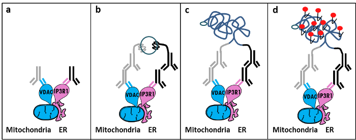
Şekil 1: Yerinde Proximity ligasyon Tahlili tarafından Endoplazmik Retikulum-mitokondri Etkileşimleri Algılama şematik İllüstrasyon. a) VDAC1 ve MAM arayüzü yakın kendi epitopuna bağlanabilir IP3R1 karşı bir tavşan primer antikora karşı yönlendirilmiş bir fare primer antikor, B) proksimite ligasyonu probları bir çift ilavesifare ve tavşan IgG karşı. Bu sondalar bağlayıcı oligo ligasyonu için şablonlar oluşturabilirler DNA ipliklerini eklenmiş. c) bağlanmasından sonra oluşturulan dairesel DNA zinciri Texas kırmızısı etiketli oligonükleotidler kullanılarak bir flüoresan nokta olarak mikroskopi ile görünür) yükseltilir ve D edilebilir. Bu rakamın büyük halini görmek için lütfen buraya tıklayınız.

In situ proksimite ligasyonu deneylerinde benzer antikorların GRP75 / IP3R1 çifti, hem de siklofilin D (CypD) ile yapılabilir / IP3R1 antikorlar CypD MAM arayüzü IP3R / GRP75 / SSVD kompleksi ile etkileştiği gösterilmiştir olduğu dikkate 12-14.

Protocol

Çözümler 1. hazırlanması PBS 14.5 ml% 37 formaldehit 5.5 mi seyreltilmesiyle PBS (tuz) içinde% 10 formaldehit hazırlayın. PBS, 50 ml glisin 3.8 g çözülmesiyle, 1 M glisin, pH 8.0, hazırlanması; 1x PBS içinde 100 mM glisin elde etmek için bu seyreltilir. 1x PBS içinde% 0.1 Triton-X100 hazırlayın. 3.0 M sodyum klorid ve 25 ° C de pH 7.0 ile, iyonu giderilmiş su içinde hazırlanan 0.30 M trisodyum sitrat ile 20x Tuzlu sodyum sitrat (SSC) hazırlayın. 1x bu tampon sulandırmak ve d…

Representative Results

Bu protokolü kullanarak bizim deneyime dayalı, biz güvenle sabit hücrelerde ER-mitokondri etkileşimleri görselleştirme ve miktar tayini için bu yöntemi tavsiye ederim. Antikorların çeşitli çiftleri kullanılarak HuH7 hepatokarsinoma hücre çizgisinde in situ proksimite ligasyonu deneyi-görüntülenmiştir ER-mitokondri etkileşimlerine temsili görüntüleri gösterilmektedir. Flüoresans mikroskopisi ile, Şekil 2'de gösterildiği g…

Discussion

Collectively, our studies indicate that the in situ proximity ligation assay is truly a relevant strategy to follow and quantify endogenous ER-mitochondria interactions in fixed cells, without the need for using organelle-specific fluorophores or fluorescent proteins. The specific use of VDAC1/IP3R1 antibodies has been adapted to study ER-mitochondria interactions in HuH7 cells. However, alternative isoforms of VDAC and IP3R may be used, depending on the cell type. In this case, antibodies need to be validated b…

Disclosures

The authors have nothing to disclose.

Acknowledgements

Biz optimize etmek ve protokol doğrulamak katkıda laboratuarımızda tüm insanlara teşekkür ederim. Bu çalışma INSERM ve ulusal araştırma kurumu (ANR-09-JCJC-0116 VE ANR-11-BSV1-033-02) tarafından desteklenmiştir. ET yüksek öğrenim ve araştırma Fransız bakanlığından bir araştırma bursu ile onu doktora sırasında desteklenmiştir.

Materials

Formaldehyde Sigma F-8775
Glycine Sigma G-8898
Triton Sigma T8532
35mm Glass bottom culture dishes MatTeK corporation P35G-0-14-C
Blocking solution Sigma DUO-92004 or DUO-92002 provided in the Duolink PLA probes, Sigma
VDAC1 antibody Abcam ab14734
IP3R1-H80 antibody Santa Cruz sc28614
CypD antibody Abcam ab110324
Grp75 antibody Santa Cruz sc13967
TBS 10X euromedex ET220 Dilute to obtain 1X
Tween 100X euromedex 2001-B dilute in TBS to obtain 0,01%
PLA Probes Mouse MINUS Sigma DUO-92004 Duolink, Sigma
PLA Probes Rabbit PLUS Sigma DUO-92002 Duolink, Sigma
Duolink detection reagents red Sigma DUO-92008 Duolink, Sigma
Ligation solution Sigma DUO-92008 Part of the Duolink detection reagents red, Sigma
Ligase Sigma DUO-92008 Part of the Duolink detection reagents red, Sigma
Amplification solution Sigma DUO-92008 Part of the Duolink detection reagents red, Sigma
Polymerase Sigma DUO-92008 Part of the Duolink detection reagents red, Sigma
Duolink Mounting Medium Sigma DUO80102 Duolink, Sigma
Softwares:
Blob-finder software BlobFinder is a freely distributed software that can perform calculations on cells from fluorescence microscopy images. This software can be downloaded for free from The Centre for Image Analysis at Uppsala University who have developed the software and the work was supported by the EU FP6 Project ENLIGHT and  Olink Bioscience. http://www.cb.uu.se/~amin/BlobFinder/index_files/Page430.htm
ImageJ software Can be downloaded for free from: http://rsb.info.nih.gov/ij/download.html

References

  1. Bravo-Sagua, R., et al. Organelle communication: signaling crossroads between homeostasis and disease. The international journal of biochemistry & cell biology. 50, 55-59 (2014).
  2. Giorgi, C., et al. Mitochondria-associated membranes: composition, molecular mechanisms, and physiopathological implications. Antioxidants & redox signaling. 22, 995-1019 (2015).
  3. Phillips, M. J., Voeltz, G. K. Structure and function of ER membrane contact sites with other organelles. Nature reviews. Molecular cell biology. 17, 69-82 (2016).
  4. Cosson, P., et al. The RTM resistance to potyviruses in Arabidopsis thaliana: natural variation of the RTM genes and evidence for the implication of additional genes. PLoS One. 7, 39169 (2012).
  5. Mannella, C. A. Structure and dynamics of the mitochondrial inner membrane cristae. Biochim Biophys Acta. 1763, 542-548 (2006).
  6. Csordas, G., et al. Structural and functional features and significance of the physical linkage between ER and mitochondria. The Journal of cell biology. 174, 915-921 (2006).
  7. Mannella, C. A., Buttle, K., Rath, B. K., Marko, M. Electron microscopic tomography of rat-liver mitochondria and their interaction with the endoplasmic reticulum. Biofactors. 8, 225-228 (1998).
  8. Rizzuto, R., et al. Close contacts with the endoplasmic reticulum as determinants of mitochondrial Ca2+ responses. Science. 280, 1763-1766 (1998).
  9. Wieckowski, M. R., Giorgi, C., Lebiedzinska, M., Duszynski, J., Pinton, P. Isolation of mitochondria-associated membranes and mitochondria from animal tissues and cells. Nat Protoc. 4, 1582-1590 (2009).
  10. Csordas, G., et al. Imaging interorganelle contacts and local calcium dynamics at the ER-mitochondrial interface. Mol Cell. 39, 121-132 (2010).
  11. Szabadkai, G., et al. Chaperone-mediated coupling of endoplasmic reticulum and mitochondrial Ca2+ channels. J Cell Biol. 175, 901-911 (2006).
  12. Tubbs, E., et al. Mitochondria-associated endoplasmic reticulum membrane (MAM) integrity is required for insulin signaling and is implicated in hepatic insulin resistance. Diabetes. 63, 3279-3294 (2014).
  13. Paillard, M., et al. Depressing Mitochondria-Reticulum Interactions Protects Cardiomyocytes From Lethal Hypoxia-Reoxygenation Injury. Circulation. 128, 1555-1565 (2013).
  14. Rieusset, J., et al. Disruption of calcium transfer from ER to mitochondria links alterations of mitochondria-associated ER membrane integrity to hepatic insulin resistance. Diabetologia. 59, 614-623 (2016).
  15. Allalou, A., Wahlby, C. BlobFinder, a tool for fluorescence microscopy image cytometry. Computer methods and programs in biomedicine. 94, 58-65 (2009).
  16. Theurey, P., et al. Mitochondria-associated endoplasmic reticulum membranes allow adaptation of mitochondrial metabolism to glucose availability in the liver. Journal of molecular cell biology. , (2016).
  17. de Brito, O. M., Scorrano, L. Mitofusin 2 tethers endoplasmic reticulum to mitochondria. Nature. 456, 605-610 (2008).
  18. Soderberg, O., et al. Direct observation of individual endogenous protein complexes in situ by proximity ligation. Nature methods. 3, 995-1000 (2006).
  19. De Pinto, V., Messina, A., Lane, D. J., Lawen, A. Voltage-dependent anion-selective channel (VDAC) in the plasma membrane. FEBS letters. 584, 1793-1799 (2010).
  20. Kaul, S. C., Taira, K., Pereira-Smith, O. M., Wadhwa, R. Mortalin: present and prospective. Experimental gerontology. 37, 1157-1164 (2002).

Play Video

Cite This Article
Tubbs, E., Rieusset, J. Study of Endoplasmic Reticulum and Mitochondria Interactions by In Situ Proximity Ligation Assay in Fixed Cells. J. Vis. Exp. (118), e54899, doi:10.3791/54899 (2016).

View Video