Summary

जालिका और माइटोकॉन्ड्रिया बातचीत का अध्ययन द्वारा<em> बगल में</em> निकटता Ligation परख तय कोशिकाओं में

Published: December 10, 2016
doi:

Summary

यहाँ, हम तय कोशिकाओं में कल्पना और जालिका और माइटोकॉन्ड्रिया के बीच बातचीत अंतर्जात उच्च संवेदनशीलता के साथ यों तो एक प्रक्रिया का वर्णन। प्रोटोकॉल एक माइटोकॉन्ड्रिया जुड़े झिल्ली इंटरफेस में inositol 1,4,5-triphosphate रिसेप्टर / ग्लूकोज विनियमित प्रोटीन 75 / वोल्टेज पर निर्भर आयनों चैनल / cyclophilin डी जटिल को निशाना सीटू निकटता बंधाव परख में अनुकूलित की सुविधा है।

Abstract

Structural interactions between the endoplasmic reticular (ER) and mitochondrial membranes, in domains known as mitochondria-associated membranes (MAM), are crucial hubs for cellular signaling and cell fate. Particularly, these inter-organelle contact sites allow the transfer of calcium from the ER to mitochondria through the voltage-dependent anion channel (VDAC)/glucose-regulated protein 75 (GRP75)/inositol 1,4,5-triphosphate receptor (IP3R) calcium channeling complex. While this subcellular compartment is under intense investigation in both physiological and pathological conditions, no simple and sensitive method exists to quantify the endogenous amount of ER-mitochondria contact in cells. Similarly, MAMs are highly dynamic structures, and there is no suitable approach to follow modifications of ER-mitochondria interactions without protein overexpression. Here, we report an optimized protocol based on the use of an in situ proximity ligation assay to visualize and quantify endogenous ER-mitochondria interactions in fixed cells by using the close proximity between proteins of the outer mitochondrial membrane (VDAC1) and of the ER membrane (IP3R1) at the MAM interface. Similar in situ proximity ligation experiments can also be performed with the GRP75/IP3R1 and cyclophilin D/IP3R1 pairs of antibodies. This assay provides several advantages over other imaging procedures, as it is highly specific, sensitive, and suitable to multiple-condition testing. Therefore, the use of this in situ proximity ligation assay should be helpful to better understand the physiological regulations of ER-mitochondria interactions, as well as their role in pathological contexts.

Introduction

माइटोकांड्रिया और जालिका (ईआर) सेल में स्वतंत्र अंगों नहीं हैं, लेकिन वे संपर्क साइटों माइटोकॉन्ड्रिया जुड़े जालिका झिल्ली (एमएएम) के रूप में परिभाषित में संरचनात्मक और कार्यात्मक बातचीत। वास्तव में, MAMS क्षेत्रों में जहां ईआर की झिल्ली और माइटोकॉन्ड्रिया बारीकी apposed कर रहे हैं के अनुरूप, दोनों पक्षों से प्रोटीन के बीच बातचीत की इजाजत दी। बहरहाल, इन अंगों की झिल्लियों इन क्षेत्रों के भीतर फ्यूज नहीं है, इसलिए वे अपनी अलग संस्थाओं बनाए रखें। MAMS, माइटोकॉन्ड्रिया के लिए ईआर से एक महत्वपूर्ण (सीए 2) कैल्शियम में भूमिका और फॉस्फोलिपिड हस्तांतरण खेलने ऊर्जा चयापचय और सेल अस्तित्व 1-3 प्रभावित।

ईआर और माइटोकॉन्ड्रिया के बीच सहयोग पहली इलेक्ट्रॉन माइक्रोस्कोपी के साथ 1970 के दशक में कल्पना की गई थी। तब से, संचरण इलेक्ट्रॉन माइक्रोस्कोपी 4,5, इलेक्ट्रॉन टोमोग्राफी 6.7 या ईआर और माइटोकॉन्ड्रिया विशेष की fluorophore इम्युनो-स्थानीयकरणएस / फ्लोरोसेंट प्रोटीन 8 प्रतिष्ठित ईआर माइटोकॉन्ड्रिया बातचीत का अध्ययन करने के लिए इस्तेमाल किया गया। एमएएम के विश्लेषण के लिए एक और उपयोगी उपकरण subcellular विभाजन के उपयोग पर आधारित है। यह अंतर ultracentrifugation एक Percoll ढाल 9 के लिए युग्मित द्वारा एमएएम अंशों के अलगाव की अनुमति देता है। हालांकि, अंतिम उत्पाद समृद्ध एमएएम भिन्न, बजाय शुद्ध अंशों में शामिल है। कुल मिलाकर, इन रणनीतियों का विशेष रूप से संवेदनशील और / या मात्रात्मक नहीं हैं, और वे आसानी से बड़ी स्क्रीनिंग के लिए उत्तरदायी नहीं हैं। वैकल्पिक रूप से, दवा-inducible फ्लोरोसेंट अंतर-organelle linkers का उपयोग कर आनुवंशिक दृष्टिकोण में उभरा है, लेकिन वे प्रोटीन 10 की अंतर्जात अभिव्यक्ति के स्तर पर organelle बातचीत के विश्लेषण की अनुमति नहीं है।

एमएएम 11 पर IP3R / GRP75 / VDAC परिसर के Szabadkai की खोज पर आधारित है, हम ईआर माइटोकॉन्ड्रिया बातचीत का विश्लेषण करने के लिए एक मात्रात्मक विधि विकसित की है। हम सीटू निकटता ligati में इस्तेमाल कियापरख का पता लगाने और VDAC1 और IP3R1 के बीच बातचीत यों तो दो organelle-सतह प्रोटीन तय कोशिकाओं 12 में एमएएम इंटरफेस में सीए 2 -channeling परिसर में शामिल किया गया। संक्षेप में, हम बाहरी mitochondrial झिल्ली (माउस विरोधी VDAC1 प्राथमिक एंटीबॉडी) और IP3R1 ईआर झिल्ली पर (खरगोश विरोधी IP3R1 प्राथमिक एंटीबॉडी) (चित्रा 1, एक पैनल) पर VDAC1 की जांच की। फिर, परख के अनुसार, हम दोनों विरोधी माउस और विरोधी खरगोश आईजीजी (माउस और खरगोश निकटता बंधाव परख जांच) है, जो पूरक oligonucleotide एक्सटेंशन संयुग्मित हैं जोड़ा। दो लक्षित प्रोटीन 40 एनएम से नीचे दूरी पर हैं, ओलईगोन्युक्लियोटाईड्स एक परिपत्र डीएनए टेम्पलेट (चित्रा 1, पैनल बी) के गठन की अनुमति देने के लिए बाद में जोड़ा कनेक्टर ओलिगोस के साथ संकरण कर सकते हैं। यह परिपत्र डीएनए अणु ligated है और प्रवर्धित, covalently निकटता जांच में से एक से जुड़ी एक एकल असहाय डीएनए उत्पाद बनाने (चित्रा 1, पैनल ग) </stroएनजी>। एमएएम इंटरफेस में ईआर और माइटोकॉन्ड्रिया के बीच की दूरी को 25 एनएम 6, निकटता बंधाव और प्रवर्धन किया जा सकता है, बाद में पता लगाने के लिए अग्रणी टेक्सास रेड लेबल ओलईगोन्युक्लियोटाईड्स जांच (चित्रा 1, पैनल डी के संकरण के कारण 10 एनएम से पर्वतमाला के बाद )। प्रत्येक फ्लोरोसेंट डॉट इस प्रकार व्यक्ति की कोशिकाओं में सीटू ईआर माइटोकॉन्ड्रिया बातचीत की मात्रा का ठहराव के लिए अनुमति देता VDAC1 / IP3R1 के बीच बातचीत का प्रतिनिधित्व करता है।

आकृति 1
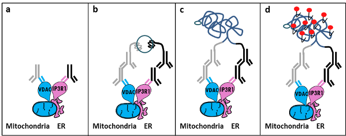
चित्रा 1: द्वारा जालिका-माइटोकॉन्ड्रिया बातचीत का पता लगाने के योजनाबद्ध चित्रण सीटू निकटता Ligation परख में। क) एक माउस प्राथमिक एंटीबॉडी VDAC1 और एक खरगोश प्राथमिक एंटीबॉडी IP3R1 के खिलाफ निर्देशित एमएएम इंटरफेस में निकटता में उनकी epitopes करने के लिए बाध्य कर सकते हैं के खिलाफ निर्देशित, ख) निकटता बंधाव जांच की एक जोड़ी के अलावामाउस और खरगोश आईजीजी के खिलाफ निर्देशित। ये जांच डीएनए किस्में है कि कनेक्टर ओलिगोस के बंधाव के लिए टेम्पलेट्स फार्म कर सकते हैं संलग्न है। ग) बंधाव के बाद गठित परिपत्र डीएनए किनारा टेक्सास रेड लेबल oligonucleotides का उपयोग कर से परिलक्षित किया जा सकता है और डी) एक फ्लोरोसेंट डॉट के रूप में माइक्रोस्कोपी द्वारा कल्पना। यह आंकड़ा का एक बड़ा संस्करण देखने के लिए यहां क्लिक करें।

सीटू निकटता बंधाव परख प्रयोगों में इसी प्रकार के एंटीबॉडी के GRP75 / IP3R1 जोड़ी, साथ ही cyclophilin डी (CypD) के साथ प्रदर्शन किया जा सकता है / IP3R1 एंटीबॉडी, विचार है कि CypD एमएएम इंटरफेस में IP3R / GRP75 / VDAC परिसर के साथ बातचीत करने के लिए दिखाया गया था 12-14।

Protocol

1. समाधान की तैयारी पीबीएस के 14.5 मिलीलीटर में 37% formaldehyde के 5.5 मिलीलीटर गिराए द्वारा पीबीएस (कम नमक) में 10% formaldehyde तैयार करें। पीबीएस के 50 मिलीलीटर में ग्लाइसिन की 3.8 ग्राम भंग करके 1 एम ग्लाइसिन, पीएच 8.0, तैयार कर?…

Representative Results

इस प्रोटोकॉल का उपयोग हमारे अनुभव के आधार पर, हम सुरक्षित रूप से दृश्य और तय कोशिकाओं में ईआर-माइटोकॉन्ड्रिया बातचीत की मात्रा का ठहराव के लिए इस विधि की सिफारिश कर सकते हैं। Huh7 हिपेटोकार्?…

Discussion

Collectively, our studies indicate that the in situ proximity ligation assay is truly a relevant strategy to follow and quantify endogenous ER-mitochondria interactions in fixed cells, without the need for using organelle-specific fluorophores or fluorescent proteins. The specific use of VDAC1/IP3R1 antibodies has been adapted to study ER-mitochondria interactions in HuH7 cells. However, alternative isoforms of VDAC and IP3R may be used, depending on the cell type. In this case, antibodies need to be validated b…

Disclosures

The authors have nothing to disclose.

Acknowledgements

हम हमारी प्रयोगशाला में सभी लोगों को जो अनुकूलन और प्रोटोकॉल को मान्य करने के लिए योगदान धन्यवाद। इस काम INSERM और राष्ट्रीय अनुसंधान एजेंसी (ANR-09-JCJC-0116 और ANR-11-BSV1-033-02) द्वारा समर्थित किया गया। ईटी उच्च शिक्षा और अनुसंधान के फ्रेंच मंत्रालय से एक शोध फैलोशिप द्वारा उसे पीएचडी के दौरान समर्थन किया था।

Materials

Formaldehyde Sigma F-8775
Glycine Sigma G-8898
Triton Sigma T8532
35mm Glass bottom culture dishes MatTeK corporation P35G-0-14-C
Blocking solution Sigma DUO-92004 or DUO-92002 provided in the Duolink PLA probes, Sigma
VDAC1 antibody Abcam ab14734
IP3R1-H80 antibody Santa Cruz sc28614
CypD antibody Abcam ab110324
Grp75 antibody Santa Cruz sc13967
TBS 10X euromedex ET220 Dilute to obtain 1X
Tween 100X euromedex 2001-B dilute in TBS to obtain 0,01%
PLA Probes Mouse MINUS Sigma DUO-92004 Duolink, Sigma
PLA Probes Rabbit PLUS Sigma DUO-92002 Duolink, Sigma
Duolink detection reagents red Sigma DUO-92008 Duolink, Sigma
Ligation solution Sigma DUO-92008 Part of the Duolink detection reagents red, Sigma
Ligase Sigma DUO-92008 Part of the Duolink detection reagents red, Sigma
Amplification solution Sigma DUO-92008 Part of the Duolink detection reagents red, Sigma
Polymerase Sigma DUO-92008 Part of the Duolink detection reagents red, Sigma
Duolink Mounting Medium Sigma DUO80102 Duolink, Sigma
Softwares:
Blob-finder software BlobFinder is a freely distributed software that can perform calculations on cells from fluorescence microscopy images. This software can be downloaded for free from The Centre for Image Analysis at Uppsala University who have developed the software and the work was supported by the EU FP6 Project ENLIGHT and  Olink Bioscience. http://www.cb.uu.se/~amin/BlobFinder/index_files/Page430.htm
ImageJ software Can be downloaded for free from: http://rsb.info.nih.gov/ij/download.html

References

  1. Bravo-Sagua, R., et al. Organelle communication: signaling crossroads between homeostasis and disease. The international journal of biochemistry & cell biology. 50, 55-59 (2014).
  2. Giorgi, C., et al. Mitochondria-associated membranes: composition, molecular mechanisms, and physiopathological implications. Antioxidants & redox signaling. 22, 995-1019 (2015).
  3. Phillips, M. J., Voeltz, G. K. Structure and function of ER membrane contact sites with other organelles. Nature reviews. Molecular cell biology. 17, 69-82 (2016).
  4. Cosson, P., et al. The RTM resistance to potyviruses in Arabidopsis thaliana: natural variation of the RTM genes and evidence for the implication of additional genes. PLoS One. 7, 39169 (2012).
  5. Mannella, C. A. Structure and dynamics of the mitochondrial inner membrane cristae. Biochim Biophys Acta. 1763, 542-548 (2006).
  6. Csordas, G., et al. Structural and functional features and significance of the physical linkage between ER and mitochondria. The Journal of cell biology. 174, 915-921 (2006).
  7. Mannella, C. A., Buttle, K., Rath, B. K., Marko, M. Electron microscopic tomography of rat-liver mitochondria and their interaction with the endoplasmic reticulum. Biofactors. 8, 225-228 (1998).
  8. Rizzuto, R., et al. Close contacts with the endoplasmic reticulum as determinants of mitochondrial Ca2+ responses. Science. 280, 1763-1766 (1998).
  9. Wieckowski, M. R., Giorgi, C., Lebiedzinska, M., Duszynski, J., Pinton, P. Isolation of mitochondria-associated membranes and mitochondria from animal tissues and cells. Nat Protoc. 4, 1582-1590 (2009).
  10. Csordas, G., et al. Imaging interorganelle contacts and local calcium dynamics at the ER-mitochondrial interface. Mol Cell. 39, 121-132 (2010).
  11. Szabadkai, G., et al. Chaperone-mediated coupling of endoplasmic reticulum and mitochondrial Ca2+ channels. J Cell Biol. 175, 901-911 (2006).
  12. Tubbs, E., et al. Mitochondria-associated endoplasmic reticulum membrane (MAM) integrity is required for insulin signaling and is implicated in hepatic insulin resistance. Diabetes. 63, 3279-3294 (2014).
  13. Paillard, M., et al. Depressing Mitochondria-Reticulum Interactions Protects Cardiomyocytes From Lethal Hypoxia-Reoxygenation Injury. Circulation. 128, 1555-1565 (2013).
  14. Rieusset, J., et al. Disruption of calcium transfer from ER to mitochondria links alterations of mitochondria-associated ER membrane integrity to hepatic insulin resistance. Diabetologia. 59, 614-623 (2016).
  15. Allalou, A., Wahlby, C. BlobFinder, a tool for fluorescence microscopy image cytometry. Computer methods and programs in biomedicine. 94, 58-65 (2009).
  16. Theurey, P., et al. Mitochondria-associated endoplasmic reticulum membranes allow adaptation of mitochondrial metabolism to glucose availability in the liver. Journal of molecular cell biology. , (2016).
  17. de Brito, O. M., Scorrano, L. Mitofusin 2 tethers endoplasmic reticulum to mitochondria. Nature. 456, 605-610 (2008).
  18. Soderberg, O., et al. Direct observation of individual endogenous protein complexes in situ by proximity ligation. Nature methods. 3, 995-1000 (2006).
  19. De Pinto, V., Messina, A., Lane, D. J., Lawen, A. Voltage-dependent anion-selective channel (VDAC) in the plasma membrane. FEBS letters. 584, 1793-1799 (2010).
  20. Kaul, S. C., Taira, K., Pereira-Smith, O. M., Wadhwa, R. Mortalin: present and prospective. Experimental gerontology. 37, 1157-1164 (2002).
check_url/54899?article_type=t

Play Video

Cite This Article
Tubbs, E., Rieusset, J. Study of Endoplasmic Reticulum and Mitochondria Interactions by In Situ Proximity Ligation Assay in Fixed Cells. J. Vis. Exp. (118), e54899, doi:10.3791/54899 (2016).

View Video