Summary

Snelle en specifieke Immunomagnetic isolatie van de primaire oligodendrocyten muis

Published: May 21, 2018
doi:

Summary

We beschrijven de immunomagnetic isolatie van primaire muisknop oligodendrocyten, waarmee het snelle en specifieke isolement van de cellen voor in vitro cultuur.

Abstract

De efficiënte en robuuste isolatie en de cultuur van primaire oligodendrocyten (OLs) is een waardevol instrument voor het in vitro onderzoek naar de ontwikkeling van oligodendroglia, alsook de biologie van demyeliniserende ziekten zoals multiple sclerose en Pelizaeus-Merzbacher-achtige ziekte (PMLD). Hier presenteren we een eenvoudige en efficiënte selectiemethode voor de isolatie van de immunomagnetic van fase drie O4+ preoligodendrocytes cellen van neonatale muizen pups. Aangezien onvolwassen OL vormen meer dan 80% van de knaagdier-hersenen witte stof op postnatale dag 7 (P7) deze methode isolatie zorgt niet alleen voor hoge cellulaire opbrengst, maar ook de specifieke Isolatievan OLs al toegezegd om de oligodendroglial afstamming, daalt het mogelijkheid van het isoleren van contaminerende cellen zoals astrocyten en andere cellen uit de hersenen van de muis. Deze methode is een wijziging van de technieken die eerder gerapporteerd en biedt Oligodendrocyt voorbereiding zuiverheid boven de 80% in ongeveer 4 uur.

Introduction

Oligodendrocyten (OLs) zijn de cellen van de myelinating van het centrale zenuwstelsel (CNS)1. De isolatie en de cultuur van primaire oligodendrocyten in een strak gereguleerde omgeving is een waardevol instrument voor het in vitro onderzoek naar de ontwikkeling van oligodendroglia, alsook de biologie van demyeliniserende ziekten zoals multiple sclerose,2 . Dit vereist een efficiënte en robuuste Oligodendrocyt isolatie en cultuur methode3. In deze studie profiteerde we van de expressie van een onderscheidende Oligodendrocyt cel oppervlakte markering om een gemodificeerde isolatie-techniek die is snelle en specifieke.

Vier verschillende fasen van Oligodendrocyt rijping zijn geïdentificeerd, elk gekenmerkt door de expressie van de oppervlakte merkers onderscheidend cel voor elke ontwikkelings fase (Figuur 1). Deze cel oppervlakte markers kunnen worden herkend door specifieke antilichamen4,5, en kunnen worden gebruikt voor het isoleren van OLs in specifieke stadia. In de eerste fase, Oligodendrocyt voorlopercellen (OPCs) de capaciteit hebben om te vermenigvuldigen, migreren en specifiek express bloedplaatjes afkomstige groei factor receptor (PDGF-Rα)6, ganglioside A2B5, Proteoglycaan NG27,8 , polysialic zuur-neurale cel adhesie molecuul9 en vetzuren-zuur-bindend-proteïne 7 (FABP7)10. Bipolaire morfologie hebben OPCs met enkele korte processen die afkomstig zijn van de tegengestelde Polen van de cel lichaam, dat is kenmerkend voor de voorloper van de neurale cellen11.

Figure 1
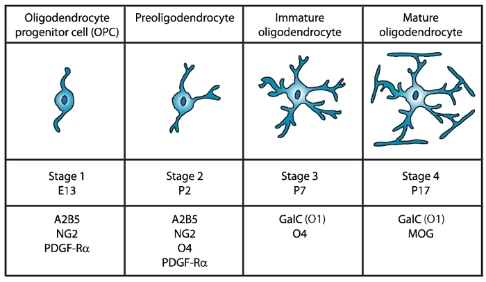
Figuur 1: expressie van cel oppervlakte markers tijdens de ontwikkeling van de muis Oligodendrocyt. OLs cel oppervlakte markeringen zoals A2B5, GalC (O1), NG2, O4 en PDGF-Rα kunnen worden gebruikt om te isoleren specifiek oligodendrocyten specifieke ontwikkelingsstoornissen stadium met behulp van specifieke antilichamen.   Klik hier voor een grotere versie van dit cijfer.

In de tweede fase, OPCs aanleiding geven tot preoligodendrocytes en express op het celmembraan niet alleen OPC markeringen, maar ook het sulfatide (een sulfated galactolipid) erkend door de O4 antilichaam12,13, en de GPR17-eiwit14, die blijft bestaan tot de fase van onrijpe Oligodendrocyt (OL). In dit stadium uitbreiden preoligodendrocytes multipolaire korte processen. Preoligodendrocytes zijn de grote OL Stadium op postnatale dag 2 (P2) in de cerebrale witte stof voor zowel rat en muis met een kleine bevolking van onrijpe OLs15.

Tijdens de derde fase blijven de onrijpe OLs express van O4, uitdrukking van A2B5 en NG2 markers verliezen en beginnen te express galactocerebroside C16. In dit stadium, OLs zich inzetten voor de oligodendroglial afkomst en post mitotische cellen met lange vertakte takken17,18geworden. Onrijpe OL vormen meer dan 80% van de knaagdier witte stof op P7 en op dit moment de eerste cellen van MBP+ 15,19,20,21worden waargenomen. Daarom kon Isolatievan OLs op P7 cellulaire hoogrentende zorgen.

In het laatste en vierde ontwikkelingsstadium OL express volwassen OLs myelinating eiwitten (myeline basic protein (MBP), proteolipid eiwitten (PLP), myeline verbonden glycoproteïne (MAG) en myeline Oligodendrocyt glycoproteïne (MOG)22,23 ,24,25,26. In dit stadium, volwassen OLs uitbreiden van membranen dat formulier compact enwrapping omhulsels rond de axonen en zijn in staat om myelinate. Dit valt samen met de waarneming dat in hersenen rat en muis, MBP+ cellen steeds meer overvloedig op P1419,20,21 worden.

Sinds de eerste Isolatievan Oligodendrocyt door Fewster en collega’s in 196727, zijn verschillende methoden voor de isolatie van OLs uit het knaagdier CNS doorgevoerd met inbegrip van de immunopanning28,29,30, fluorescentie-geactiveerde cel sorteren (FACS) exploiteren cel oppervlak-specifieke antigenen28,31, differentiële kleurovergang centrifugeren32,33,34,35 en een schudden methode gebaseerd op differentiële hechting van verschillende CNS glia36,37. Bestaande cultuur methoden hebben echter beperkingen, met name wat betreft zuiverheid, de opbrengst en de tijd die nodig is voor het uitvoeren van de procedures38. Meer efficiënte isolatie methoden voor oligodendrocyten zijn dus vereist.

In deze paper presenteren we een eenvoudige en efficiënte selectiemethode voor de isolatie van de immunomagnetic van fase drie O4+ preoligodendrocytes cellen van neonatale muizen pups. Deze methode is een wijziging van de technieken die zijn gemeld door Emery et al. 39 en Dincman et al. 40 en biedt een Oligodendrocyt voorbereiding zuiverheid boven de 80% in ongeveer 4 uur.

Protocol

De muizen gebruikt in deze studie werden verzorgd volgens de richtlijnen van het protocolnummer SUNY Downstate Medical Center divisie van laboratorium dier middelen (DLAR) 15-10492. Opmerking: Primaire oligodendrocyten werden geïsoleerd van neonatale (P5-P7 wild-type C57Bl/6N) muizen. In dit stadium vormen onvolwassen OLs meer dan 80% van de knaagdier witte stof zorgen voor hoge cellulaire opbrengst. Alle buffer en reagens composities zijn beschikbaar aan het einde van de Tabel van ma…

Representative Results

Het doel van deze studie was om een verbeterde isolatie methode voor O4+ primaire muisknop oligodendrocyten waarvoor de minste mogelijke manipulatie van de doelcellen. De hele procedure van euthanasie voor de pups aan de beplating van de cellen in coverslips duurt ongeveer 4 uur en gegevens hieronder vertegenwoordigen drie onafhankelijke experimenten. Na weefsel dissociatie, gemiddeld 4,3 ± 0,46 x 10⁷ cellen waren geïsoleerd voor elke onafhankelijke experiment met …

Discussion

In deze mededeling presenteren we een methode voor de efficiënte isolatie van hoogst gezuiverde onvolwassen muis Oligodendrocyt culturen. In vergelijking met eerder gepubliceerde protocollen39,40, leverde deze methode een hogere zuiverheid met een veel lager niveau van GFAP-positieve astrocyten en een zeer laag percentage van andere cellen niet-gekenmerkt. Het is belangrijk om erop te wijzen dat deze onrijpe OLs al toegezegd om de oligodendroglial afstamming. Du…

Disclosures

The authors have nothing to disclose.

Acknowledgements

Deze studie werd ondersteund door subsidies van de National Multiple Sclerosis Society (RG4591A1/2) en de National Institutes of Health (R03NS06740402). De auteurs bedanken Dr Ivan Hernandez en zijn leden van de lab voor het verstrekken van laboratorium ruimte, apparatuur en advies.

Materials

10ml serological pipets Fisher Scientific 13-676-10J
10ml syringe Luer-Loc tip BD, Becton Dickinson 309604
15ml conical tubes Falcon 352097
24-well tissue culture plates Falcon 353935
40µm cell strainer Fisher Scientific 22368547
50ml conical tubes Falcon 352098
5ml serological pipets Fisher Scientific 13-676-10H
60mm tissue culture plates Falcon 353002
70µm cell strainer Fisher Scientific 22363548
Alexa Fluor 488 goat anti-mouse IgG (H+L) secondary antibody Invitrogen A11001
Alexa Fluor 488 goat anti-rabbit IgM (H+L) secondary antibody Invitrogen A21042
Alexa Fluor 488 goat anti-rabbit IgM (H+L) secondary antibody Invitrogen A11008
Alexa Fluor 594 goat anti-chicken IgG (H+L) secondary antibody Invitrogen A11042
Anti-O4 beads- Anti-O4MicroBeads Miltenyi Biotec 130-094-543
Apo-Transferrin human Sigma T1147
Autofil complete bottle top filter assembly, 0.22um filter, 250ml USA Scientific 6032-1101
Autofil complete bottle top filter assembly, 0.22um filter, 250ml USA Scientific 6032-1102
B27 Supplement Invitrogen 17504-044
Boric acid Sigma B7660
Bovine Growth Serum (BGS) GE Healthcare Life Sciences SH30541.03
BSA Fisher Scientific BP-1600-100
CNTF Peprotech 450-50
d-Biotin Sigma B4639
Desoxyribonuclease I (DNAse I) Worthington LS002007
EDTA Fisher Scientific S311
Epifluorescence microscope with an Olympus DP70 camera Olympus Bx51
Feather disposable scalpels Andwin Scientific EF7281C
Forskolin Sigma F6886
German glass coverslips, #1 thickness, 12mm diameter round NeuVitro GG-12-oz
GFAP antibody Aves GFAP
Glucose Fisher Scientific D16-1
GlutaMAX Invitrogen 35050-61
Insulin Invitrogen 12585-014
Magnetic separator stand – MACS multistand Miltenyi Biotec 130-042-303
Magnetic separator-MiniMACS separator Miltenyi Biotec 130-042-302
Millex PES 0.22µm filter unit Millipore SLG033RS
Mounting media- Prolong Gold with DAPI Thermo Fisher P36930
N-acetyl-cysteine (NAC) Sigma A8199
Natural mouse laminin Invitrogen 23017-015
Neurobasal Medium A Invitrogen 10888-022
Neurotrophin-3 (NT-3) Peprotech 450-03
NG2 antibody Millipore AB5320
Papain Worthington LS003126
PBS without Ca2+ and Mg2+ Sigma D5652
PDGF Peprotech 100-13A
Petri dishes Falcon 351029
Poly-D-Lysine Sigma P6407
Primocin Invivogen ant-pm-2
Progesterone Sigma P8783
Putrescine Sigma P5780
Selection column-LS columns Miltenyi Biotec 130-042-401
Sodium Selenite Sigma S5261
Trace elements B Corning 25-000-CI
Triiodothyronine (T3) Sigma T6397
Triton-X Sigma T8787
Trypan Blue Solution Corning 25-900-CI
Tween 20 Sigma P1379
B27NBMA 487.75 mL Neurobasal Medium A; 10 mL B27 Supplement; 1 mL Primocin; 1.25 mL Glutamax; Filter sterilize and store at 4 °C until use.
B27NBMA + 10% BGS 27 mL B27NBMA; 3 mL Bovine growth serum
CNTF solution stock (10 µg/ml; 1000X) Order from Peprotech (450-50). Make up at 0.1 to 1 mg/ml according to Manufacturer’s instruction (may vary from lot to lot) in buffer (e.g. DPBS + 0.2% BSA). Store at -80 °C.
Working solution (10 µg/ml, 1000X)
1. Make on 0.2% BSA (Fisher scientific BP-1600-100) in DPBS solution and filter sterilize.
2. Dilute master stock aliquot to 10µg/ml in sterile, chilled 0.2% BSA/DPBS.
3. Aliquot (20µl/tube) and snap freeze in liquid nitrogen.
4. Store aliquots at -80 °C.
d-Biotin stock solution (50 µg/ml; 5000X) Resuspend d-Biotin (Sigma-B4639) in double-distilled H2O at 50 µg/ml (e.g. 2.5 mg in 50 ml of ddH2O). Resuspension might take fair amount of agitation/vortexing, or mild warming briefly at 37°C. If the d-Biotin still will not solubilize, it is fine to make up a less concentrated (e.g. 10µg/ml), and to add a higher volume to the B27NBMA (1/1000), instead of 1/5000). Store at 4°C.
DNase I stock solution 1. Dissolve at 12,500 U Deoxyribonuclease I / ml in HBSS chilled on ice.
2. Filter sterilize on ice
3. Aliquot at 200 µl and freeze overnight at -20°C.
4. Store aliquots at -20 to -30°C.
Dulbecco’s Phosphate Buffered Saline (w/o Ca2+ and Mg2+) Dissolve pouch in 1 Liter of water to yield 1 liter of medium at 9.6 grams of powder per liter of medium. Store at 2-8 °C.
Forskolin stock solution (4.2 mg/ml; 1000X) Add 1 ml of sterile DMSO to 50 mg Forskolin in bottle (Sigma-F6886) and pipette until resuspended. Transfer to a 15 ml centrifuge tube and add 11 ml of sterile DMSO to bring to 4.2 mg/ml. Aliquot (e.g. 20 µl) and store at -20°C.
Hank’s balanced salts (HBSS) (Sigma 1. Measure 900 ml of water (temperature 15-20 °C) in a cylinder and stir gently.
2. Add the power and stir until dissolved.
3. Rinse original package with a small amount of water to remove all traces of the powder.
4. Add to the solution in step 2.
5. Add 0.35 gr of sodium bicarbonate (7.5% w/v) for each liter of final volume.
6. Keep stirring until dissolved.
7. Adjust the pH of the buffer while stirring to 0.1-0.3 units below pH= 7.4 since it may rise during filtration. The use of 1N HCl or 1N NaOH is recommended to adjust the pH.
8. Add additional water to bring the final volume to 1L.
9. Sterilize by filtration using a membrane with a porosity of 0.22 microns.
10. Store at 2-8 °C.
Insulin stock solution (4000 µg/ml) Thaw the bottle and aliquot 25 µl per microcentrifuge tube and store at -20°C.
Laminin solution Slowly thaw laminin in the cold (2°C to 8°C) to avoid gel formation. Then, aliquot into polypropylene tubes. Store at 5° C to -20° C in aliquots (e.g. 20 µl) and do not freeze/thaw repeatedly. Laminin may be stored at these temperatures for up to six months.
Magnetic Cell Sorting (MCS) Buffer Prepare the solution containing phosphate-buffered saline (PBS), pH 7.2, and 0.5% bovine serum albumin (BSA), 0.5 mM EDTA, 5µg/ml Insulin, 1 g/L Glucose. Sterilize and degas by filtration the buffer by passing it through a 0.22 µm Millex filter. Store the buffer at 4°C until use
N-Acetyl-L-cysteine (NAC) stock solution (5mg/ml; 1000X) Dissolve N-Acetyl-L-cysteine (Sigma-A8199) at 5 mg/ml in DMEM (e.g. 50 mg NAC in 10 ml B27NBMA). Filter sterilize and aliquot (e.g. 20 µl). Store at -20°C.
NT3 stock solution (1 µg/ml; 1000X) Master stock:
Order from Peprotech (450-03). Make up at 0.1 to 1 mg/ml according to manufacturer’s instructions (may vary from lot to lot), in buffer (e.g. DPBS + 0.2% BSA). Store at -80°C.

Working stock (1µg/ml; 1000X):
1. Make on 0.2% BSA in DPBS solution and filter sterilize.
2. Dilute master stock aliquot to 1 µg/ml in sterile, chilled 0.2% BSA/DPBS.
3. Aliquot (e.g. 20µl/tube) and snap freeze in liquid nitrogen.
4. Store aliquots at -80°C.
PDGF stock solution (10 µg/ml; 1000X) Master stock:
Order from Peprotech (100-13A). Make up at 0.1 to 1 mg/ml according to manufacturer’s instructions (may vary from lot to lot) in buffer (e.g. DPBS) + 0.2% BSA). Store at -80°C.

Working stock (1µg/ml; 1000X):
1. Make on 0.2% BSA in DPBS solution and filter sterilize.
2. Dilute master stock aliquot to 1µg/ml in sterile, chilled 0.2% BSA/DPBS.
3. Aliquot (e.g. 20µl/tube) and snap freeze in liquid nitrogen.
4. Store aliquots at -80°C.
Poly-D-lysine (1mg/ml; 100X) Resuspend poly-D-lysine, molecular weight 70-150 kD (Sigma P6407) at 0.5mg/ml in 0.15M boric acid pH 8.4 (e.g. 50mg in 50ml borate buffer). Filter sterilize and aliquot (e.g. 100µl/tube). Store at -20°C. Prior to use, dilute the 100X stock (1mg/ml) to 50 µg/ml in sterile water.
Oligodendrocyte proliferation media see Supplementary Table 1
Oligodendrocyte differentiation media see Supplementary Table 1
Sato supplement (100X) see Supplementary Table 1
References: the list of reagents and recipes were adopted from the protocols previously described by Emery et. al. 2013 (Emery, B. & Dugas, J. C. Purification of oligodendrocyte lineage cells from mouse cortices by immunopanning. Cold Spring Harb Protoc. 2013 (9), 854-868, doi:10.1101/pdb.prot073973, (2013)) and Dincman et. al. (Dincman, T. A., Beare, J. E., Ohri, S. S. & Whittemore, S. R. Isolation of cortical mouse oligodendrocyte precursor cells. J Neurosci Methods. 209 (1), 219-226, doi:10.1016/j.jneumeth.2012.06.017, (2012))

References

  1. Emery, B. Regulation of oligodendrocyte differentiation and myelination. Science. 330 (6005), 779-782 (2010).
  2. Yang, Z., Watanabe, M., Nishiyama, A. Optimization of oligodendrocyte progenitor cell culture method for enhanced survival. J Neurosci Methods. 149 (1), 50-56 (2005).
  3. Niu, J., et al. An efficient and economical culture approach for the enrichment of purified oligodendrocyte progenitor cells. J Neurosci Methods. 209 (1), 241-249 (2012).
  4. Zhang, S. C. Defining glial cells during CNS development. Nat Rev Neurosci. 2 (11), 840-843 (2001).
  5. Pfeiffer, S. E., Warrington, A. E., Bansal, R. The oligodendrocyte and its many cellular processes. Trends Cell Biol. 3 (6), 191-197 (1993).
  6. Hart, I. K., Richardson, W. D., Heldin, C. H., Westermark, B., Raff, M. C. PDGF receptors on cells of the oligodendrocyte-type-2 astrocyte (O-2A) cell lineage. Development. 105 (3), 595-603 (1989).
  7. Nishiyama, A., Lin, X. H., Giese, N., Heldin, C. H., Stallcup, W. B. Interaction between NG2 proteoglycan and PDGF alpha-receptor on O2A progenitor cells is required for optimal response to PDGF. J Neurosci Res. 43 (3), 315-330 (1996).
  8. Pringle, N. P., Mudhar, H. S., Collarini, E. J., Richardson, W. D. PDGF receptors in the rat CNS: during late neurogenesis, PDGF alpha-receptor expression appears to be restricted to glial cells of the oligodendrocyte lineage. Development. 115 (2), 535-551 (1992).
  9. Grinspan, J. B., Franceschini, B. Platelet-derived growth factor is a survival factor for PSA-NCAM+ oligodendrocyte pre-progenitor cells. J Neurosci Res. 41 (4), 540-551 (1995).
  10. Sharifi, K., et al. Differential expression and regulatory roles of FABP5 and FABP7 in oligodendrocyte lineage cells. Cell Tissue Res. 354 (3), 683-695 (2013).
  11. Chittajallu, R., Aguirre, A., Gallo, V. NG2-positive cells in the mouse white and grey matter display distinct physiological properties. J Physiol. 561 (Pt 1), 109-122 (2004).
  12. Bansal, R., Warrington, A. E., Gard, A. L., Ranscht, B., Pfeiffer, S. E. Multiple and novel specificities of monoclonal antibodies O1, O4, and R-mAb used in the analysis of oligodendrocyte development. J Neurosci Res. 24 (4), 548-557 (1989).
  13. Sommer, I., Schachner, M. Monoclonal antibodies (O1 to O4) to oligodendrocyte cell surfaces: an immunocytological study in the central nervous system. Dev Biol. 83 (2), 311-327 (1981).
  14. Boda, E., et al. The GPR17 receptor in NG2 expressing cells: focus on in vivo cell maturation and participation in acute trauma and chronic damage. Glia. 59 (12), 1958-1973 (2011).
  15. Dean, J. M., et al. Strain-specific differences in perinatal rodent oligodendrocyte lineage progression and its correlation with human. Dev Neurosci. 33 (3-4), 251-260 (2011).
  16. Yu, W. P., Collarini, E. J., Pringle, N. P., Richardson, W. D. Embryonic expression of myelin genes: evidence for a focal source of oligodendrocyte precursors in the ventricular zone of the neural tube. Neuron. 12 (6), 1353-1362 (1994).
  17. Armstrong, R. C., Dorn, H. H., Kufta, C. V., Friedman, E., Dubois-Dalcq, M. E. Pre-oligodendrocytes from adult human CNS. J Neurosci. 12 (4), 1538-1547 (1992).
  18. Gard, A. L., Pfeiffer, S. E. Oligodendrocyte progenitors isolated directly from developing telencephalon at a specific phenotypic stage: myelinogenic potential in a defined environment. Development. 106 (1), 119-132 (1989).
  19. Bjelke, B., Seiger, A. Morphological distribution of MBP-like immunoreactivity in the brain during development. Int J Dev Neurosci. 7 (2), 145-164 (1989).
  20. Hardy, R. J., Friedrich, V. L. Progressive remodeling of the oligodendrocyte process arbor during myelinogenesis. Dev Neurosci. 18 (4), 243-254 (1996).
  21. Hartman, B. K., Agrawal, H. C., Kalmbach, S., Shearer, W. T. A comparative study of the immunohistochemical localization of basic protein to myelin and oligodendrocytes in rat and chicken brain. J Comp Neurol. 188 (2), 273-290 (1979).
  22. Wei, Q., Miskimins, W. K., Miskimins, R. Stage-specific expression of myelin basic protein in oligodendrocytes involves Nkx2.2-mediated repression that is relieved by the Sp1 transcription factor. J Biol Chem. 280 (16), 16284-16294 (2005).
  23. Stolt, C. C., et al. Terminal differentiation of myelin-forming oligodendrocytes depends on the transcription factor Sox10. Genes Dev. 16 (2), 165-170 (2002).
  24. Emery, B., et al. Myelin gene regulatory factor is a critical transcriptional regulator required for CNS myelination. Cell. 138 (1), 172-185 (2009).
  25. Reynolds, R., Wilkin, G. P. Development of macroglial cells in rat cerebellum. II. An in situ immunohistochemical study of oligodendroglial lineage from precursor to mature myelinating cell. Development. 102 (2), 409-425 (1988).
  26. Scolding, N. J., et al. Myelin-oligodendrocyte glycoprotein (MOG) is a surface marker of oligodendrocyte maturation. J Neuroimmunol. 22 (3), 169-176 (1989).
  27. Fewster, M. E., Scheibel, A. B., Mead, J. F. The preparation of isolated glial cells from rat and bovine white matter. Brain Res. 6 (3), 401-408 (1967).
  28. Gard, A. L., Williams, W. C., Burrell, M. R. Oligodendroblasts distinguished from O-2A glial progenitors by surface phenotype (O4+GalC-) and response to cytokines using signal transducer LIFR beta. Dev Biol. 167 (2), 596-608 (1995).
  29. Gard, A. L., Pfeiffer, S. E. Glial cell mitogens bFGF and PDGF differentially regulate development of O4+GalC- oligodendrocyte progenitors. Dev Biol. 159 (2), 618-630 (1993).
  30. Barres, B. A., Raff, M. C. Proliferation of oligodendrocyte precursor cells depends on electrical activity in axons. Nature. 361 (6409), 258-260 (1993).
  31. Behar, T., McMorris, F. A., Novotny, E. A., Barker, J. L., Dubois-Dalcq, M. Growth and differentiation properties of O-2A progenitors purified from rat cerebral hemispheres. J Neurosci Res. 21 (2-4), 168-180 (1988).
  32. Vitry, S., Avellana-Adalid, V., Lachapelle, F., Baron-Van Evercooren, A. Migration and multipotentiality of PSA-NCAM+ neural precursors transplanted in the developing brain. Mol Cell Neurosci. 17 (6), 983-1000 (2001).
  33. Duncan, I. D., Paino, C., Archer, D. R., Wood, P. M. Functional capacities of transplanted cell-sorted adult oligodendrocytes. Dev Neurosci. 14 (2), 114-122 (1992).
  34. Goldman, J. E., Geier, S. S., Hirano, M. Differentiation of astrocytes and oligodendrocytes from germinal matrix cells in primary culture. J Neurosci. 6 (1), 52-60 (1986).
  35. Althaus, H. H., Montz, H., Neuhoff, V., Schwartz, P. Isolation and cultivation of mature oligodendroglial cells. Naturwissenschaften. 71 (6), 309-315 (1984).
  36. McCarthy, K. D., de Vellis, J. Preparation of separate astroglial and oligodendroglial cell cultures from rat cerebral tissue. J Cell Biol. 85 (3), 890-902 (1980).
  37. Szuchet, S., Yim, S. H. Characterization of a subset of oligodendrocytes separated on the basis of selective adherence properties. J Neurosci Res. 11 (2), 131-144 (1984).
  38. Chew, L. J., DeBoy, C. A., Senatorov, V. V. Finding degrees of separation: experimental approaches for astroglial and oligodendroglial cell isolation and genetic targeting. J Neurosci Methods. 236, 125-147 (2014).
  39. Emery, B., Dugas, J. C. Purification of oligodendrocyte lineage cells from mouse cortices by immunopanning. Cold Spring Harb Protoc. 2013 (9), 854-868 (2013).
  40. Dincman, T. A., Beare, J. E., Ohri, S. S., Whittemore, S. R. Isolation of cortical mouse oligodendrocyte precursor cells. J Neurosci Methods. 209 (1), 219-226 (2012).
  41. Buttery, P. C., ffrench-Constant, C. Laminin-2/integrin interactions enhance myelin membrane formation by oligodendrocytes. Mol Cell Neurosci. 14 (3), 199-212 (1999).
  42. Chun, S. J., Rasband, M. N., Sidman, R. L., Habib, A. A., Vartanian, T. Integrin-linked kinase is required for laminin-2-induced oligodendrocyte cell spreading and CNS myelination. J Cell Biol. 163 (2), 397-408 (2003).
  43. Colognato, H., Ramachandrappa, S., Olsen, I. M., ffrench-Constant, C. Integrins direct Src family kinases to regulate distinct phases of oligodendrocyte development. J Cell Biol. 167 (2), 365-375 (2004).
  44. ffrench-Constant, C., Colognato, H. Integrins: versatile integrators of extracellular signals. Trends Cell Biol. 14 (12), 678-686 (2004).
  45. Oh, L. Y., Yong, V. W. Astrocytes promote process outgrowth by adult human oligodendrocytes in vitro through interaction between bFGF and astrocyte extracellular matrix. Glia. 17 (3), 237-253 (1996).
  46. Besnard, F., Perraud, F., Sensenbrenner, M., Labourdette, G. Effects of acidic and basic fibroblast growth factors on proliferation and maturation of cultured rat oligodendrocytes. Int J Dev Neurosci. 7 (4), 401-409 (1989).
  47. Armstrong, R., Friedrich, V. L., Holmes, K. V., Dubois-Dalcq, M. In vitro analysis of the oligodendrocyte lineage in mice during demyelination and remyelination. J Cell Biol. 111 (3), 1183-1195 (1990).
  48. Grinspan, J. B., Stern, J. L., Franceschini, B., Pleasure, D. Trophic effects of basic fibroblast growth factor (bFGF) on differentiated oligodendroglia: a mechanism for regeneration of the oligodendroglial lineage. J Neurosci Res. 36 (6), 672-680 (1993).
  49. Mason, J. L., Goldman, J. E. A2B5+ and O4+ Cycling progenitors in the adult forebrain white matter respond differentially to PDGF-AA, FGF-2, and IGF-1. Mol Cell Neurosci. 20 (1), 30-42 (2002).
  50. Schildge, S., Bohrer, C., Beck, K., Schachtrup, C. Isolation and culture of mouse cortical astrocytes. J Vis Exp. (71), (2013).
check_url/57543?article_type=t

Play Video

Cite This Article
Flores-Obando, R. E., Freidin, M. M., Abrams, C. K. Rapid and Specific Immunomagnetic Isolation of Mouse Primary Oligodendrocytes. J. Vis. Exp. (135), e57543, doi:10.3791/57543 (2018).

View Video