Summary

지역화를 Autometallography 및 Cetacean 조직에 실버 반 계량의 사용

Published: October 04, 2018
doi:

Summary

프로토콜 cetacean 간 및 신장 조직에 Ag를 지역화 하려면 autometallography에 의해 제공 됩니다. 또한, cetacean 조직학 Ag 분석 결과 (욕설) 라는 새로운 분석 결과 그 조직에 Ag 농도 견적 하기 위하여 개발 된다.

Abstract

나노 (AgNPs) 섬유, 화장품, 그리고 그들의 강력한 항균 효과 인해 의료 항목을 포함 하 여 상용 제품에 광범위 하 게 사용 되었습니다. 그들은 또한 환경에 공개 될 수 있습니다 하 고 바다에 축적. 따라서, AgNPs는 Ag 오염의 주요 원천 및 Ag의 환경 독성에 대 한 대중의 인식을 증가. Ag의 (에서 소비자/육 식 동물) 확대 및 bioaccumulation (생산자)에 이전 학문은 설명 했다. 고래, 바다의 정점 육 식으로 수 있습니다 부정적인 영향 되었습니다 Ag/Ag 화합물에 의해. Cetacean 조직에 Ag/Ag 화합물의 농도 유도 결합된 플라즈마 질량 분광학 (ICP-MS)에 의해 측정 될 수 있다, 비록 ICP MS를 사용 하 여 높은 자본 비용 및 조직 스토리지/준비에 대 한 요구 사항에 의해 제한 됩니다. 따라서, autometallography (AMG) 메서드를 사용 하 여 이미지 정량 분석 포 르 말린 고정, 파라핀 끼워 넣어진 (FFPE) 조직 suborgan 수준에서 Ag 배포를 지역화 하 고 cetacean에 Ag 농도 추정 하는 보조 방법 있을 수 있습니다 조직입니다. AMG 긍정적인 신호 근 신장 관 상피, hepatocytes, Kupffer 세포의 세포질에 다양 한 크기의 검은 알갱이를 주로 갈색입니다. 때때로, 일부 비정 질 황금 노란색 갈색 AMG 긍정적인 신호는 루멘과 일부 근 신장 tubules의 지하실 막에 설명 되어 있습니다. Ag 농도 추정에 대 한 분석 결과 Cetacean 조직학 Ag 분석 결과 (욕설), AMG 방법 및 ICP-MS의 이미지 정량 분석에서 데이터에 의해 설립 하는 회귀 모델은 이름은입니다. AMG 지역화 CHAA와 반 계량 중 금속 사용 spatio 시간적 및 크로스-종 연구에 대 한 편리한 방법론을 제공 합니다.

Introduction

나노 (AgNPs) 섬유, 화장품, 그리고 그들의 위대한 항균 효과1,2인 의료 항목을 포함 하 여 상용 제품에 광범위 하 게 사용 되었습니다. 따라서, AgNPs와 AgNP 포함 된 제품의 생산 시간이3,4증가 됩니다. 그러나, AgNPs 환경으로 공개 될 수 있습니다 그리고 바다5,6에 축적. 그들은 Ag 오염의 주요 소스 되 고 Ag의 환경 독성에 대 한 대중의 인식을 증가.

해양 환경에서 AgNPs Ag의 상태는 복잡 하 고 끊임없이 변화입니다. 이전 연구 AgNPs 입자, 집계, 분해, 반응 화학 종 또는+ Ag 이온7,8에서 다시 생성으로 남아 있을 수 있는 표시 했습니다. 여러 종류의 Ag, AgCl, 등 그들은 생물이 유기 체에 의해 섭취 될 수 있다 하 고 먹이 사슬9,10입력 해양 퇴적 물에서 발견 되었습니다. 대만의 남서 해안을 따라 치 구 라군 지역에서 실시 이전 연구, 해양 퇴적 물의 Ag 농도 매우 낮고 crustal 풍요에 유사한 그리고 그 생선 간 조직의 일반적으로 검출 아래 제한 (< 0.025 μ g/g 습식/습식)11. 그러나, 다른 국가에서 이전 연구는 간의 상대적으로 높은 Ag 농도 고래12,13의 설명 했다. 고래는 간은에서 Ag 농도 연령에 따라, 그들의 시체에 Ag의 소스는 대부분 그들의 먹이12제안 이다. 이 결과 더 높은 영양 수준에서 동물에 Ag의 biomagnification을 좋습니다. 고래, 바다에서 정점 육 식으로 수 있습니다 고통을 할 Ag/Ag 화합물12,,1314로 인 한 부정적인 건강에 미치는 영향. 가장 중요 한 것은, 고래, 처럼 인간은 포유동물, 및 부정적인 건강 영향 고래에 Ag/Ag 화합물으로 인 한 인간에서도 발생할 수 있습니다. 즉, 고래 해양 환경 및 인간 건강에 대 한 센 티 넬 동물 수 있습니다. 따라서, 건강 효과, 조직 분포 및 고래에 Ag의 농도 큰 관심사.

ICP-MS를 사용 하 여 높은 자본 비용 (악기 및 유지 관리) 및 조직 스토리지에 대 한 요구 사항에 의해 제한 cetacean 조직에 Ag/Ag 화합물의 농도 유도 결합된 플라즈마 질량 분광학 (ICP-MS)에 의해 측정 될 수 있다, 비록 /preparation12,15. 또한, 그것은 일반적으로의 물류 어려움, 부족 인력, 관련된 리소스12의 부족으로 좌초 cetacean 경우 모든 조사에서 포괄적인 조직 샘플을 수집 어렵습니다. 냉동된 조직 샘플 ICP MS 분석에 대 한 제한 된 냉동 공간 때문에 쉽게 저장 되지 않습니다 그리고 냉동된 조직 샘플 깨진된 냉장 장비12인해 삭제 될 수 있습니다. 이러한 상기 장애물 ICP MS 분석 냉동된 조직 샘플을 사용 하 여 오염 수준 cetacean 조직에서의 수사를 방해. 반면, 포 르 말린 고정 조직 샘플은 상대적으로 죽은 좌초 고래의 검 시 동안 수집 하기 쉽다. 따라서, 그것은 포 르 말린 고정 조직 샘플을 사용 하 여 cetacean 조직에서 중 금속 검출/측정 하는 사용 하기 편한 하 고 저렴 한 방법을 개발 하는 데 필요한.

Suborgan 분포 및 농도의 알칼리 그리고 알칼리 성 지구 금속 포 르 말린 고정 하는 동안 변경 될 수 있습니다, 비록 파라핀 포함 (FFPE) 과정, 전이 금속, Ag에 낮은 효과 지적된16있다 합니다. 따라서, FFPE 직물 금속 현지화 및 측정16,17에 대 한 이상적인 샘플 자원으로 간주 되었습니다. Autometallography (AMG), 조직화 학적인 과정 FFPE 직물 단면도에 검은 AMG 긍정적인 신호를 변함없이 크기의 황금 노란색으로 중 금속을 증폭 수 있습니다 및 이러한 증폭 된 중 금속은 가벼운 현미경 검사 법18, 에서 구상 될 수 있다 19 , 20 , 21. 그러므로, AMG 메서드 중 금속의 suborgan 배포판에 정보를 제공 합니다. 그것은 ICP MS만 기관 레벨18에 중 금속의 농도 측정할 수 있기 때문에 생물 학적 시스템에서 중 금속의 대사 경로 공부에 대 한 중요 한 추가 정보를 제공할 수 있습니다. 또한, 디지털 이미지 분석 소프트웨어 ImageJ, 같은 조직학 조직 섹션22,23의 정량 분석에 적용 되었습니다. FFPE 직물 단면도의 검은 AMG 긍정적인 신호를 변함없이 크기의 황금 노란색 정량 고 중 금속의 농도 추정 하는 데 사용 될 수 있습니다. 회귀 모델 이미지 정량 분석 및 ICP-MS, cetacean 라는에서 얻은 데이터를 기반으로 예상할 수 있는 절대 Ag 농도 이미지 정량 분석 AMG 메서드에서 직접 결정 될 수 없다, 조직학 Ag 분석 결과 (욕설)입니다. ICP MS 분석 가장 좌초 고래에 의해 Ag 농도 측정에 어려움을 고려 하면 CHAA는 Ag 농도 cetacean 조직, ICP MS 분석의 부족에 의해 결정 될 수 없다 추정 하 귀중 한 보조 방법 냉동 조직 샘플입니다. 이 종이 suborgan 수준 및 고래의 간 및 신장 조직에 Ag 농도 추정 하는 욕설 이라는 분석 결과에서 Ag 지역화에 대 한 조직화 학적인 기법 (AMG 방법)의 프로토콜을 설명 합니다.

Figure 1
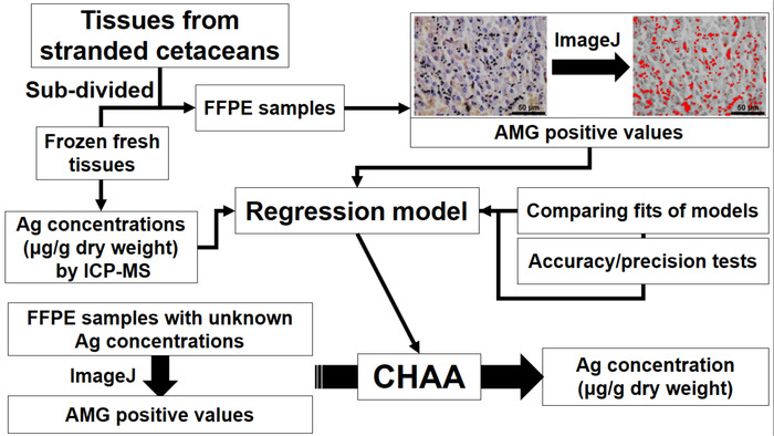
그림 1: Ag 농도 추정에 대 한 설립 및 cetacean 조직학 Ag 분석 결과 (욕설)의 응용 프로그램을 묘사 하는 순서도. CHAA cetacean 조직학 Ag 분석 결과, FFPE = = 포 르 말린 고정 파라핀 포함, ICP MS = 유도 결합된 플라즈마 질량 분광학. 이 그림의 더 큰 버전을 보려면 여기를 클릭 하십시오.

Protocol

연구 국제 지침에 따라 수행 하 고 cetacean 조직 샘플의 사용은 위원회의 농업의 대만 (연구 허용 104-07.1-SB-62)에 의해 허용 되었다. 1. 조직 샘플 준비 ICP MS 분석 참고: 간, 신장 조직 수집에서 갓 죽은 고 적당히 autolyzed 좌초 고래24, 4 종, 1 그램퍼스 griseus (Gg), 6 좌초 된 고래를 포함 하 여 2 Kogia 종 (코), 2 Lagenodelphis 호세이 …

Representative Results

Cetacean 간 및 신장 조직에 AMG 긍정적인 신호의 대표 이미지는 그림 5에 나와 있습니다. AMG 긍정적인 신호 근 신장 관 상피, hepatocytes, Kupffer 세포의 세포질에 다양 한 크기의 검은 알갱이 변함없이 크기의 갈색을 포함합니다. 때때로, 아 몰 퍼스 황금 노란색 갈색 AMG 긍정적인 신호 루멘과 일부 근 신장 tubules의 지하실 막에 설명 되어 있습니다. ICP-MS의 ?…

Discussion

문서 연구의 목적은 suborgan 수준에서 Ag 배포를 평가 하 고 cetacean 조직에 Ag 농도 추정 하는 보조 메서드를 설정 하는 것입니다. 현재 프로토콜 ICP-MS, 2) AMG 분석 알려진된 Ag 농도와 일치 하는 쌍 조직 샘플의 3) Ag 농도 추정에 대 한 회귀 모델 (욕설)의 설립에 의해 cetacean 조직에 Ag 농도의 결정 1) 포함 AMG 양수 값, 4)에 의해 평가의 정확성과 정밀도 CHAA, 및 5의) 욕설에 의해 Ag의 추정 농도.

<p class="jo…

Disclosures

The authors have nothing to disclose.

Acknowledgements

우리는 대만 Cetacean 좌초 네트워크 샘플 수집 및 저장, 대만 Cetacean 사회, 타이페이;를 포함 하 여에 대 한 감사 Cetacean 연구 실험실 (교수 선취 Siang Chou), 연구소의 생태학 및 진화 생물학, 국립 대만 대학, 타이 페이; 자연 과학 (박사 Chiou-주 야 오), 타이중;의 국립 박물관 그리고 해양 생물학 & Cetacean 연구 센터, 국립 쳉 쿵 대학. 우리 또한 그들의 허용에 대 한 임업 국, 농업 위원회, 행정원을 감사합니다.

Materials

HQ Silver enhancement kit Nanoprobes #2012
Surgipath Paraplast Leica Biosystems 39601006 Paraffin
100% Ethanol Muto Pure Chemical Co., Ltd 4026
Non-Xylene Muto Pure Chemical Co., Ltd 4328
Silane coated slide Muto Pure Chemical Co., Ltd 511614
Cover glass (25 x 50 mm) Muto Pure Chemical Co., Ltd 24501
Malinol Muto Pure Chemical Co., Ltd 20092
GM Haematoxylin Staining Muto Pure Chemical Co., Ltd 3008-1
10% neutral buffered formalin solution Chin I Pao Co., Ltd
Tip (1000 μL) MDBio, Inc. 1000
PIPETMAN Classic P1000 Gilson, Inc. F123602
15 ml Centrifuge Tube GeneDireX, Inc. PC115-0500
Dogfish liver National Research Council of Canada DOLT-2
Dogfish muscle National Research Council of Canada DORM-2
Inductively coupled plasma mass spectrometry (ICP-MS) PerkinElmer Inc. PE-SCIEX ELAN 6100 DRC
FreeZone 6 liter freeze dry system Labconco 7752030 For freeze drying
BRAND® SILBERBRAND volumetric flask Merck Z326283
30 mL standard vial, flat interior with 33 mm closure Savillex Corporation 200-030-12 For diagestion
Nitric acid, superpur®, 65.0% Merck 1.00441 For diagestion
Hot Plate/Stirrers Corning® PC-220 For diagestion
High Shear lab mixer Silverson SL2T For homogenization
Sterile polypropylene sample jar (250mL) Thermo Scientific™ 6186L05 For homogenization
Digital camera Nikon Corporation DS-Fi2
Light microscope Nikon Corporation ECLIPSE Ni-U
Shandon™ Finesse™ 325 manual microtome Thermo Scientific™ A78100001H
Accu-Cut® SRM™ 200 rotary microtome Sakura 1429
Microtome blade S35 FEATHER® 207500000
Slide staining dish and cover Brain Research Laboratories #3215
Steel staining rack Brain Research Laboratories #3003
Shandon embedding center Thermo Scientific™ S-EC
Shandon Citadel® tissue processor Thermo Scientific™ 69800003
Slide warmer Lab-Line Instruments 26005
Water bath Shandon Capshaw 3964
Filter paper Merck 1541-070
Prism 6.01 for windows GraphPad Software Statistic software
ImageJ National Institutes of Health
Stainless steel tissue embedding mould Shenyang Roundfin Trade Co., Ltd RD-TBM003 For paraffin emedding

References

  1. McGillicuddy, E., et al. Silver nanoparticles in the environment: Sources, detection and ecotoxicology. Science Total Environment. 575, 231-246 (2017).
  2. Yu, S. J., Yin, Y. G., Liu, J. F. Silver nanoparticles in the environment. Environmental Science: Processes and Impacts. 15 (1), 78-92 (2013).
  3. Hansen, S. F., et al. Nanoproducts- what is actually available to European consumers?. Environmental Science: Nano. 3 (1), 169-180 (2016).
  4. Vance, M. E., et al. Nanotechnology in the real world: Redeveloping the nanomaterial consumer products inventory. Beilstein Journal of Nanotechnology. 6, 1769-1780 (2015).
  5. Farre, M., Gajda-Schrantz, K., Kantiani, L., Barcelo, D. Ecotoxicity and analysis of nanomaterials in the aquatic environment. Analytical and Bioanalytical Chemistry. 393 (1), 81-95 (2009).
  6. Walters, C. R., Pool, E. J., Somerset, V. S. Ecotoxicity of silver nanomaterials in the aquatic environment: a review of literature and gaps in nano-toxicological research. Journal of Environmental Science and Health. Part A, Toxic/hazardous Substances & Environmental Engineering. 49 (13), 1588-1601 (2014).
  7. Levard, C., Hotze, E. M., Lowry, G. V., Brown, G. E. Environmental transformations of silver nanoparticles: impact on stability and toxicity. Environmental Science & Technology. 46 (13), 6900-6914 (2012).
  8. Massarsky, A., Trudeau, V. L., Moon, T. W. Predicting the environmental impact of nanosilver. Environmental Toxicology and Pharmacology. 38 (3), 861-873 (2014).
  9. Wang, H., et al. Toxicity, bioaccumulation, and biotransformation of silver nanoparticles in marine organisms. Environmental Science and Technology. 48 (23), 13711-13717 (2014).
  10. Buffet, P. E., et al. A marine mesocosm study on the environmental fate of silver nanoparticles and toxicity effects on two endobenthic species: the ragworm Hediste diversicolor and the bivalve mollusc Scrobicularia plana. Science of the Total Environment. 470, 1151-1159 (2014).
  11. Chen, M. H. Baseline metal concentrations in sediments and fish, and the determination of bioindicators in the subtropical Chi-ku Lagoon, S W Taiwan. Marine Pollution Bulletin. 44 (7), 703-714 (2002).
  12. Li, W. T., et al. Investigation of silver (Ag) deposition in tissues from stranded cetaceans by autometallography (AMG). Environmental Pollution. , 534-545 (2018).
  13. Chen, M. H., et al. Tissue concentrations of four Taiwanese toothed cetaceans indicating the silver and cadmium pollution in the western Pacific Ocean. Marine Pollution Bulletin. 124 (2), 993-1000 (2017).
  14. Li, W. T., et al. Immunotoxicity of silver nanoparticles (AgNPs) on the leukocytes of common bottlenose dolphins (Tursiops truncatus). Scientific Reports. , (2018).
  15. Bornhorst, J. A., Hunt, J. W., Urry, F. M., McMillin, G. A. Comparison of sample preservation methods for clinical trace element analysis by inductively coupled plasma mass spectrometry. American Journal of Clinical Pathology. 123 (4), 578-583 (2005).
  16. Bonta, M., Torok, S., Hegedus, B., Dome, B., Limbeck, A. A comparison of sample preparation strategies for biological tissues and subsequent trace element analysis using LA-ICP-MS. Analytical and Bioanalytical Chemistry. 409 (7), 1805-1814 (2017).
  17. Bischoff, K., Lamm, C., Erb, H. N., Hillebrandt, J. R. The effects of formalin fixation and tissue embedding of bovine liver on copper, iron, and zinc analysis. Journal of Veterinary Diagnostic Investigation. 20 (2), 220-224 (2008).
  18. Miller, D. L., Yu, I. J., Genter, M. B. Use of Autometallography in Studies of Nanosilver Distribution and Toxicity. International Journal of Toxicology. 35 (1), 47-51 (2016).
  19. Anderson, D. S., et al. Influence of particle size on persistence and clearance of aerosolized silver nanoparticles in the rat lung. Toxicological Sciences. 144 (2), 366-381 (2015).
  20. Kim, W. Y., Kim, J., Park, J. D., Ryu, H. Y., Yu, I. J. Histological study of gender differences in accumulation of silver nanoparticles in kidneys of Fischer 344 rats. Journal of Toxicology and Environmental Health, Part A. 72 (21-22), 1279-1284 (2009).
  21. Danscher, G. Applications of autometallography to heavy metal toxicology. Pharmacology Toxicology. 68 (6), 414-423 (1991).
  22. Deroulers, C., et al. Analyzing huge pathology images with open source software. Diagnostic Pathology. 8, 92 (2013).
  23. Shu, J., Dolman, G. E., Duan, J., Qiu, G., Ilyas, M. Statistical colour models: an automated digital image analysis method for quantification of histological biomarkers. BioMedical Engineering Online. 15, 46 (2016).
  24. Geraci, J. R., Lounsbury, V. J. Specimen and data collection. Marine mammals ashore: a field guide for strandings. , 167-230 (2005).
  25. Shih, C. -. C., Liu, L. -. L., Chen, M. -. H., Wang, W. -. H. . Investigation of heavy metal bioaccumulation in dolphins from the coastal waters off Taiwan. , (2001).
  26. Liang, C. S., et al. The relationship between the striatal dopamine transporter and novelty seeking and cognitive flexibility in opioid dependence. Progress in Neuro-Psychopharmacology and Biological Psychiatry. 74, 36-42 (2017).
  27. Spiess, A. N., Neumeyer, N. An evaluation of R2 as an inadequate measure for nonlinear models in pharmacological and biochemical research: a Monte Carlo approach. BMC Pharmacology. 10, 6 (2010).
  28. Stoltenberg, M., Danscher, G. Histochemical differentiation of autometallographically traceable metals (Au, Ag, Hg, Bi, Zn): protocols for chemical removal of separate autometallographic metal clusters in Epon sections. Histochemical Journal. 32 (11), 645-652 (2000).
  29. Dimitriadis, V. K., Domouhtsidou, G. P., Raftopoulou, E. Localization of Hg and Pb in the palps, the digestive gland and the gills in Mytilus galloprovincialis (L.) using autometallography and X-ray microanalysis. Environmental Pollution. 125 (3), 345-353 (2003).
  30. Loumbourdis, N. S., Danscher, G. Autometallographic tracing of mercury in frog liver. Environmental Pollution. 129 (2), 299-304 (2004).
  31. Stoltenberg, M., Larsen, A., Kemp, K., Bloch, D., Weihe, P. Autometallographic tracing of mercury in pilot whale tissues in the Faroe Islands. International Journal of Circumpolar Health. 62 (2), 182-189 (2003).

Play Video

Cite This Article
Li, W., Liou, B., Yang, W., Chen, M., Chang, H., Chiou, H., Pang, V. F., Jeng, C. Use of Autometallography to Localize and Semi-Quantify Silver in Cetacean Tissues. J. Vis. Exp. (140), e58232, doi:10.3791/58232 (2018).

View Video