The goal of this article is to provide a primer for the development and use of the VX2 carcinoma rabbit model for liver cancer.
The rabbit VX2 tumor is an animal model commonly utilized for translational research regarding hepatocellular carcinoma (HCC) in the field of Interventional Radiology. This model employs an anaplastic squamous cell carcinoma that is easily and reliably propagated in the skeletal muscle of donor rabbits for eventual harvest and allograft implantation into the liver of naïve recipients. This tumor graft rapidly grows within the liver of recipient rabbits into an angiographically identifiable tumor characterized by a necrotic core surrounded by a viable hypervascular capsule. The physical size of the rabbit anatomy is sufficient to facilitate vascular instrumentation allowing for the application and testing of various interventional techniques. Despite these benefits, there exists a paucity of technical resources to act as a concrete reference for researchers working with the model. Herein, we present a comprehensive visual outline for the technical aspects of development, growth, propagation, and angiographic utilization of the rabbit VX2 tumor model for use by novice and experienced researchers alike.
The rabbit VX2 tumor model has played a role in experimental oncology since its development in 19351,2. This tumor is a virus-induced anaplastic squamous cell carcinoma characterized by hypervascularity, rapid growth, and easy propagation in skeletal muscle3,4. While the rabbit VX2 tumor model has been used to investigate a multitude of cancers5,6,7,8; the focus of this paper is liver cancer9.
The purpose of the method described is to present a model for primary liver cancer, or hepatocellular carcinoma (HCC), that can be used by Interventional Radiologists for translational research. It can be used for pharmacokinetic studies, therapeutic investigations, and ablative method testing10,11,12,13,14,15.
The method detailed herein yields multiple advantages over other models within the same sphere such as rodent models like rats, mice, and woodchucks, or larger models like primates16. One of the primary benefits is the rapid and reliable tumor growth which allows researchers to establish an active tumor line within a month of first hind limb propagation17. Additionally, this tumor has straightforward sonographic visibility and a hypervascular periphery which allows for both transarterial locoregional treatments and ablative therapies. Finally, and most importantly, the size of the rabbit vasculature permits feasible and technically easy utilization of vascular instrumentation18.
The following protocol follows all requirements and guidelines mandated by the University of Illinois – Chicago. It was reviewed and approved by the local Institutional Animal Care and Use Committee prior to execution.
1. VX2 Hind Limb Tumor Development
2. VX2 Hind Limb Tumor Growth and Harvesting
Note: Assuming successful inoculation, there should be a palpable (3‒4 cm) tumor nodule at the injection site within 2 weeks. Usually, this nodule will be palpable around 1 week; however, it is better to let the tumor grow to allow for sufficient tissue collection. Typically, this nodule will be firm, indurated, and elevated above the level of the muscle. If
3. Liver Tumor Implantation via Laparotomy
4. VX2 Tumor Suspension Preparation
5. Angiographic Utilization of the VX2 Liver Tumor
When looking at Figure 1, it is clear that the quadricep of the rabbit is enlarged. Additionally, multiple small discrete nodules, typically correlating with tumor growth through the fascia, are visible. Upon palpation, the injected limb should appear than the non-injected limb. If a researcher requires more definitive assurance of tumor presence, ultrasound imaging can be used to identify the tumor embedded in the muscle. If a tumor is not detected, the hind limb should be re-injected with a tumor cell suspension.
In order to confirm successful vascular access, blood return into the sheath is observed on aspiration as seen in Figure 13D. If vascular access was unsuccessful, attempted aspiration will yield air in the sheath or present with significant resistance when pulling the plunger of the syringe.
For liver tumor growth, there are two ways to confirm successful propagation: angiographically and on necropsy. On angiography, identification of the tumor may occur immediately as is the case in Figure 14A where the tumor draws blood supply directly from the common hepatic artery. It may also take some time in cases where the tumor is lateral as is the case in Figure 14D. If the tumor is not readily visible after injection of contrast into the common hepatic artery, the researcher should attempt to inject contrast into left and right hepatic arteries in order to improve the chances of highlighting the tumor. It may also help to look for aberrant arteries traveling laterally towards the distal edge of the liver as seen in Figure 14C. On necropsy, the tumor should be readily visible as seen in Figure 15B (compare to Figure 15A).
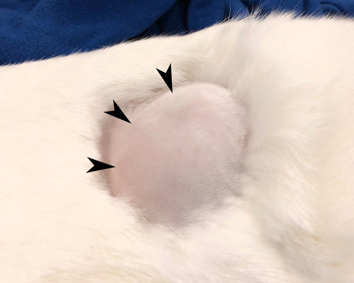
Figure 1: Rabbit hind limb. Shaved rabbit hind limb with mass indicative of tumor growth. Please click here to view a larger version of this figure.
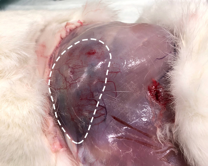
Figure 2: Exposed hind limb. The same limb as shown Figure1 with overlying skin reflected revealing a large area of hypervascularity and discoloration distinct from the surrounding muscle representing the location of the tumor (white dotted line). Please click here to view a larger version of this figure.
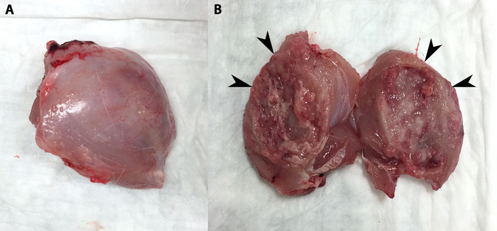
Figure 3: Tumor removed en bloc and bisected. (A) Tumor and surrounding muscle removed en bloc. (B) Tumor has been bisected to reveal its capsular wall (arrowheads) and necrotic core. Tumor process can be seen in both halves as well as some necrotic debris. Please click here to view a larger version of this figure.
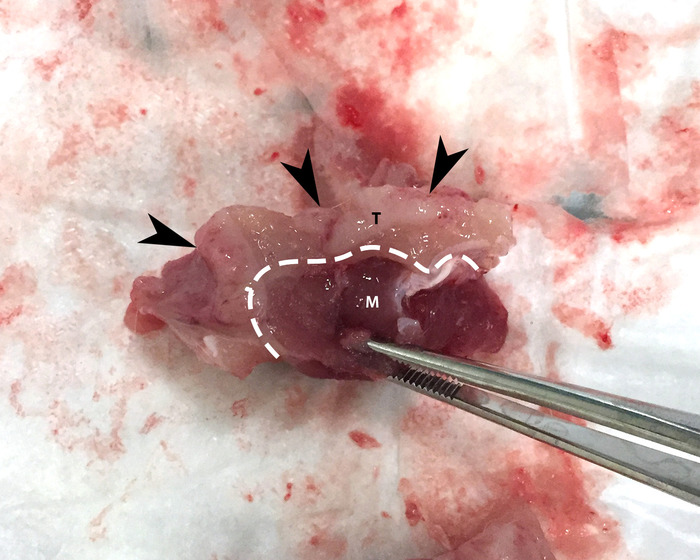
Figure 4: Tumor capsule. Arrowheads denote a piece of tumor (T), which is delineated from adjacent muscle (M) by the tumor capsule (dashed white line). Please click here to view a larger version of this figure.
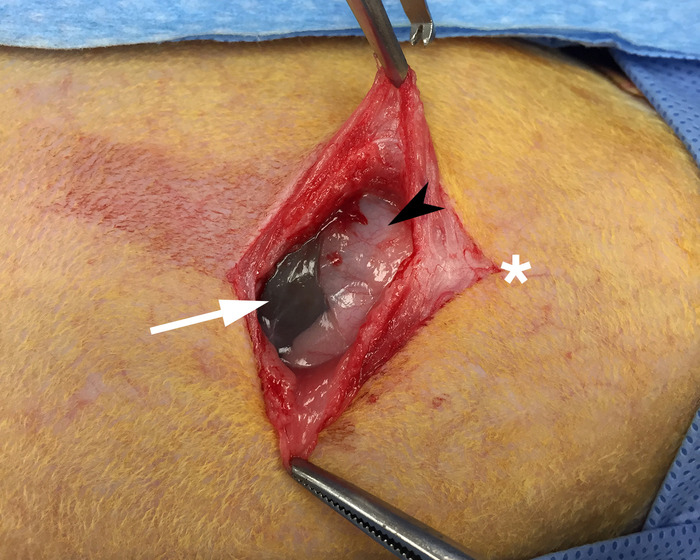
Figure 5: Exposed xiphoid process. Skin and underlying muscle have been reflected to allow for visualization of the xiphoid process (black arrow) and gut (white arrow). The white star denotes the cranial direction. Please click here to view a larger version of this figure.
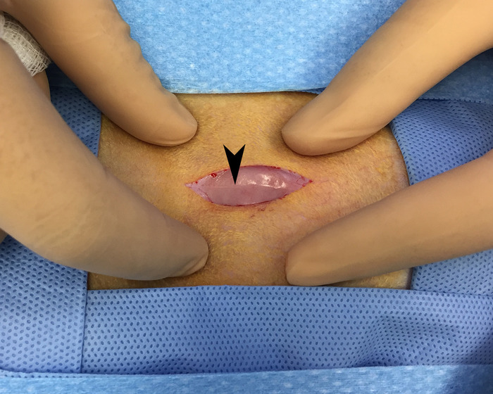
Figure 6: Linea alba. Overlying skin and fascia have been reflected to allow for visualization of the linea alba (black arrow) running in a cranial to caudal direction. This area is avascular and provides for blood-loss free access of the peritoneal space. Please click here to view a larger version of this figure.
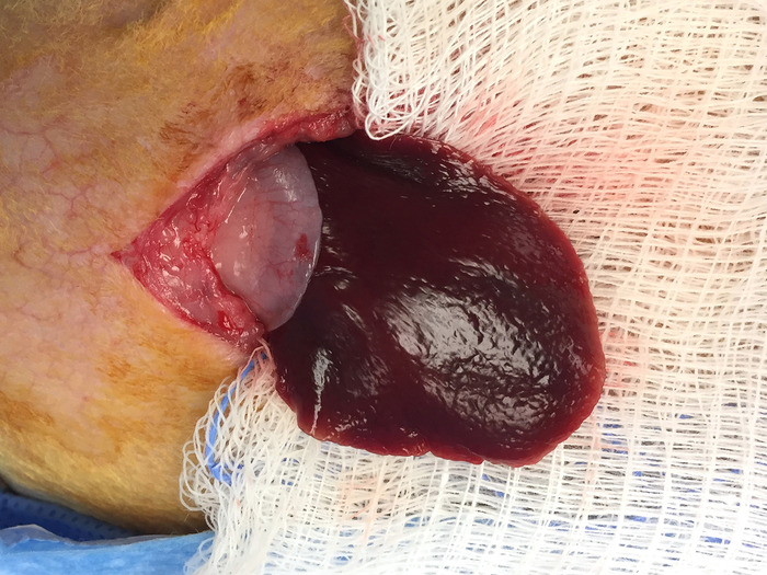
Figure 7: Lobe of liver outside peritoneum. This image shows a lobe of the liver that was gently extracted from the peritoneal space and placed on a piece of gauze. Please click here to view a larger version of this figure.
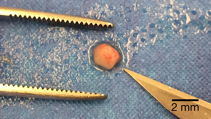
Figure 8: Post-processed tumor piece for implantation. A piece of tumor processed to the appropriate size for implantation placed next to the tip of a #11-blade for scale. Please click here to view a larger version of this figure.
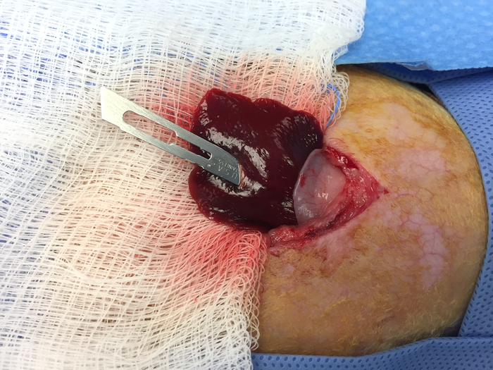
Figure 9: Creating a pocket in the liver for tumor implantation. An #11-blade is inserted to the appropriate depth in the extracted lobe of the liver. This will create an appropriately sized pocked for the implantation of the tumor piece from Figure 8. Please click here to view a larger version of this figure.
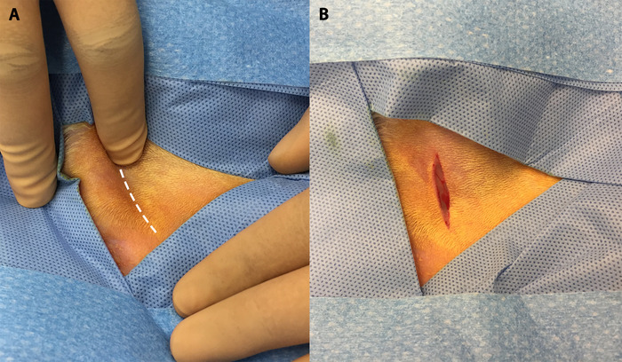
Figure 10: The femoral groove and initial incision. (A) Palpation of the hind limb allows for visualization of the femoral groove (white dotted line). (B) Initial incision in the hind limb made along the femoral groove. Please click here to view a larger version of this figure.
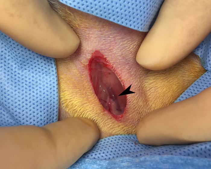
Figure 11: Identification of the femoral bundle. Blunt dissection of the initial incision reveals femoral vein (black arrow). Please click here to view a larger version of this figure.
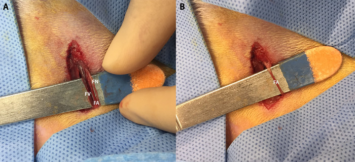
Figure 12: Dissection of femoral bundle and isolation of femoral artery. (A) Dissection of the femoral bundle allows us to individually distinguish (from left to right) the femoral vein (FV), femoral artery (FA), and femoral nerve (FN). (B) The femoral artery isolated on a scalpel handle. Note the blood column allowing for distinction from the femoral nerve. Please click here to view a larger version of this figure.
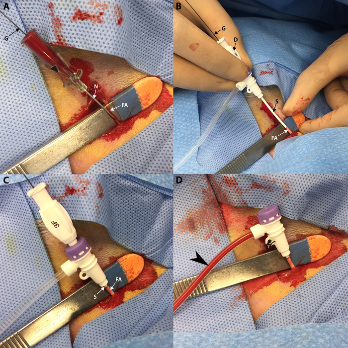
Figure 13: Vascular access. (A) A guidewire (G) is advanced into the femoral artery (FA) through the access needle (N) which was previously inserted into the femoral artery. (B) A sheath (S) and dilator (D) are advanced over the guidewire (G) into the femoral artery (FA). (C) Sheath (S) and dilator are advanced fully into the femoral artery (FA) up to the sheath hub. (D) Sheath is secured with silk after the dilator and guidewire have been removed. Aspiration yields blood (black arrow) in the sheath. Please click here to view a larger version of this figure.
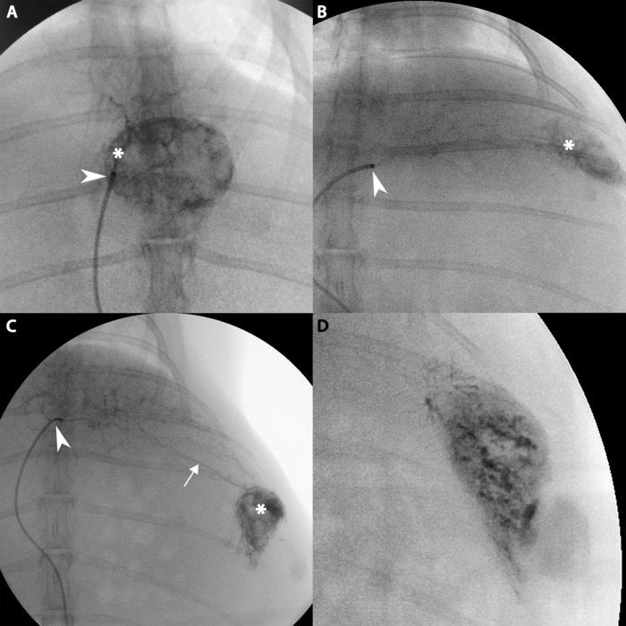
Figure 14: Angiographic imaging of hepatic tumor. (A) Catheter tip (white arrow) delivering contrast directly into artery feeding the tumor (white star). (B) Catheter tip (white arrow) delivering contrast into distal left hepatic artery and moderate contrast uptake by lateral tumor (white star). (C) Further contrast injection into tumor from B demonstrating an aberrant artery (white line) traveling from the catheter (white arrow) to the tumor (white star). (D) The tumor from B after further contrast uptake. Please click here to view a larger version of this figure.
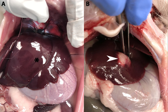
Figure 15: Rabbit liver. (A) A healthy rabbit liver showing the left medial lobe (white star) overlying the left lateral lobe (black star). (B) A rabbit liver with a fully developed hepatic tumor (white arrow). Please click here to view a larger version of this figure.
The first critical step in the VX2 tumor methodology is successful propagation of a tumor in the hind limb of a donor rabbit. Refer to the first paragraph in the "Representative Results" section for more information regarding this step.
The next critical step is ensuring that the viable tumor capsule is properly identified. Not only will this be necessary for tumor suspension preparation, but it is also important for selecting and generating tumor pieces for hepatic implantation. The demarcation between viable tumor and surrounding muscle tissue is annotated in Figure 4. If the incorrect tissue sample is scraped during suspension preparation, subsequent hind limb propagation will fail. If this occurs during hepatic implantation, tumor growth in the liver will not occur. This will not be apparent until angiography.
During the hepatic implantation process, care should be taken when approaching the left lobe of the liver. Oftentimes, if the liver is readily apparent when entering the peritoneum, it is actually the medial lobe of the liver that operators are observing. Implantation into the medial lobe of the liver presents a handful of issues for angiographic use. The first is the medial lobe's anatomic relationship with the spine. A medial lobe tumor can often be obfuscated by the spine on fluoroscopy making confirmation and treatment of the tumor difficult. Additionally, the gastroduodenal artery is more often associated with the vasculature supplying the medial lobe. This increases the risk of non-target embolization of the gut and can potentially lead to bowel ischemia/infarction and possible death of the animal. As stated earlier, this will not qualify as a failure; however, it does warrant more care during visualization and treatment.
The final critical step is successful and stable femoral artery access. As seen in Figure 12, the femoral artery should be isolated atop a scalpel blade handle. While this is mostly done to allow the researcher to more accurately perform the Seldinger technique20, it should be maintained throughout the procedure. This is because removing the scalpel handle once the sheath has been introduced can cause unintended motion of the sheath within the vasculature leading to sheath occlusion and possible damage of the vasculature and surrounding structures. If the sheath becomes dislodged during the procedure, apply pressure in the femoral groove proximal to the access site in order to stop the bleeding, and the artery can then be ligated. Do not attempt to reinsert the sheath. The researcher can attempt to gain access from the contralateral femoral artery.
While the VX2 platform is a robust model in current use for translational research regarding HCC, it does have relevant shortcomings. The primary weakness of this model is that its disease state does not mimic that of a human HCC. The tumor induced is not pathologically similar to human HCC nor is the non-cirrhotic liver parenchymal microenvironment. Moreover, the VX2 tumor shows substantial internal necrosis, which excludes this model from use for treatment efficacy studies. Some alternative models include rodent models such as mice, rats, and woodchucks or larger animal models such as pigs and primates.16,21 These alternatives all provide different advantages and disadvantages; however, in the opinion of the authors, for angiographic utilization and cost efficacy, the VX2 rabbit remains dominant.
The authors have nothing to disclose.
We would like to acknowledge the veterinary staff at the University of Illinois – Chicago's Biological Resources Laboratory.
MethoCult (Methycellulose) | Stemcell Technologies | M3134 | |
VX2 Cell Line | NCI | VX-2 | |
5 mL Syringe | BD | 309646 | |
16-Gauge Needle | BD | 305197 | |
22-Gauge Needle | BD | 305155 | |
Hair Clippers | Wahl | 41870-0438 | |
Foam Insulated Box | Mr. Box Online | 10 x 10 x 4 | |
Acepromazine | Henry Schein | 003845 | |
Buprenorphine | Par | 42023-179-05 | |
Meloxicam | Henry Schein | 049755 | |
Alcohol Pads | Covidien | 5033 | |
Ketamine | Henry Schein | 056344 | |
Xylazine | Akorn | 59399-110-20 | |
Pentobarbital (Fatal-Plus) | Vortech | 9373 | |
Sterile Petri Dish | Thermo Fisher | 172931 | |
DMEM | Gibco | 11965092 | |
Saline | Baxter | 2F7124 | |
15-Blade | Steris | 02-050-015 | |
Scalpel Handle x 2 | Steris | 22-2381 | |
Curved Hemostat | WPI | 501288 | |
Atraumatic Forceps | Sklar | 52-5077 | |
Gauze | Medline | NON21430LF | |
11-Blade | Steris | 02-050-011 | |
Surgicel | Ethicon | 1951 | |
3-0 PDS / Taper | Ethicon | Z305H | |
4 – 0 Vicryl / Cutting | Ethicon | J392H | |
40 micron strainer | BD | 352340 | |
50 mL conical tube | Thermo Fisher | 339652 | |
plastic pipette | Thomas Scientific | HS206371B | |
Centrifuge | Sorvall | 75004240 | |
1.40mL Tubes (Internal Thread) | Micronic | MP32131-Z20 | |
3-F VSI Micro-HV Introducer Kit | Vascular Solutions | Custom Order (P15180391) | |
.018 45-degree angle glidewire | Terumo | RG*GA1818SA | |
Direxion bern-shape microcatheter | Boston Scientific | M001195230 | |
Omnipaque | GE | Y510 |