We present a protocol for selective chemical lumbar sympathectomy (CLS), which inactivates only gray rami communicantes and not the sympathetic trunk. Selective CLS can help achieve therapeutic efficacy in vasodilation, sweat reduction, and pain relief that are comparable to conventional CLS, and serious complications, particularly ureteropelvic damages, can be reduced.
Chemical lumbar sympathectomy (CLS) is a commonly used, minimally invasive procedure for the treatment of conditions including ischemic diseases of the lower extremities, hyperhidrosis, etc. It is commonly practiced to position the puncture needle tip in front of the anterior fascia of the psoas major muscle and inject the inactivating agent around the sympathetic trunk, which is defined as conventional CLS. Although relatively rare, ureteropelvic damage is the most frequently reported complication of conventional CLS and can cause serious harm to patients. We found that injecting the inactivating agent behind the anterior fascia, which only targets gray rami communicantes, helped achieve therapeutic efficacy in vasodilation, sweat reduction, and pain relief comparable to conventional CLS, and serious complications were largely reduced. We define this procedure as selective CLS. Here, we present a protocol of selective CLS. The precise needle tract and accurate evaluation of the spreading of the contrast agent are critical to ensure that the drug is injected behind the anterior fascia of the psoas major muscle. The needle tip is at approximately one-third the dividing line of the vertebral body in the lateral view of a lumbar X-ray. The contrast is mainly confined around the needle tip and spreads outward and downward along the psoas muscle fibers. In this way, the anterior fascia provides a natural barrier for the ureteropelvic area, and the psoas major muscle provides a natural barrier for the lumbar nerve root. There are several highlights of this article, including 1) a detailed description of the selective CLS procedures, 2) an explanation of the anatomical basis for the implementation of selective CLS, and 3) an explanation of the differences between selective and conventional CLS.
Chemical lumbar sympathectomy (CLS) has been shown to be an effective treatment for ischemic diseases1,2,3,4,5 including thromboangiitis obliterans (sometimes called Buerger's disease), ischemic diabetic foot, Raynaud's disease, parmoplantar hyperhidrosis6, erythromelalgia7,8,9, and livedo reticularis10. It has replaced open surgery due to a number of advantages. It is minimally invasive and economically attractive, it does not require general anesthesia and hospitalization, and it can be performed repeatedly. However, exquisite precision is critical. For example, paraplegia has been reported in CLS using blind technique11. The precision of the procedure is greatly improved with radiographical guidance to control the exact position of the puncture needle tip and puncture path and depth, through the use of contrast media to visualize treatment areas. However, even with radiographical guidance, damages to adjacent organs, particularly ureteropelvic organs12,13,14,15,16,17,18,19, have still been reported.
It is commonly practiced to position the needle tip beyond the anterior fascia of the psoas major muscle and inject the drug around the sympathetic trunk8,12,13,14,15, which we define as conventional CLS. However, because the location of the sympathetic trunk and ureter are both in front of the anterior fascia, inactivating agents may spread to the ureter and cause damage. In reports of ureteropelvic damage that presented radiographic images12,13,14,15, the drug was injected in front of the anterior fascia, according to contrast spreading.
Based on our prior experience performing CLS procedures, we have found that targeting gray rami communicantes comparably effective and safer compared to targeting the sympathetic trunk. Gray rami communicantes are postganglionic sympathetic fibers located behind the sympathetic trunk and in front of the lumber nerve root, running along the lateral edge of the vertebral body, mostly within the psoas major muscle. Thus, the anterior fascia provides a good barrier for the ureteropelvic area, and the psoas major muscle provides a good barrier for the lumbar nerve root. We define this procedure as selective CLS, and the technical details of selective CLS are described in this protocol.
The protocol has been approved by the local medical and ethics committees. The procedure lasts approximately 15 min per side.
1. Indications
2. Contraindications
3. Preoperative Preparation
4. Treatment Procedure
5. Postoperative Care
The needle tip should be in the optimal area as shown in Figure 1. In selective CLS, the contrast is injected behind the anterior fascia of the psoas major muscle, targeting gray rami communicantes. A comparison of contrast spreading in selective CLS (A) and conventional CLS (B) is shown in Figure 2.
The expected clinical effects are vasodilation (for treatment of Raynaud's syndrome, livedo reticularis, thromboangiitis obliterans, ischemic diabetic foot, and erythromelalgia; Figure 3), sweat reduction (for treatment of hyperhidrosis), and pain relief (for treatment of erythromelalgia and phantom limb pain).
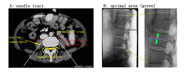
Figure 1: Relationship between puncture needle and surrounding organs/tissues. (A) A diagram of an axial lumber CT scan at the level of the needle tract. The white line represents the needle tract. It should be noted that the needle tip reaches close proximity to, but does not break through, the anterior fascia of the psoas muscle (red arc). A = abdominal aorta, V = inferior vena cava. (B) Lateral view of a lumbar X-ray. The green area illustrates the optimal area, located beside the lumbar vessels (usually located in the middle of the vertebral body horizontally), behind the anterior fascia of the psoas muscle and away from the lumbar nerve root behind the vertebral body. This is a safe location, closest to the gray rami communicantes. A = lumbar artery, V = lumbar vein. Please click here to view a larger version of this figure.
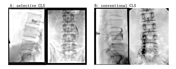
Figure 2: Comparison of contrast agent spreading in X-ray views between selective CLS (A) and conventional CLS (B). (A) Selective CLS. The contrast agent was injected behind the anterior fascia of the psoas muscle. The lateral view showed that the contrast agent spread around and behind the needle tip. The anterior-posterior (AP) view showed that the contrast agent diffused outward and downward along the spaces among psoas muscle fibers and formed thin strips, outlining the contour of the muscle fibers. (B) Conventional CLS. The contrast agent was injected in front of the anterior fascia of the psoas muscle. The lateral view showed that the contrast was concentrated in loose tissue in front of the needle tip. The AP view showed that the contrast agent was concentrated along the vertebral body, thus overlapping with the vertebral body in X ray view, and did not diffuse outward. Please click here to view a larger version of this figure.
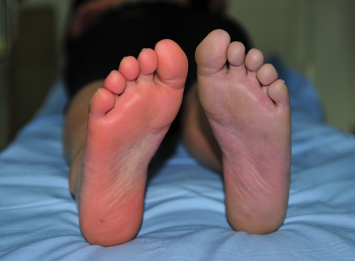
Figure 3: Clinical observation after selective CLS on the right side of a patient with erythromelalgia. The patient's blood vessels dilated, skin temperature increased, and skin was rosy and dry on the right limb. In contrast, skin was damp and cold with a slight violaceous skin color on the left side without treatment. Please click here to view a larger version of this figure.
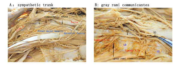
Figure 4: Anatomic illustration of sympathetic trunk and gray rami communicantes. (A) Anatomically, the psoas muscle is attached to the lateral side of the vertebral body and the lumbar disc (LD). In this illustration, most of the anterior fascia of the psoas muscle is removed, exposing the psoas muscle. The lumbar sympathetic trunk, located in front of the anterior fascia and adjacent to the abdominal aorta (right lumbar sympathetic trunk adjacent to inferior vena cava, not shown). The enlarged lumbar sympathetic ganglia (SG) are also visible. (B) With the psoas muscle removed from the outside of the vertebral body, the lumbar blood vessels and gray rami communicantes covered in the deep side of the psoas muscle are exposed. The gray rami communicantes originate from the sympathetic ganglia, extend through the anterior fascia, extend backward along the lateral edge of the vertebral body, surround the lumbar blood vessels, and finally join the lumbar nerve roots. Please click here to view a larger version of this figure.
Here we classify CLS as either conventional or selective CLS, with the major difference being the targeting of sympathetic trunk vs. gray rami communicantes (injection of phenol in front of vs. behind the anterior fascia of the psoas major muscle). We have implemented selective CLS in young, female patients with livedo reticularis10, children with erythromelalgia7,9, and senile patients with end-stage arteriosclerosis obliterans and high-risk comorbidities, and we have confirmed its long-lasting, beneficial effects and safety.
Ureteropelvic damage is the most frequently reported complication of CLS. There have been more than 10 cases reported12,13,14,15,16,17,18,19. In one report, the incidence was reported as 1.24% (3 fibrotic ureter stenoses out of 241 CT-guided CLS)16, and the author concluded that damage to the ureter may follow even when the procedure is technically satisfactory. Although relatively rare, this complication is serious and causes great harm to the patient. In selective CLS, anterior fascia can provide a natural protective barrier of important organs such as the ureter, abdominal aorta, and inferior vena cava. In our hospital, ureteropelvic damage has never occurred in more than 300 CLS procedures.
The risks of sympathetic trunk inactivation have been recognized in thoracic sympathectomy, and selective sympathicotomy (ramicotomy) has been reported under video-assisted thoracoscopic surgery20,21,22. The anatomical distribution of lumber sympathetic nerves is different from thoracic nerves, which makes implementation of selective CLS possible (see Figure 4 and explanation below).
The lumbar sympathetic trunk is located in front of the anterior fascia of the psoas muscle, and this location is relatively fixed (Figure 1, Figure 4). However, the lumbar sympathetic ganglia have large variations in their relative positions to the vertebral body and in numbers (2–6 in the general population, most commonly 4, maximum of 8) among different individuals. Even within the same individual, distribution of the ganglia is not necessarily symmetrical. In some cases, the ganglia are located beside the vertebral bodies, and in others, they are located beside the lumbar discs. The gray rami communicantes originate from the sympathetic ganglia, and the relative positions to the vertebral body from where they originate are highly variable, too. The relative positions to the vertebral bodies from where communicantes merge into the lumbar nerve roots tend to be consistent, because the locations of lumbar nerve roots are fixed. This makes it possible to target gray rami communicantes without open surgery.
The optimal injection area (Figure 1) also successfully avoids the lumbar arteries and veins across the vertebral body, reducing the risk of operative and postoperative bleeding. More importantly, the psoas muscle attached to the lateral side of the vertebral body becomes a natural protective barrier of the lumbar nerve root, protecting the lumbar nerve root behind the psoas muscle and ensuring that no major nerve damage occurs.
There have been no complications other than mild transient genitofemoral neuralgia, which occurred in less than 20% of all cases treated by selected CLS, and all spontaneously resolved within a few days at our hospital. The incidence, intensity, and duration reported in the literature were highly variable, with ranges between 5 and 66%, from mild to significant, and lasting days to years2,23,24,25,26,27,28. The genitofemoral nerve originates from the L1/L2 segments of the lumbar plexus, then passes downwards and forwards along the psoas muscle and through the anterior fascia of the psoas muscle at approximately the L3 level. Injecting a small amount of drug behind the anterior fascia at L3/L4 would theoretically decrease the chance and degree of genitofemoral nerve damage.
Kuntz's fibers have been blamed for surgical failures of endothoracic sympathectomy29. There are no published reports on Kuntz's fibers in lumber sympathetic nerve. To ensure satisfactory and persistent sympathetic interruption in CLS, it is recommended to inject the 2 mL of phenol in the optimal area (Figure 1B) in the directions of both the head and feet. Moreover, if sympathetic interruption is not satisfactory, it is recommended to inject supplemental phenol to another optimal area or at the L5, which is preferred over injection of a higher dose in the same area. In this regard, the chances of inactivating gray rami communicantes may be highly increased, and a higher degree of safety can be achieved.
Gray rami communicantes are unmyelinated nerves, which are more vulnerable to phenol compared to myelinated nerves. In our experience, 2 mL of 5% phenol is usually sufficent for each injection site. We have used a 4 mL injection in an earlier case, and we gradually lowered the amount to 2 mL.
In summary, we describe the procedure of selective CLS, which targets gray rami communicantes and not the sympathetic trunk, and we emphasize the anterior fascia of the psoas major muscle as a protective barrier when performing selective CLS. The precise needle tract, accurate evaluation of the contrast agent spreading, and multiple injections with small doses of phenol are all critical in ensuring the success of selective CLS.
The authors have nothing to disclose.
This work is funded by grants from Peking University Third Hospital Scientific Research Foundation for the Returned Overseas Scholars (Grant No: 77434-01, Long Zhang) and Beijing Higher Education Young Elite Teacher Project (Grant No: YETP0072, Wen-Hui Wang).
0.9% Saline solution | Chinese Dazhong Pharmaceutical Co.,Ltd. | 8F84D7 | 10 mL ⅹ2 |
10-cm 9# puncture needle | Peking University Third Hospital | Not Applicable | 7# for children |
2% Lidocaine Hydrochloride Injection solution | Hubei Tiansheng Pharmaceutical Co.,Ltd. | 20180434 | 5 mL ⅹ2 |
5% Phenol diluted in aqueous solution | Peking University Third Hospital | Not Applicable | 10 mL |
Angiography system (DSA) | Philips | FD20 | have X-ray fluoroscopy function |
Aseptic puncture kit | Peking University Third Hospital | Not Applicable | 6 pieces of surgical towel, gauze, cotton balls, blood vessel forceps, etc. |
Contrast media(iodixanol injection; 320) | GE Healthcare | 14160155 | 320 mg I/mL |
Infrared frontal thermometer | OMRON health care (China) Co.,Ltd. | MC-720 | |
Iodophors skin disinfectants | Shanghai Likang Disinfectant Hi-Tech Co., Ltd. | 20180404 | |
Sterile surgical gloves | Beijing Ruijing Latex products Co., Ltd. | 2018070352 | |
Syringes and needles 10 mL | Shandong Weigao Group Medical Plolymer Co.,Ltd. | 20180711 | 10 mL ⅹ2 |
Syringes and needles 5 mL | Shandong Weigao Group Medical Plolymer Co.,Ltd. | 20180721 | 5 mL ⅹ1 |