We describe a protocol for the chemical conjugation of the model antigen ovalbumin to an endocytosis receptor-specific antibody for in vivo dendritic cell targeting. The protocol includes purification of the antibody, chemical conjugation of the antigen, as well as purification of the conjugate and the verification of efficient conjugation.
Targeted antigen delivery to cross-presenting dendritic cells (DC) in vivo efficiently induces T effector cell responses and displays a valuable approach in vaccine design. Antigen is delivered to DC via antibodies specific for endocytosis receptors such as DEC-205 that induce uptake, processing, and MHC class I- and II-presentation.
Efficient and reliable conjugation of the desired antigen to a suitable antibody is a critical step in DC targeting and among other factors depends on the format of the antigen. Chemical conjugation of full-length protein to purified antibodies is one possible strategy. In the past, we have successfully established cross-linking of the model antigen ovalbumin (OVA) and a DEC-205-specific IgG2a antibody (αDEC-205) for in vivo DC targeting studies in mice. The first step of the protocol is the purification of the antibody from the supernatant of the NLDC (non-lymphoid dendritic cells)-145 hybridoma by affinity chromatography. The purified antibody is activated for chemical conjugation by sulfo-SMCC (sulfosuccinimidyl 4-[N-maleimidomethyl] cyclohexane-1-carboxylate) while at the same time the sulfhydryl-groups of the OVA protein are exposed through incubation with TCEP-HCl (tris (2-carboxyethyl) phosphine hydrochloride). Excess TCEP-HCl and sulfo-SMCC are removed and the antigen is mixed with the activated antibody for overnight coupling. The resulting αDEC-205/OVA conjugate is concentrated and freed from unbound OVA. Successful conjugation of OVA to αDEC-205 is verified by western blot analysis and enzyme-linked immunosorbent assay (ELISA).
We have successfully used chemically crosslinked αDEC-205/OVA to induce cytotoxic T cell responses in the liver and to compare different adjuvants for their potential in inducing humoral and cellular immunity following in vivo targeting of DEC-205+ DC. Beyond that, such chemically coupled antibody/antigen conjugates offer valuable tools for the efficient induction of vaccine responses to tumor antigens and have been proven to be superior to classical immunization approaches regarding the prevention and therapy of various types of tumors.
Dendritic cells (DC) are central players of the immune system. They are a diverse group of cells specialized in antigen-presentation and their major function is to bridge innate and adaptive immunity1,2. Importantly, DC not only play an important role in efficient and specific pathogen-directed responses but are also involved in many aspects of antitumor immunity1,3.
Due to their exclusive role in host immunity, DC came into focus as target cells for vaccination4. One approach is to target antigens to DC in vivo to induce antigen-specific immune responses and over the last years, a large number of studies have been dedicated to defining suitable receptors and targeting strategies1,4. One example is the C-type lectin receptor DEC-205, which can be targeted by DEC-205-specific antibodies to induce endocytosis. Importantly, DEC-205 targeting in the combination with suitable adjuvants has been shown to efficiently induce long-lived and protective CD4+ and CD8+ T cells, as well as antibody responses, also against tumor antigens3,5,6,7,8,9.
There are a number of studies showing conjugated antigens targeted to DC to be superior to free un-conjugated antigen3,5,10,11,12. This makes the conjugation of the antigen to the respective DC targeting moiety a central step in DC targeting approaches. In the case of DC targeting via antibodies or antibody fragments, antigens can be either chemically or genetically linked and either strategy provides its own (dis)advantages1. On the one hand, in genetically engineered antibody-antigen constructs there is a control over the antigen dose as well as the location providing superior comparability between lots1. At the same time however, chemical conjugation needs less preparation and provides more flexibility especially when attempting to test and compare different antigens and/or vaccination strategies in experimental and pre-clinical models.
Here, we present a protocol for the efficient and reliable chemical conjugation of ovalbumin (OVA) as a model protein antigen to a DEC-205-specific IgG2a antibody (αDEC-205) suitable for in vivo DC targeting in mice. First, αDEC-205 is purified from NLDC-145 hybridoma cells13. For chemical conjugation, the heterobifunctional crosslinker sulfosuccinimidyl 4-[N-maleimidomethyl] cyclohexane-1-carboxylate (sulfo-SMCC), which contains NHS (N-hydroxysuccinimide) ester and maleimide groups, is used, allowing covalent conjugation of amine- and sulfhydryl-containing molecules. Specifically, the primary amines of the antibody initially react with sulfo-SMCC and the resulting maleimide-activated αDEC-205 then reacts with the sulfhydryl-containing OVA protein reduced through TCEP-HCl (Tris(2-carboxyethyl) phosphine hydrochloride). The final product is chemically conjugated αDEC-205/OVA (Figure 1). Beyond chemical conjugation itself, our protocol describes removal of excess OVA from the conjugate as well as the verification of successful conjugation through western blot analysis and a specific enzyme-linked immunosorbent assay. We have successfully employed this approach in the past to chemically conjugate OVA and other proteins or immunogenic peptides to αDEC-205. We demonstrate efficient binding to CD11c+ cells in vitro as well as the efficient induction of cellular and humoral immunity in vivo.
Certainly, there are drawbacks to this method such as in lot-to-lot comparability and in the exact dosing of the antigen within the final conjugate. Nevertheless, chemical conjugation provides experimental flexibility in the choice of the antibody and the protein antigen as compared to genetically engineered constructs. Therefore, we believe this approach is especially valuable in evaluating different antigens for their efficiency in DC targeting in pre-clinical mouse models, importantly also in the context of specific antitumor immune responses.
All of the described animal experiments were approved by the local government agency (Niedersächsisches Landesamt für Verbraucherschutz und Lebensmittelsicherheit; file number 33.12-42502-04-10/0108) and were performed according to the national and institutional guidelines.
1. Production of αDEC-205 from the hybridoma cell line NLDC-145
2. Purification of the αDEC-205 antibody from the NLDC-145 cell supernatant
NOTE: From the NLDC-145 cell supernatant, αDEC-205 is purified using a protein G Sepharose column (reusable). The column dimensions are 15 mm x 74 mm and 5 mL protein G Sepharose are packed per column.
3. Chemical conjugation of OVA to αDEC-205
NOTE: A ratio of 0.5 mg OVA protein to 2.5 mg αDEC-205 (1:5) is required for optimal chemical conjugation. However, this ratio can vary for other proteins and antibodies and needs to be optimized for alternative conjugates. Reduction of the disulfide bonds of the OVA protein is performed through incubation with 30 mM TCEP-HCl, which exposes the sulfhydryl-groups for chemical conjugation to αDEC-205 and 240 µl of TCEP-HCl are needed in step 3.2. Both steps, TCEP-induced reduction of OVA (step 3.1.) and sulfo-SMCC activation of αDEC-205 (step 3.2.), should preferably be performed in parallel.
4. Verification of the chemical conjugation by western blot
NOTE: For verification of successful chemical conjugation, western blot analysis detecting either OVA (4.2) or αDEC-205 (4.10) is performed. Detection of OVA (4.2.) or αDEC-205 (4.10.) should be performed in parallel. An orbital platform shaker should preferably be used for all incubation steps of the western blot membranes to allow uniform distribution of the respective solutions.
5. Verification of the chemical conjugation by ELISA
Chemical conjugation of αDEC-205 to OVA protein using this protocol will typically allow efficient generation of αDEC-205/OVA for in vivo DC targeting approaches. There are different strategies to verify the technique itself and to test the functionality of the yielded conjugate. Western blot analysis and ELISA are used to verify successful conjugation and at the same time detect potentially left free OVA (Figure 2). In vitro binding studies (Figure 3) and in vivo immunizations (Figure 4) confirm binding of the conjugate to DEC-205 and targeting of DC.
Parallel western blot analysis is used to detect both the conjugated OVA (Figure 2A) as well as conjugated αDEC-205 (Figure 2B). Specifically, the positive signal for OVA at the level of the antibody's molecular weight in the blot confirms association of OVA and the antibody (Figure 2A). Furthermore, staining for OVA in the western blot analysis allows the detection of excess free OVA potentially still present next to the αDEC-205/OVA yielded in step 3.6., which is not the case for the blot shown (Figure 2A). In case large amounts of free OVA are detected, steps 3.4.1. to 3.4.8. of the protocol should be repeated. Complementary to the staining for OVA (Figure 2A), staining for αDEC-205 in western blot analysis verifies successful conjugation through an increase in the molecular weight between "naked" αDEC-205 and the conjugate as shown in Figure 2B.
Next to western blotting, also a specific ELISA allows verification of the successful conjugation of αDEC-205 to OVA. In contrast to the western blot analyses however, this ELISA does not allow the detection of free and un-conjugated αDEC-205 or OVA. Due to the assay setup (Figure 2C), a positive signal is only produced if conjugation was efficient. The positive association between the detected signal (absorption at 450 nm) and the analyzed amount of protein verifies the successful generation of αDEC-205/OVA through chemical conjugation as shown in Figure 2D. At the same time, the positive signal already yielded from 9.38 ng of the αDEC-205/OVA conjugate demonstrates the strong sensitivity of this method (Figure 2D). In case there is no increase in adsorption for increasing amounts of the conjugate, the conjugation was presumably not successful. In this case, also the western blot analyses would yield negative results, i.e., no detection of the conjugate in the blot stained for OVA and no increase in the molecular weight in the blot stained for αDEC-205.
While the western blot and ELISA assays are used to evaluate the conjugation and the removal of free antigen as such, subsequent functional analyses are needed to confirm binding to DEC-205 and targeting of DC. To this end, we have performed in vitro binding studies (Figure 3) and in vivo immunizations (Figure 4). For these experiments, female 6-8 week C57BL/6 and 8-12 week Balb/c mice were obtained from commercial sources or bred at the animal facility of the Helmholtz Centre for Infection Research (HZI) and were housed under specific pathogen-free conditions. Figure 3 demonstrates functional assays for the binding of a conjugate of αDEC-205 and the HCV Core protein (αDEC-205/Core) to CD11c+ cells in vitro. Flow-cytometry clearly showed αDEC-205/Core to efficiently bind bone-marrow derived CD11c+ cells (Figure 3A,B) as well as freshly isolated mouse CD11c+ splenocytes (data not shown). These assays demonstrate the chemical conjugation not to interfere with the binding capacity of αDEC-205. This is further confirmed by immunofluorescence analyses showing binding of αDEC-205/Core to MHC-II+ CD11c+ cells sorted from in vitro generated bone-marrow derived dendritic cells (BMDC) (Figure 3C).
In the past, we have shown the αDEC-205/OVA conjugates produced by the demonstrated protocol to efficiently induce OVA-specific immune responses in vivo in mice, confirming successful generation of the conjugate as well as functional targeting of DC (Figure 4)12,14. Specifically, subcutaneous vaccination with αDEC-205/OVA efficiently induced humoral and cellular OVA-specific immune responses. Importantly, in a recombinant adenovirus challenge model we detected antiviral CD8+ T cells capable of eliminating virus-infected hepatocytes, which has strong implications for vaccines directed at hepatotropic viruses12. Moreover, the highly effective induction of antigen-specific cytotoxic T cells underlines the potential of this approach for the in vivo priming of antitumor immunity. Also, we have successfully used αDEC-205/OVA to test and compare different adjuvants in the context of in vivo DC targeting14. In vaccination with αDEC-205/OVA together with the adjuvant combination Poly(I:C) (polyinosinic-polycytidylic acid) and CpG (synthetic oligodeoxynucleotides containing unmethylated CpG motifs) we observed generally (Figure 4A) and for some time-points significantly higher OVA-specific IgG levels as compared to vaccination with OVA alone (Figure 4B). Furthermore, αDEC-205/OVA efficiently induced OVA-specific CD4+ as well as CD8+ T cell responses (Figure 4C,D) and the αDEC-205/OVA-induced CD8+ T cell response significantly exceeded that induced by OVA alone (Figure 4D).
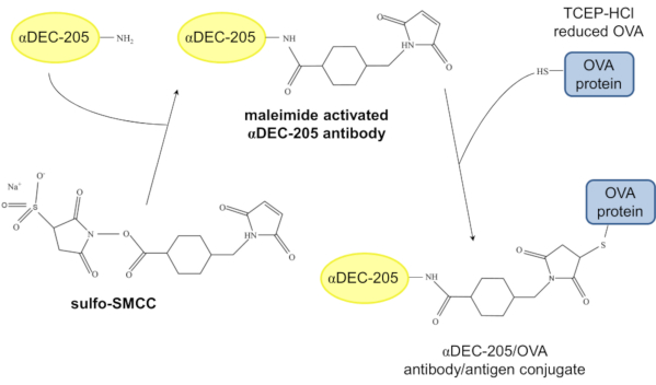
Figure 1: Model of the chemical conjugation of αDEC-205 and OVA. In a first step, the primary amine of αDEC-205 reacts with the NHS ester of the crosslinker sulfo-SMCC resulting in maleimide activated αDEC-205. Following reduction of the disulfide bonds of the OVA protein through incubation with TCEP-HCl, the maleimide activated αDEC-205 reacts with the TCEP-HCl-reduced OVA protein to form the αDEC-205/OVA antibody/antigen conjugate. Abbreviations: N-hydroxysuccinimide ester (NHS ester); sulfosuccinimidyl 4-[N-maleimidomethyl]cyclohexane-1-carboxylate (sulfo-SMCC); Tris(2-carboxysethyl)phosphine hydrochloride (TCEP-HCl). Please click here to view a larger version of this figure.
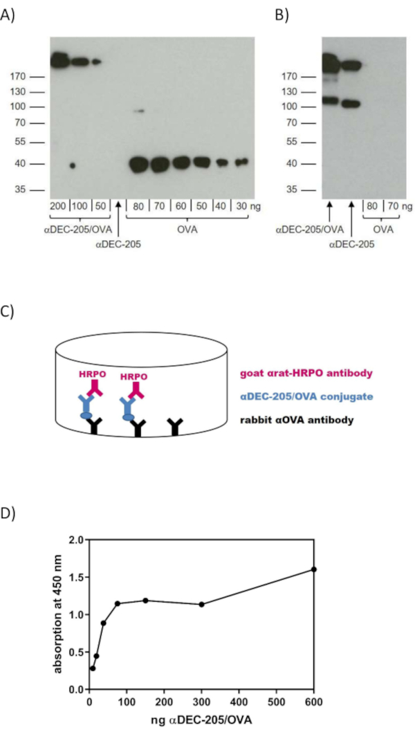
Figure 2: Verification of the chemically conjugated αDEC-205/OVA. To verify effective chemical conjugation of αDEC-205 and the OVA protein, western blot analysis (A,B) and ELISA (C,D) are performed. Samples of αDEC-205/OVA, αDEC-205 and different concentrations of OVA protein were subjected to SDS-PAGE (10%) and subsequent western blot analysis utilizing a rabbit αOVA primary antibody and a goat αrabbit-IgG-HRPO secondary antibody to detect OVA protein (A) or a goat αrat-IgG(H+L)-HRPO antibody to detect αDEC-205 (B). (C) Schematic representation of the ELISA for the verification of the αDEC-205/OVA conjugate. The rabbit αOVA coating antibody binds αDEC-205/OVA via the conjugated OVA. Goat αrat-IgG(H+L)-HRPO recognizes the αDEC-205 fraction of the bound conjugate and a positive signal thus confirms effective conjugation. (D) ELISA was performed as described in (C). Serially diluted amounts (1:2) of αDEC-205/OVA (600 ng to 9.38 ng) were analyzed. Data are shown as the mean of triplicates of a representative assay. Abbreviations: enzyme-linked immunosorbent assay (ELISA); horse radish peroxidase (HRPO); ovalbumin (OVA); sodium dodecyl sulfate polyacrylamide gel electrophoresis (SDS-PAGE). Panels A and B have been modified from Volckmar et al.12. http://creativecommons.org/licenses/by/4.0/. Please click here to view a larger version of this figure.
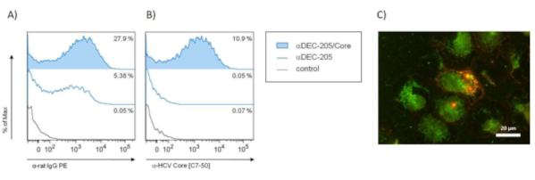
Figure 3: Binding of αDEC-205/Core to bone-marrow derived dendritic cells (BMDC) by flow-cytometry and immunofluorescence microscopy. To analyze the capacity of αDEC-205/Core to bind its target molecule DEC-205 on BMDCs in vitro, fluorescence-activated cell sorting (FACS) analysis (A,B) and immunofluorescence microscopy (C) were performed. In brief, bone marrow cells were isolated from the hind legs of female Balb/c mice (n=3) 8-12 weeks of age and cultured in RPMI medium supplemented with 1% penicillin/streptomycin, 1% glutamine, 0.25 mM mercaptoethanol and 5 ng/mL GM-CSF (granulocyte-macrophage colony stimulating factor). On day 6, the non-adherent BMDCs were carefully harvested and used for binding analysss. (A,B) In vitro generated BMDCs were incubated with 10 µg/mL αDEC-205/Core, αDEC-205 or medium (control) for 1 h at 4 °C followed by staining with APC-labeled αCD11c [clone HL3]. Detection of bound αDEC-205/Core on the surface of BMDCs was performed by additionally staining of the cells with either PE-labeled goat αrat (A) or mouse αHCV Core [clone C7-50] followed by secondary αmouse-IgG1-PE staining (B). Representative histograms show the PE signal and % PE-positive cells of the gated CD11c+ cells. (C) BMDCs generated in vitro from naïve Balb/c mice were sorted for MHC-II+ and CD11c+ cells and were incubated with 10 µg/mL αDEC-205/Core for 1 h at 4 °C. Cell-bound αDEC-205/Core was stained with Alexa 594-coupled αrat-IgG or with mouse αHCV Core [clone C7-50] and Alexa 488-coupled αmouse-IgG for 30 min at 4 °C after washing. The cells were visualized by immunofluorescence microscopy (scale bar = 20 µm). The binding capacity of αDEC-205/Core to BMDCs was confirmed by an overlay of both stainings (double positive = orange). Abbreviations: bone-marrow derived dendritic cells (BMDC); fluorescence-activated cell sorting (FACS); granulocyte-macrophage colony stimulating factor (GM-CSF); hepatitis C virus (HCV). Please click here to view a larger version of this figure.
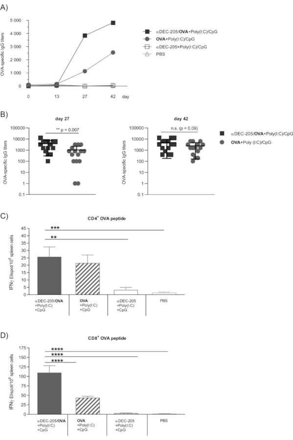
Figure 4: OVA-specific humoral and cellular immune responses following immunization with αDEC-205/OVA. The functionality of αDEC-205/OVA to target DC in vivo was proven through immunization experiments as previously published in Volckmar et al.12. Briefly, female 6-8 week old C57BL/6 mice (n=5) were subcutaneously immunized on days 0, 14 and 28 with 30 µg αDEC-205/OVA conjugate together with the adjuvants 50 µg Poly(I:C)/50 µg CpG, 30 µg αDEC-205 alone or 7 µg OVA protein alone in a total volume of 50 µL PBS per animal. Further controls were treated with PBS alone. (A,B) To monitor the humoral immune response, vaccinated mice were lightly anesthetized through isoflurane inhalation and blood samples were collected from the retro-orbital sinus on day 0, 13 and 27 and by cardiac puncture on day 42. Sera were prepared as described and assayed for the presence of OVA-specific IgG by ELISA12. Endpoint titers were expressed as the reciprocal value of the last serum dilution that yielded an absorbance two times above the values of negative controls. Results are compiled from three independent experiments. (A) Kinetic of OVA-specific total serum IgG titers shown as the group mean. (B) OVA-specific IgG titer on day 27 and day 42 shown for individual mice together with the group mean. Statistics: unpaired two-sided t-test. (C,D) The induction of cellular immune responses was analyzed by enzyme-linked immunosorbent spot (ELISPOT) assays using the murine IFNγ detection kit on day 42 as previously published12. Isolated splenocytes from immunized mice were pooled for the experimental groups and the number of IFNγ spot forming units/106 cells following stimulation with 5 mg/mL CD4+ (C) or CD8+ OVA peptide (D) was analyzed (OVA peptides: CD4323–339 (ISQAVHAAHAEINEAGR) and CD8257–264 (SIINFEKL)). Bars represent the mean ± SEM (n=5, triplicates from pooled samples). Statistics: one-way ANOVA with Dunnett's multiple comparisons test (**p<0.01, ***p<0.001, ****p<0.0001). Abbreviations: cytosine-phosphate-guanine oligonucleotide sequences (CpG), enzyme-linked immunosorbent assay (ELISA), enzyme-linked immunosorbent spot (ELISPOT), ovalbumin (OVA), phosphate-buffered saline (PBS), polyinosinic-polycytidylic acid (Poly(I:C)). This figure has been modified from Volckmar et al.12 http://creativecommons.org/licenses/by/4.0/. Please click here to view a larger version of this figure.
Chemical conjugation of an endocytosis receptor-specific antibody and a protein antigen provides an efficient and, importantly, also flexible approach for in vivo DC targeting in pre-clinical mouse models. With our protocol we provide an efficient approach for the successful conjugation of the model antigen OVA to a DEC-205-specific IgG antibody.
In our protocol, αDEC-205 is purified from a hybridoma cellline and in the past, we have purified the antibody using protein G sepharose as described. Of note, we have also used FPLC to purify αDEC-205 in later studies and subsequent chemical conjugation was likewise efficient. The critical steps for efficient chemical conjugation are the priming of both the antibody and the protein. We have optimized these steps from different available protocols to finally perform incubation of the antibody with the crosslinker sulfo-SMCC and reduction of the protein through TCEP-HCl. Specifically, at this point the critical steps are the fresh preparation of TCEP-HCl (step 3.1.) and the duration of the incubation of TCEP-HCl with the protein, which should not be extended (step 3.1.2.). Moreover, the protein buffer used for the reduction of the disulfide bonds through TCEP-HCl is critical, as it can negatively affect reduction by TCEP-HCl. Also, it is important to immediately mix the activated antibody and the reduced protein (step 3.3.6.) to ensure optimal reaction conditions for the conjugation. In our hands, this approach yielded the most efficient and reliable results regarding conjugation. A further critical step for subsequent in vivo approaches is the removal of unbound OVA from the αDEC-205/OVA conjugate. To verify this step, we have optimized western blot analyses and recommend detection of both the conjugated αDEC-205 as well as the conjugated OVA protein (Figure 2A and B). In case unbound OVA is present in the final conjugate solution, blotting and detecting known concentrations of OVA (as in Figure 2A) will help estimate the amount of unbound OVA in relation to the total amount of protein loaded for the SDS-PAGE. If conjugation was inefficient, troubleshooting should address the antigen/antibody ratio used for the conjugation. In case conjugation was effective as detected by ELISA, but cannot be detected by western blot analysis, we have experienced the antibody concentrations in the western blot analyses (steps 4.6., 4.8., 4.14.) the most critical factor.
The main limitation of our approach is an at times varying conjugation efficiency in which there is no fixed correlation between the number of antibody molecules and the amount of coupled protein. Nevertheless, we believe and have experienced that the demonstrated protocol reliably allows subsequent in vivo studies of DC targeting that yield reproducible results. Furthermore, while in principle in our protocol both αDEC-205 as well as the OVA protein can be exchanged for alternative antibodies and antigens for in vivo studies of various interests, individual steps of the chemical conjugation will need to be newly optimized for the new components. This applies mainly to the optimal antibody/antigen ratio and can depend e.g. on the accessibility of reducible Cys residues. In our approaches, the optimal ratios resulting in successful conjugation reactions were 1:5 for OVA to αDEC-205 and 1.35:1 for the HCV proteins NS3 (nonstructural protein 3) and Core to αDEC-205. For each conjugate, negative effects of the crosslinker or the conjugation as such on the antigen binding capacity of the antibody need to be excluded. Of note, conjugation of immunogenic peptides instead of full-length proteins is less problematic in this regard. However, proteins provide higher antigen diversity in subsequent immunization. Genetic fusion approaches display an alternative to chemical conjugation and also have clear advantages1. Ultimately, the choice of the strategy to link antibodies and antigens for DC targeting will depend on resources, the research focus and anticipated applications of the conjugates. We believe the relative flexibility regarding antibodies and antigens displays a major advantage of chemical conjugation and we have indeed used the protocol described here for the efficient chemical conjugation of αDEC-205 to different proteins of the hepatitis C virus (HCV) (Figure 3 and data not shown).
Overall, we believe that chemical conjugation of endocytosis-receptor specific antibodies to protein antigens such as in αDEC-205/OVA displays a flexible and reliable tool to generate antibody/antigen conjugates of exceptional value in studying DC targeting approaches, also including those aiming as inducing antitumor immunity, especially in preclinical animal models.
The authors have nothing to disclose.
The authors thank S. Prettin for expert technical assistance. This work was supported by a grant of the Helmholtz Association of German Research Centers (HGF) that was provided as part of the Helmholtz Alliance ''Immunotherapy of Cancers" (HCC_WP2b).
antibody buffer 2 % | 2 % (w/v) Slim-Fast Chocolate powder in TBS-T | ||
antibody buffer 5 % | 5 % milk powder (w/v) in TBS-T | ||
blocking buffer (ELISA) | 10 % FBS in PBS | ||
blocking buffer 4 % | 4 % (w/v) Slim-Fast Chocolate powder in TBS-T | ||
blocking buffer 10 % | 10 % milk powder (w/v) in TBS-T | ||
cell culture flask T25 | Greiner Bio-One | 690175 | we use standard CELLSTAR filter cap cell culture flasks; alternatively use suspension culture flask (690195 ) |
cell culture flask T75 | Greiner Bio-One | 658175 | we use standard CELLSTAR filter cap cell culture flasks; alternatively use suspension culture flask (658195) |
cell culture flask T175 | Greiner Bio-One | 661175 | we use standard CELLSTAR filter cap cell culture flasks; alternatively use suspension culture flask (661195) |
centrifugal concentrator MWCO 10 kDa | Sartorius | VS2001 | Vivaspin 20 centrifugal concentrator |
centrifugal protein concentrator MWCO 100 kDa, 5 – 20 ml | Thermo Fisher Scientific | 88532 | Pierce Protein Concentrator, PES 5 -20 ml; we use the Pierce Concentrator 150K MWCO 20mL (catalog number 89921), which is however no longer available |
centrifuge bottles | Nalgene | 525-2314 | PPCO (polypropylene copolymer) with PP (polypropylene) screw closure, 500 ml; obtained from VWR, Germany |
coating buffer (ELISA) | 0.1 M sodium bicarbonate (NaHCO3) in H2O (pH 9.6) | ||
desalting columns MWCO 7 kDa | Thermo Fisher Scientific | 89891 | Thermo Scientific Zeba Spin Desalting Columns, 7K MWCO, 5 mL |
detection reagent ELISA (HRPO substrate) | Sigma-Aldrich/Merck | T8665-100ML | 3,3′,5,5′-Tetramethylbenzidine (TMB) liquid substrate system |
detection reagent western blot (HRPO substrate) | Roche/Merck | 12 015 200 01 | Lumi-Light Western Blotting Substrate (Roche) |
dialysis tubing MWCO 12 – 14 kDa | SERVA Electrophoresis | 44110 | Visking dialysis tubing, 16 mm diameter |
ELISA 96-well plate | Thermo Fisher Scientific | 442404 | MaxiSorp Nunc-Immuno Plate |
fetal calf serum | PAN-BIOtech | P40-47500 | FBS Good forte |
ISF-1 medium | Biochrom/bioswisstec | F 9061-01 | |
milk powder | Carl Roth | T145.2 | powdered milk, blotting grade, low in fat; alternatively we have also used conventional skimmed milk powder from the supermarket |
NLDC-145 hybridoma | ATCC | HB-290 | if not already at hand, the hybridoma cells can be acquired from ATCC |
non-reducing SDS sample buffer 4 x | for 12 ml: 4 ml of 10 % SDS, 600 µl 0.5 M Tris-HCl (ph 6.8), 3.3 ml sterile H2O, 4 ml glycerine, 100 µl of 5 % Bromphenol Blue | ||
ovalbumin | Hyglos (via BioVendor) | 321000 | EndoGrade OVA ultrapure with <0.1 EU/mg |
Penicillin/Streptomycin | Thermo Fisher Scientific | 15140122 | Gibco Penicillin/Streptomycin 10.000 U/ml; alternatively Gibco Penicillin/Streptomycin 5.000 U/ml (15070-063) can be used |
PETG polyethylene terephthalate glycol cell culture roller bottles | Nunc In Vitro | 734-2394 | standard PDL-coated, vented (1.2X), 1050 cm², 100 – 500 ml volume; obtained from VWR, Germany |
pH indicator strips | Merck | 109535 | pH indicator strips 0-14 |
polyclonal goat αrat-IgG(H+L)-HRPO (western blot) | Jackson ImmunoResearch | 112-035-062 | obtained from Dianova, Germany; used at 1:5000 for western blot |
polyclonal goat αrat-IgG+IgM-HRPO antibody (ELISA) | Jackson ImmunoResearch | 112-035-068 | obtained from Dianova, Germany; used at 1:2000 for ELISA |
polyclonal goat αrabbit-IgG-HRPO (western blot) | Jackson ImmunoResearch | 111-035-045 | obtained from Dianova, Germany; used at 1:2000 for western blot |
polyclonal rabbit αOVA (ELISA) | Abcam | ab181688 | used at 3 ng/µl |
polyclonal rabbit αOVA antibody (western blot) | OriGene | R1101 | used at 1:3,000 for western blot |
Protein G Sepharose column | Merck/Millipore | P3296 | 5 ml Protein G Sepharose, Fast Flow are packed onto an empty column PD-10 (Merck, GE 17-0435-01) |
protein standard | Thermo Fisher Scientific | 26616 | PageRuler Prestained Protein ladder 10 – 180 kDa |
PVDF (polyvinylidene difluoride) membrane | Merck/Millipore | IPVH00010 | immobilon-P PVDF (polyvinylidene difluoride) membrane |
rubber plug | Omnilab | 5230217 | DEUTSCH & NEUMANN rubber stoppers (lower Φ 17 mm; upper Φ 22 mm) |
silicone tube | Omnilab | 5430925 | DEUTSCH & NEUMANN (inside Φ 1 mm; outer Φ 3 mm) |
Slim-Fast | we have used regular Slim-Fast Chocolate freely available at the pharmacy as in this western blot approach it yielded better results than milk powder | ||
stopping solution (ELISA) | 1M H2SO4 | ||
sulfo-SMCC | Thermo Fisher Scientific | 22322 | Pierce Sulfo-SMCC Cross-Linker; alternatively use catalog number A39268 (10 x 2 mg) |
syringe filter unit 0.22 µm | Merck/Millipore | SLGV033RS | Millex-GV Syringe Filter Unit, 0.22 µm, PVDF, 33 mm, gamma sterilized |
syringe 10 ml | Omnilab | Disposable syringes Injekt® Solo B.Braun | |
Sterican® cannulas | B. Braun | Sterican® G 20 x 1 1/2""; 0.90 x 40 mm; yellow | |
TBS-T | Tris-buffered saline containing 0.1 % (v/v) Tween 20 | ||
TCEP-HCl | Thermo Fisher Scientific | A35349 | |
tubing connector | Omnilab | Kleinfeld miniature tubing connectors for silicone tube |