The adult mosquito salivary gland (SG) is required for the transmission of all mosquito-borne pathogens to their human hosts, including viruses and parasites. This video demonstrates efficient isolation of the SGs from the larval (L4) stage Anopheles gambiae mosquitoes and preparation of the L4 SGs for further analysis.
Mosquito salivary glands (SGs) are a requisite gateway organ for the transmission of insect-borne pathogens. Disease-causing agents, including viruses and the Plasmodium parasites that cause malaria, accumulate in the secretory cavities of SG cells. Here, they are poised for transmission to their vertebrate hosts during a subsequent blood meal. As adult glands form as an elaboration of larval SG duct bud remnants that persist beyond early pupal SG histolysis, the larval SG is an ideal target for interventions that limit disease transmission. Understanding larval SG development can help develop a better understanding of its morphology and functional adaptations and aid in the assessment of new interventions that target this organ. This video protocol demonstrates an efficient technique for isolating, fixing, and staining larval SGs from Anopheles gambiae mosquitoes. Glands dissected from larvae in a 25% ethanol solution are fixed in a methanol-glacial acetic acid mixture, followed by a cold acetone wash. After a few rinses in phosphate-buffered saline (PBS), SGs can be stained with a broad array of marker dyes and/or antisera against SG-expressed proteins. This method for larval SG isolation could also be used to collect tissue for in situ hybridization analysis, other transcriptomic applications, and proteomic studies.
Malaria is a major public health threat causing almost 230 million infections and an estimated 409,000 deaths in 20191. The majority of deaths are in sub-Saharan Africa and are caused by the parasite Plasmodium falciparum, whose insect vector is Anopheles gambiae, the subject of this video demonstration. Although the numbers indicate a significant drop in annual death rate since the turn of the century (>300,000 fewer annual deaths), the promising decreases in disease rates observed from 2000 to 2015 are tapering, suggesting the need for new approaches to limiting disease transmission2. Among promising additional strategies for controlling and possibly eliminating malaria is targeting mosquito vector capacity using CRISPR/Cas9-based gene-editing and gene-drive3,4,5. Indeed, it is the targeting of the mosquito vector (through the expanded use of long-lasting insecticide-treated bed nets) that has had the greatest impact on reducing disease transmission6.
Female mosquitoes acquire Plasmodium gametocytes from an infected human during a blood meal. Following fertilization, maturation, midgut epithelium traversal, population expansion, and hemocoel navigation in their obligate mosquito hosts, hundreds to tens of thousands of Plasmodium sporozoites invade the mosquito SGs and fill the secretory cavities of the constituent secretory cells. Once inside the secretory cavities, the parasites have direct access to the salivary duct and are thus poised for transmission to a new vertebrate host upon the next blood meal. Because SGs are critical for the transmission of malaria-causing sporozoites to their human hosts, and laboratory studies suggest that SGs are not essential for blood-feeding, mosquito survival, or fecundity7,8,9, they represent an ideal target for transmission-blocking measures. Adult mosquito SGs form as an elaboration of "duct bud" remnants in the larval SGs that persist beyond early pupal SG histolysis10, making the larval SG an ideal target for interventions to limit adult-stage disease transmission.
Characterizing the larval stage of SG development can help develop not only a better understanding of its morphology and functional adaptations but can also aid in assessing new interventions that target this organ through gene editing of key SG regulators. Because all previous studies of larval salivary gland architecture predate immunostaining and modern imaging techniques10,11, we have developed a protocol for isolating and staining salivary glands with a variety of antibodies and cell markers12. This video demonstrates this approach to the extraction, fixation, and staining of larval SGs from Anopheles gambiae L4 larvae for confocal imaging.
1. Preparation of solutions and tools
2. Gland dissection (Figure 1A)
3. Fixation for antibody staining (Figure 1B)
4. Immunostaining (Figure 1B)
5. Mounting stained glands for microscopy (Figure 1C)
Salivary glands are relatively easy to dissect from all stage 4 larvae. Male and female larvae can be distinguished at the late L4 larval stage by a red stripe along the dorsal thorax of females but not males (Figure 2). We also observe that antennal morphology is much more elaborate in male than in female L4 larvae (Figure 2), similar to the differences observed in this structure in adult mosquitoes. Along with the considerable overall growth during the L4 stage, the salivary glands also form a lumen during L412. Salivary glands isolated from early L4 stage larvae stained with Hoechst reveal the proximal and distal lobes separated by a narrow constriction (Figure 3B,C). The forming lumen will extend all the way from the salivary duct at the proximal-most end (not shown) through the distal lobe. The forming lumen can be seen in the immunostained distal salivary gland of a mid-to-late L4 larva (Figure 4). The apical domains of the secretory cells surrounding the forming lumen have intense Nile Red staining (Figure 4B,C) suggestive of microvilli-like structures. Also observed close to the apical surface is Rab11 staining (Figure 4C,D, arrows). Rab11 localizes to apical recycling endosomes. The Rab11 staining that also accumulates along the basal surface of the gland is an artefact due to the stickiness of the basal membrane. Similar background staining is common with immunostaining of both larval and adult salivary glands and has been mistaken for bona fide signal.
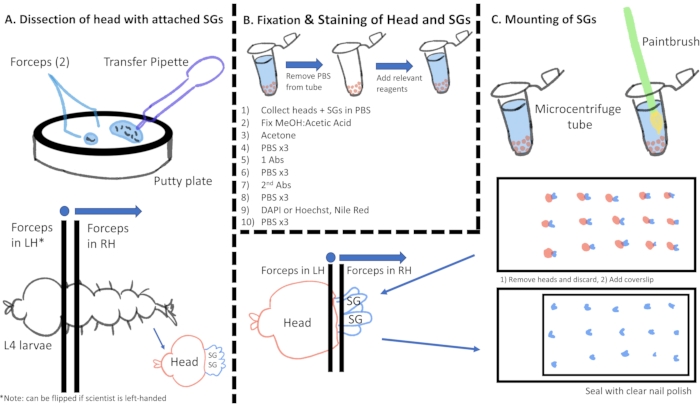
Figure 1: Cartoon visualization of the immunostaining process from gland dissection through slide preparation. Abbreviations: SGs = salivary glands; PBS = phosphate-buffered saline; MeOH = methanol; DAPI = 4′,6-diamidino-2-phenylindole; Abs = antibodies; LH = left hand; RH = right hand. Please click here to view a larger version of this figure.
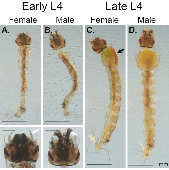
Figure 2: Male and female L4 stage Anopheles gambaie larvae. (A, B) Early L4 larvae. (C, D) Late L4 Larvae. There is considerable growth during the L4 stage. Females (A, C) have been described as having a distinguishing red stripe down the dorsal thorax (C; black arrow) that is not present in males (B, D), but also their antennae are much less elaborate (frilly) than those of male larvae from both early and late L4 (white arrows in enlarged images). Scale bars = 1 mm. Please click here to view a larger version of this figure.
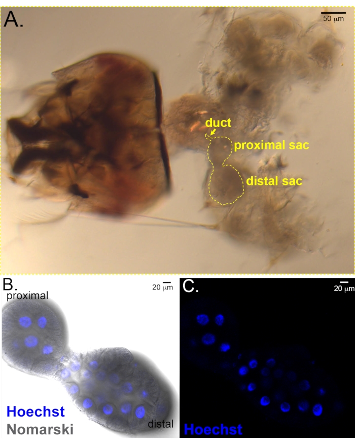
Figure 3: Early L4 salivary glands. (A) Early L4 female larval head with salivary glands and other internal organs still attached. Salivary gland is outlined. Scale bar = 50 µm. (B, C) Isolated larval glands stained with Hoechst. (B) Merge of fluorescent and Nomarski images showing the shape of the proximal and distal lobes and the blue Hoechst staining in nuclei. (C) Hoechst fluorescent image highlights nuclei only. Scale bar in bottom panels = 20 µm. Please click here to view a larger version of this figure.
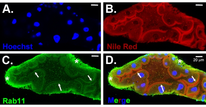
Figure 4: Immunostained distal L4 salivary gland. (A) Gland has been stained with Hoechst (nuclear DNA, blue) and (B) Nile Red (membrane marker, red); immunostained for Rab11 (apical recycling vesicles, green) (C). (D) The merged image. Note that a lumen is present at the stage shown (B–D). The arrows in C and D point to the expected Rab11 staining of vesicles near the apical surface that overlaps the Nile Red-positive vesicular staining in the merge (white arrows). Non-specific background staining around the basal surface (asterisks) is also observed, a common type of background observed in larval and adult salivary glands. Scale bars = 20 µm. Please click here to view a larger version of this figure.
The protocol described herein was adapted from a Drosophila SG dissection protocol and an adult mosquito dissection protocol14,15,16. However, most markers did not penetrate the basement membrane (data not shown) when using the adult dissection and SG staining methods. Adaptations of the adult protocol included dissecting the glands in a 25% EtOH solution, washing the glands with a combination of MeOH and glacial acetic acid, and having a 90 s acetone wash. In the original adult protocol, adult SGs were dissected in 1x PBS14,15,16. Dissecting in a 25% EtOH solution helped preserve the SGs and prevented damage during the staining periods. When performing the initial dissection, it was easiest to orient the larval SGs with the head facing the non-dominant hand and having the dominant hand gently pull outwards (because the larvae are very actively moving). The improved tissue penetration achieved by washing the glands with a MeOH: glacial acetic acid solution suggests that the lipid content in the larval SG basement membrane and/or cellular plasma membranes is distinct from that of the adult SG. The 90 s acetone wash improved the clarity of glands for imaging. Although the fixation period was recommended to be at least 19 h (overnight), longer periods worked just as effectively.
The morphology of the SGs can vary widely depending on when the larvae are dissected in the 2-day long L4 stage. In early L4 dissections (day 1), the lumen appears much smaller12. In late L4 dissections (second half of day 2), the lumen is large, and the cells are elongated. We found that the optimal period for working with larvae was the L4; L1-L3 SGs are simply too small for manual dissections. In imaging results, glands dissected at mid-to-late-L4 showed smaller cells mixed with larger, laterally elongated cells, particularly in the distal sac.
Previous reports on Anopheles SG development have provided much guidance for the current studies. These reports have included illustrations of dissected embryonic and larval SGs17,18, detailed microscopic analysis of dissected glands19,20, and transcriptomic studies20,21,22. The features of the Anopheles stephensi SG were reported during the 1950s by two different groups10,11. Many of the morphological features of the larval glands were described, including the overall organization of the SG, the relative positions of distinct cell types within the organ (duct, adult SG precursors, and the larval proximal and distal secretory sacs)10,11. Both groups also reported additional morphometric data, including the number and distribution of secretory cells within each sac and their nuclear size10,11. Rishikesh showed that, like the Drosophila larval SGs, the larval SG chromosomes from Anopheles stephensi are polytenized10. These important foundational overviews described broad biological aspects of larval Anopheles SGs.
The method described herein should be useful for studying not only larval SGs of An. gambiae but can also apply to those studying other species of mosquitos. Indeed, the same protocol has been successfully utilized (unpublished) with An. stephensi, a species frequently studied in the laboratory. One limitation to the protocol is the limited number of antibodies that have been specifically generated against mosquito proteins. Although using Drosophila antibodies for highly conserved proteins has circumnavigated this limitation12, the field could benefit from more mosquito protein-specific antibodies. Understanding larval SG cellular and molecular biology can contribute to new control or target strategies and allow for the discovery of new candidate target genes for SG disruption.
The authors have nothing to disclose.
We would like to thank the Johns Hopkins Malaria Research Institute for access to and rearing of An. gambiae larvae.
KH2PO4 | Millipore Sigma | P9791 | |
Na2HPO4 • 2H2O | Millipore Sigma | 71643 | |
NaCl | Millipore Sigma | S7653 | |
Acetone | Millipore Sigma | 179124 | |
Brush with soft bristles | Amazon (SN NJDF) Detail Paint Brush Set | B08LH63D89 | |
Cover slips (22 x 50 mm) | VWR | 48393-195 | |
DAPI (DNA) | ThermoFisher Scientific | D1306 | |
Ethyl alcohol 200 proof | Millipore Sigma | EX0276 | |
Gilson Pipetman P200 Pipette | Gilson | P200 | |
Glacial Acetic Acid | Sigma Aldrich | 695092 | |
Jewelers forceps, Dumont No. 5 | Millipore Sigma | F6521 | |
KCl | Millipore Sigma | 58221 | |
Methanol | Millipore Sigma | 1414209 | |
Nail polish | Amazon (Sally Hansen) | B08148YH9M | |
Nile Red (lipid) | ThermoFisher Scientific | N1142 | |
Paper towels/wipes | ULINE | S-7128 | |
Petri plate (to make putty plate) | ThermoFisher Scientific | FB0875712 | |
Pipette Tips | Gilson | Tips E200ST | |
Plastic Transfer Pipette | Fisher Scientific | S304671 | |
Primary antibodies (e.g., Crb, Rab11) | Developmental Studies Hybridoma Bank (DSHB); Andrew Lab | Mouse anti-Crb (Cq4) or Rabbit anti-Rab11 | |
Secondary antibodies with fluorescent tags (e.g., Alexa Fluor 488 Goat-anti Rabbit) | ThermoFisher Scientific | A11008 | |
Silicone resin and curing agent for putty plate | Dow Chemicals – Ximeter Silicone | PMX-200 | |
Slides, frosted on one end for labelling | VWR 20 X 50 mm | 48393-195 | |
Wheat Germ Agglutinin | ThermoFisher Scientific | W834 |