Summary

Visualisatie van inflammatoire caspasen geïnduceerde nabijheid in menselijke monocyt-afgeleide macrofagen

Published: April 06, 2022
doi:

Summary

Dit protocol beschrijft de workflow om monocyten-afgeleide macrofagen (MDM) te verkrijgen uit menselijke bloedmonsters, een eenvoudige methode om inflammatoire caspase Bimolecular Fluorescence Complementation (BiFC) -verslaggevers efficiënt in menselijke MDM te introduceren zonder de levensvatbaarheid en het gedrag van cellen in gevaar te brengen, en een op beeldvorming gebaseerde benadering om inflammatoire caspase-activering in levende cellen te meten.

Abstract

Inflammatoire caspasen omvatten caspase-1, -4, -5, -11 en -12 en behoren tot de subgroep van initiator caspasen. Caspase-1 is vereist om een correcte regulatie van inflammatoire signalering te garanderen en wordt geactiveerd door nabijheidsgeïnduceerde dimerisatie na rekrutering voor inflammasomen. Caspase-1 is overvloedig aanwezig in de monocytische cellijn en induceert rijping van de pro-inflammatoire cytokines interleukine (IL)-1β en IL-18 tot actieve uitgescheiden moleculen. De andere inflammatoire caspasen, caspase-4 en -5 (en hun murine homolog caspase-11) bevorderen il-1β afgifte door pyroptose te induceren. Caspase Bimolecular Fluorescence Complementation (BiFC) is een hulpmiddel dat wordt gebruikt om inflammatoire caspase geïnduceerde nabijheid te meten als een uitlezing van caspase-activering. Het caspase-1, -4 of -5 prodomein, dat het gebied bevat dat zich bindt aan het inflammasoom, is gefuseerd met niet-fluorescerende fragmenten van het gele fluorescerende eiwit Venus (Venus-N [VN] of Venus-C [VC]) die zich associëren om het fluorescerende Venus-complex te hervormen wanneer de caspasen geïnduceerde nabijheid ondergaan. Dit protocol beschrijft hoe deze verslaggevers kunnen worden geïntroduceerd in primaire menselijke monocyt-afgeleide macrofagen (MDM) met behulp van nucleofection, de cellen kunnen worden behandeld om inflammatoire caspase-activering te induceren en caspase-activering te meten met behulp van fluorescentie en confocale microscopie. Het voordeel van deze aanpak is dat het kan worden gebruikt om de componenten, vereisten en lokalisatie van het inflammatoire caspase-activeringscomplex in levende cellen te identificeren. Er moeten echter zorgvuldige controles worden overwogen om te voorkomen dat de levensvatbaarheid en het gedrag van cellen in gevaar komen. Deze techniek is een krachtig hulpmiddel voor de analyse van dynamische caspase-interacties op inflammasoomniveau en voor de ondervraging van de inflammatoire signaalcascades in levende MDM en monocyten afgeleid van menselijke bloedmonsters.

Introduction

De caspasen zijn een familie van cysteïne aspartaat proteasen die kunnen worden gegroepeerd in initiator caspasen en beul caspasen. Beul caspasen bestaan uit caspase-3, -6 en -7. Ze komen van nature voor in cellen als dimeren en worden door de initiator caspasen gesplitst om apoptose1 uit te voeren. Initiator caspasen omvatten human caspase-1, -2, -4, -5, -8, -9, -10 en -12. Ze worden gevonden als inactieve zymogenen (pro-caspasen) die worden geactiveerd door nabijheidsgeïnduceerde dimerisatie en gestabiliseerd door auto-proteolytische splitsing 2,3. De inflammatoire caspasen zijn een subset van de initiator caspasen2 en omvatten caspase-1, -4, -5 en -12 bij mensen, en caspase-1, -11 en -12 bij muis 4,5. In plaats van een apoptotische rol, spelen ze een centrale rol bij ontstekingen. Ze bemiddelen proteolytische verwerking en secretie van pro-interleukine (IL)-1β en pro-IL-18 6,7, de eerste cytokines die vrijkomen als reactie op pathogene indringers 8,9. Caspase-1 wordt geactiveerd bij werving op zijn activeringsplatform; een eiwitcomplex met een groot molecuulgewicht dat het inflammasoom wordt genoemd (figuur 1A)10. Dimerisatie van caspase-4, -5 en -11 vindt onafhankelijk van deze platforms plaats via een niet-canonieke inflammasoomroute11,12.

Canonieke inflammasomen zijn cytosolische multimere eiwitcomplexen die bestaan uit een inflammasoomsensoreiwit, het adaptoreiwit ASC (apoptose-geassocieerd speck-achtig eiwit dat een CARD bevat) en het effectoreiwit caspase-110. De best bestudeerde canonieke inflammasomen zijn de NOD-achtige receptorfamilie met een pyrinedomein (NLRP), NLRP1 en NLRP3, de NLR-familie met een CARD (NLRC), NLRC4 en de afwezige in melanoom 2 (AIM2). Ze bevatten elk een pyrinedomein, een CARD of beide domeinen. Het CARD-domein bemiddelt de interactie tussen CARD-bevattende caspasen en hun upstream activators. Daarom is het steigermolecuul ASC, dat bestaat uit een N-terminal pyrinedomein (PYD) en een C-terminal CARD-motief13,14, vereist voor de rekrutering van caspase-1 voor deNLRP1 10, NLRP315 en AIM216 inflammasomen.

Elk inflammasoom is vernoemd naar zijn unieke sensoreiwit dat verschillende pro-inflammatoire stimuli herkent (figuur 1B). Activatoren van deze route worden canonieke stimuli genoemd. Inflammasomen dienen als sensoren voor microbiële componenten en weefselstress en assembleren om een robuuste ontstekingsreactie te veroorzaken door activering van de inflammatoire caspasen17. Inflammasoomassemblage initieert caspase-1-activering om rijping en secretie van de belangrijkste substraten pro-IL-1β en pro-IL-18 te bemiddelen. Dit proces verloopt via een mechanisme in twee stappen. Ten eerste upreguleert een priming stimulus de expressie van bepaalde inflammasoomeiwitten en pro-IL-1β door activering van de NF-κB-route. Ten tweede induceert een intracellulaire (canonieke) stimulus inflammasoomassemblage en rekrutering van procaspase-1 6,7.

Caspase-4 en caspase-5 zijn de menselijke orthologen van murine caspase-1111. Ze worden op een inflammasoomonafhankelijke manier geactiveerd door intracellulair lipopolysaccharide (LPS), een molecuul dat wordt aangetroffen in het buitenmembraan van Gram-negatieve bacteriën 18,19,20, en door extracellulair heem, een product van hemolyse van rode bloedcellen 21. Er is voorgesteld dat LPS zich rechtstreeks bindt aan het CARD-motief van deze eiwitten en hun oligomerisatie induceert20. Activering van caspase-4 of caspase-5 bevordert de afgifte van IL-1β door het induceren van een inflammatoire vorm van celdood die pyroptose wordt genoemd door splitsing van het porievormende eiwit gasdermin D (GSDMD)18,19. Bovendien induceert de efflux van kaliumionen als gevolg van caspase-4 en GSDMD-gemedieerde pyroptotische dood activering van het NLRP3-inflammasoom en daaropvolgende activering van caspase-1 22,23. Daarom worden caspase-4, -5 en -11 beschouwd als intracellulaire sensoren voor LPS die pyroptose en caspase-1-activering kunnen induceren als reactie op specifieke stimuli11,24.

Figure 1
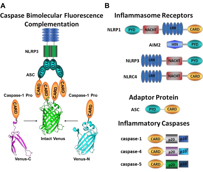
Figuur 1: Inflammatoire caspasen en caspase-bimoleculaire fluorescentiecomplementatie (BiFC) assay. (A) Diagram met het caspase-BiFC-systeem, waarbij twee caspase-1 prodomeinen (C1-pro) gekoppeld aan elk niet-fluorescerend fragment van Venus (Venus-C of Venus-N) worden gerekruteerd naar het NLRP3-activeringsplatform, waardoor Venus wordt gedwongen om opnieuw te vouwen en te fluoresceren. Dit complex verschijnt als een groene vlek onder de microscoop en dient als een uitlezing voor inflammatoire caspase-geïnduceerde nabijheid, wat de eerste stap is in initiator caspase-activering. (B) Schema dat de domeinorganisatie van inflammasoomcomponenten en inflammatoire caspasen weergeeft. Klik hier om een grotere versie van deze figuur te bekijken.

Het meten van specifieke initiator caspases activering is moeilijk, en er zijn niet veel methoden beschikbaar om dit te doen door middel van imaging benaderingen. Caspase Bimolecular Fluorescence Complementation (BiFC) kan worden gebruikt om inflammatoire caspase-activering direct in levende cellen te visualiseren (figuur 1A)25. Deze techniek is onlangs aangepast voor gebruik in menselijke monocyten-afgeleide macrofagen (MDM)21. Caspase BiFC meet de eerste stap in inflammatoire caspase-activering, geïnduceerde nabijheid om dimerisatie te vergemakkelijken. Expressie van plasmiden die coderen voor het CARD-bevattende caspase-prodomein gefuseerd tot niet-fluorescerende fragmenten van het fotostabiele gele fluorescerende eiwit Venus (Venus-C [VC]) en Venus-N [VN]) worden gebruikt. Wanneer de twee caspase-prodomeinen worden gerekruteerd naar hun activeringsplatform of geïnduceerde nabijheid ondergaan, worden de twee helften van Venus in de nabijheid gebracht en gedwongen om opnieuw te vouwen en te fluoresceren (zie figuur 1A,B). Dit biedt een real-time uitlezing van specifieke inflammatoire caspase-activering.

Menselijke MDM brengt overvloedig inflammasoomgenen en patroonherkenningsreceptoren tot expressie die gevaarsignalen en pathogene producten identificeren. Dit biedt een ideaal celtype voor de ondervraging van inflammatoire caspase-routes. Bovendien kunnen ze worden afgeleid uit perifeer bloed en zelfs uit patiëntmonsters om inflammatoire caspase-activering in een specifieke ziektetoestand te beoordelen. Dit protocol beschrijft hoe de BiFC caspase-verslaggevers in MDM kunnen worden geïntroduceerd met behulp van nucleofection, een op elektroporatie gebaseerde transfectiemethode, hoe de cellen moeten worden behandeld om inflammatoire caspase-activering te induceren en hoe de actieve caspase-complexen kunnen worden gevisualiseerd met behulp van microscopiebenaderingen. Bovendien kan deze methodologie worden aangepast om de moleculaire samenstelling van deze complexen, subcellulaire lokalisatie, kinetiek en grootte van deze zeer geordende structuren te bepalen 25,26,27.

Protocol

Dit protocol volgt de richtlijnen van de ethische commissie voor menselijk onderzoek van Baylor College of Medicine voor de manipulatie van menselijke monsters. Bloedmonsters worden behandeld volgens de institutionele veiligheidsrichtlijnen voor menselijke monsters. Bloedmonsters worden verkregen bij een regionale bloedbank, waar ze worden verzameld met citraatfosfaat dextrose (CPD) oplossing. Bloed verzameld met andere anticoagulantia zoals natriumheparine, lithiumheparine of EDTA kan echter ook worden gebruikt voor dit…

Representative Results

Het schema in figuur 2 geeft een overzicht van hoe menselijke MDM kan worden verkregen, getransfecteerd en in beeld gebracht. Na incubatie van de geselecteerde CD14+ monocyten met GM-CSF gedurende 7 dagen, verandert de celmorfologie in de loop van de differentiatieperiode (figuur 3A), gaande van bolvormige suspensiecellen tot spichtig en volledig gehecht (dag 3 en 4), en ten slotte naar meer verspreide cellen wanneer volledig gedifferentieerd (dag 7). Volledig …

Discussion

Dit protocol beschrijft de workflow om macrofagen te verkrijgen uit monocyten geïsoleerd uit menselijke bloedmonsters en een methode om de inflammatoire caspase BiFC-verslaggevers efficiënt in menselijke MDM te introduceren zonder de levensvatbaarheid en het gedrag van cellen in gevaar te brengen.

Dit protocol maakt gebruik van de BiFC-techniek35 om de inflammatoire caspasen op het caspase recruitment domein (CARD) te taggen met niet-fluorescerende fragmenten van het …

Disclosures

The authors have nothing to disclose.

Acknowledgements

We bedanken de leden van LBH’s lab uit heden en verleden die hebben bijgedragen aan de ontwikkeling van deze techniek. Dit lab wordt ondersteund door NIH/NIDDK T32DK060445 (BEB), NIH/NIDDK F32DK121479 (BEB), NIH/NIGMS R01GM121389 (LBH). Figuur 2 is getekend met behulp van Biorender software.

Materials

48 well tissue culture2:34 plates Genesee Scientific 25-108
10 cm Tissue Culture Dishes VWR 25382-166
2 Mercaptoethanol 1000x Thermo Fisher Scientific 21985023
8 well chambered coverglass with 1.5 HP coverglass Cellvis c8-1.5H-N
AutoMACS columns Miltenyi (Biotec) 130-021-101 For automated separation using AutoMACS Pro Separator only
AutoMACS Pro Separator Miltenyi (Biotec) 130-092-545 For automated separation using AutoMACS Pro Separator only
AutoMACS Pro Washing Solution Miltenyi (Biotec) 130-092-987 For automated separation using AutoMACS Pro Separator only
AutoMACS Rinsing Solution Miltenyi (Biotec) 130-091-222 For automated separation using AutoMACS Pro Separator only
AutoMacs running buffer Miltenyi (Biotec) 130-091-221 For manual or automated separation using QuadroMACS or AutoMACS pro Separator
Axio Observer Z1 motorized inverted microscope equipped with a CSU-X1A 5000 spinning disk unit Zeiss Any confocal microscope equipped with a laser module fitted with laser lines of 568 nm (RFP) and 488 or 512 nm (GFP or YFP) wavelengths can be used
AxioObserver A1, Research Grade Inverted Microscope Zeiss Any epifluorescence microscope with fluorescence filters capable of exciting 568 nm (RFP) and 488 or 512 nm (GFP or YFP) wavelengths can be used
CD14+ MICROBEADS Miltenyi (Biotec) 130-050-201 For manual or automated separation using QuadroMACS or AutoMACS pro Separator
DPBS without calcium chloride and magnesium chloride Sigma D8537-6x500ML
DsRed mito plasmid Clontech 632421 Similar plasmids that can be used as fluorescent reporters can be found on Addgene
Fetal Bovine Serum Thermo Fisher Scientific 10437028
Ficoll-Paque PLUS 6 x 100 mL Sigma GE17-1440-02
GlutaMAX Supplement (100x) Thermo Fisher Scientific 35050079
GM-CSF Thermo Fisher Scientific PHC2011
Hemin BioXtra, from Porcine, ≥96.0% (HPLC) Sigma 51280-1G
HEPES Thermo Fisher Scientific 15630106
Inflammatory caspase BiFC plasmids Available by request from LBH lab
LPS-EB Ultrapure Invivogen TLRL-3PELPS
LS Columns Miltenyi (Biotec) 130-042-401 For manual separation using QuadroMACS Separator only
MACS 15 mL Tube Rack Miltenyi (Biotec) 130-091-052 For manual separation using QuadroMACS Separator only
MACS MultiStand Miltenyi (Biotec) 130-042-303 For manual separation using QuadroMACS Separator only
mCherry plasmid Yungpeng Wang Lab Similar plasmids that can be used as fluorescent reporters can be found on Addgene
Neon Transfection System Thermo Fisher Scientific MPK5000 Includes Neon electroporation device, pipette and pipette station
Neon Transfection System 10 µL Kit Thermo Fisher Scientific MPK1096 Includes resuspension buffer R, resuspension buffer T, electrolytic buffer E, 96 x 10 µL Neon tips and Neon electroporation tubes
Nigericin sodium salt, ready made solution Sigma SML1779-1ML
Penicillin-Streptomycin (10,000 U/mL) Thermo Fisher Scientific 15140122
Poly-D-Lysine Hydrobromide Sigma P7280-5mg
QuadroMACS Separator Miltenyi (Biotec) 130-090-976 For manual separation using QuadroMACS Separator only
qVD-OPh Fisher (ApexBio) 50-101-3172
RPMI 1640 Medium Thermo Fisher Scientific 11875119
Trypsin-EDTA (0.25%), phenol red Thermo Fisher Scientific 25200072
UltraPure 0.5 M EDTA, pH 8.0 Thermo Fisher Scientific 15575020
Zeiss Zen 2.6 (blue edition) software Zeiss Any software used to operate the confocal microscope of choice

References

  1. Boatright, K. M., et al. A unified model for apical caspase activation. Molecular Cell. 11 (2), 529-541 (2003).
  2. Pop, C., Salvesen, G. S. Human caspases: activation, specificity, and regulation. Journal of Biological Chemistry. 284 (33), 21777-21781 (2009).
  3. Boice, A., Bouchier-Hayes, L. Targeting apoptotic caspases in cancer. Biochimica et Biophysica Acta (BBA) – Molecular Cell Research. 1867 (6), 118688 (2020).
  4. Bolívar, B. E., Vogel, T. P., Bouchier-Hayes, L. Inflammatory caspase regulation: maintaining balance between inflammation and cell death in health and disease. The FEBS Journal. 286 (14), 2628-2644 (2019).
  5. Martinon, F., Tschopp, J. Inflammatory caspases: linking an intracellular innate immune system to autoinflammatory diseases. Cell. 117 (5), 561-574 (2004).
  6. Lamkanfi, M., Vishva, M. D. Mechanisms and functions of inflammasomes. Cell. 157 (5), 1013-1022 (2014).
  7. Viganò, E., et al. Human caspase-4 and caspase-5 regulate the one-step noncanonical inflammasome activation in monocytes. Nature Communications. 6, 8761 (2015).
  8. Cerretti, D. P., et al. Molecular cloning of the interleukin-1 beta converting enzyme. Science. 256 (5053), 97 (1992).
  9. van de Veerdonk, F. L., Netea, M. G., Dinarello, C. A., Joosten, L. A. Inflammasome activation and IL-1β and IL-18 processing during infection. Trends in Immunology. 32 (3), 110-116 (2011).
  10. Martinon, F., Burns, K., Tschopp, J. The inflammasome: a molecular platform triggering activation of inflammatory caspases and processing of proIL-beta. Molecular Cell. 10 (2), 417-426 (2002).
  11. Kayagaki, N., et al. Noncanonical inflammasome activation targets caspase-11. Nature. 479 (7371), 117-121 (2011).
  12. Kayagaki, N., et al. Noncanonical inflammasome activation by intracellular LPS independent of TLR4. Science. 341 (6151), 1246-1249 (2013).
  13. Masumoto, J., et al. ASC, a novel 22-kDa protein, aggregates during apoptosis of human promyelocytic leukemia HL-60 cells. The Journal of Biological Chemistry. 274 (48), 33835-33838 (1999).
  14. Bertin, J., DiStefano, P. S. The PYRIN domain: a novel motif found in apoptosis and inflammation proteins. Cell Death and Differentiation. 7 (12), 1273-1274 (2000).
  15. Agostini, L., et al. NALP3 forms an IL-1beta-processing inflammasome with increased activity in Muckle-Wells autoinflammatory disorder. Immunity. 20 (3), 319-325 (2004).
  16. Bürckstümmer, T., et al. An orthogonal proteomic-genomic screen identifies AIM2 as a cytoplasmic DNA sensor for the inflammasome. Nature Immunology. 10 (3), 266-272 (2009).
  17. Latz, E., Xiao, T. S., Stutz, A. Activation and regulation of the inflammasomes. Nature Reviews Immunology. 13 (6), 397-411 (2013).
  18. Kayagaki, N., et al. Caspase-11 cleaves gasdermin D for noncanonical inflammasome signalling. Nature. 526 (7575), 666-671 (2015).
  19. Shi, J., et al. Cleavage of GSDMD by inflammatory caspases determines pyroptotic cell death. Nature. 526 (7575), 660-665 (2015).
  20. Shi, J., et al. Inflammatory caspases are innate immune receptors for intracellular LPS. Nature. 514 (7521), 187-192 (2014).
  21. Bolívar, B. E., et al. Noncanonical Roles of Caspase-4 and Caspase-5 in Heme-Driven IL-1β Release and Cell Death. The Journal of Immunology. 206 (8), 1878-1889 (2021).
  22. Schmid-Burgk, J. L., et al. Caspase-4 mediates noncanonical activation of the NLRP3 inflammasome in human myeloid cells. European Journal of Immunology. 45 (10), 2911-2917 (2015).
  23. Baker, P. J., et al. NLRP3 inflammasome activation downstream of cytoplasmic LPS recognition by both caspase-4 and caspase-5. European Journal of Immunology. 45 (10), 2918-2926 (2015).
  24. Cullen, S. P., Kearney, C. J., Clancy, D. M., Martin, S. J. Diverse activators of the NLRP3 inflammasome promote IL-1β secretion by triggering necrosis. Cell Reports. 11 (10), 1535-1548 (2015).
  25. Sanders, M. G., et al. Single-cell imaging of inflammatory caspase dimerization reveals differential recruitment to inflammasomes. Cell Death & Disease. 6, 1813 (2015).
  26. Charendoff, C. I., Bouchier-Hayes, L. Lighting up the pathways to caspase activation using bimolecular fluorescence complementation. Journal of Visualized Experiments: JoVE. (133), e57316 (2018).
  27. Bouchier-Hayes, L., et al. Characterization of cytoplasmic caspase-2 activation by induced proximity. Molecular Cell. 35 (6), 830-840 (2009).
  28. Riedhammer, C., Halbritter, D., Weissert, R. Peripheral blood mononuclear cells: Isolation, Freezing, thawing, and culture. Methods in Molecular Biology. 1304, 53-61 (2016).
  29. Betsou, F., Gaignaux, A., Ammerlaan, W., Norris, P. J., Stone, M. Biospecimen science of blood for peripheral blood mononuclear cell (PBMC) functional applications. Current Pathobiology Reports. 7 (2), 17-27 (2019).
  30. Perregaux, D., et al. IL-1 beta maturation: evidence that mature cytokine formation can be induced specifically by nigericin. The Journal of Immunology. 149 (4), 1294-1303 (1992).
  31. Cheneval, D., et al. Increased mature interleukin-1β (IL-1β) secretion from THP-1 cells induced by nigericin is a result of activation of p45 IL-1β-converting enzyme processing. Journal of Biological Chemistry. 273 (28), 17846-17851 (1998).
  32. Fernandes-Alnemri, T., et al. The pyroptosome: a supramolecular assembly of ASC dimers mediating inflammatory cell death via caspase-1 activation. Cell Death and Differentiation. 14 (9), 1590-1604 (2007).
  33. Lu, A., et al. Unified polymerization mechanism for the assembly of ASC-dependent inflammasomes. Cell. 156 (6), 1193-1206 (2014).
  34. Muller-Eberhard, U., Javid, J., Liem, H. H., Hanstein, A., Hanna, M. Brief report: Plasma concentrations of hemopexin, haptoglobin and heme in patients with various hemolytic diseases. Blood. 32 (5), 811-815 (1968).
  35. Shyu, Y. J., Liu, H., Deng, X., Hu, C. D. Identification of new fluorescent protein fragments for bimolecular fluorescence complementation analysis under physiological conditions. Biotechniques. 40 (1), 61-66 (2006).
  36. Thornberry, N. A., et al. A novel heterodimeric cysteine protease is required for interleukin-1βprocessing in monocytes. Nature. 356 (6372), 768-774 (1992).
  37. Jensen, K., Anderson, J. A., Glass, E. J. Comparison of small interfering RNA (siRNA) delivery into bovine monocyte-derived macrophages by transfection and electroporation. Veterinary Immunology and Immunopathology. 158 (3-4), 224-232 (2014).
  38. Tada, Y., Sakamoto, M., Fujimura, T. Efficient gene introduction into rice by electroporation and analysis of transgenic plants: use of electroporation buffer lacking chloride ions. Theoretical and Applied Genetics. 80 (4), 475-480 (1990).
  39. Bhowmik, P., et al. Targeted mutagenesis in wheat microspores using CRISPR/Cas9. Scientific Reports. 8 (1), 6502 (2018).
  40. Sansom, D. M., Manzotti, C. N., Zheng, Y. What’s the difference between CD80 and CD86. Trends in Immunology. 24 (6), 314-319 (2003).
  41. Kurokawa, M., Kornbluth, S. Caspases and kinases in a death grip. Cell. 138 (5), 838-854 (2009).
check_url/63162?article_type=t

Play Video

Cite This Article
Bolívar, B. E., Bouchier-Hayes, L. Visualization of Inflammatory Caspases Induced Proximity in Human Monocyte-Derived Macrophages. J. Vis. Exp. (182), e63162, doi:10.3791/63162 (2022).

View Video