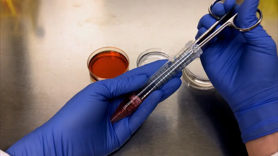
The present protocol describes a simple method for isolating preadipocytes from adipose tissue in broiler embryos. This method enables isolation with high yield, primary culture, and adipogenic differentiation of preadipocytes. Oil Red O staining and lipid/DNA stain measured the adipogenic ability of isolated cells induced with differentiation media.
Method Article
Isolation of Preadipocytes from Broiler Chick Embryos
In This Article
Summary
Abstract
Primary preadipocytes are a valuable experimental system for understanding the molecular pathways that control adipocyte differentiation and metabolism. Chicken embryos provide the opportunity to isolate preadipocytes from the earliest stage of adipose development. This primary cell can be used to identify factors influencing preadipocyte proliferation and adipogenic differentiation, making them a valuable model for studies related to childhood obesity and control of excess fat deposition in poultry. The rapid growth of postnatal adipose tissue effectively wastes feed by allocating it away from muscle growth in broiler chickens. Therefore, methods to understand the earliest stages of adipose tissue development may provide clues to regulate this tendency and identify ways to limit adipose expansion early in life. The present study was designed to develop an efficient method for isolation, primary culture, and adipogenic differentiation of preadipocytes isolated from developing adipose tissue of commercial broiler (meat-type) chick embryos. The procedure has been optimized to yield cells with high viability (~98%) and increased capacity to differentiate into mature adipocytes. This simple method of embryonic preadipocyte isolation, culture, and differentiation supports functional analyses of fat growth and development in early life.
Introduction
Obesity is a global health threat to both adults and children. Children who are overweight or obese are approximately five times more likely to be obese as adults, placing them at significantly increased risk for cardiovascular disease, diabetes, and many other comorbidities. About 13.4% of US children aged 2-5 have obesity1, illustrating that the tendency to accumulate excess body fat can be set in motion very early in life. For very different reasons, the accumulation of excess adipose tissue is a concern for broiler (meat-type) chickens. Modern broilers are incredibly efficient but still accumulate more lipid than is physiologically necessary2,3. This tendency begins soon after hatch and effectively wastes feed, the most expensive production component, by allocating it away from muscle growth. Therefore, for both children and broiler chickens, albeit for very different reasons, there is a need to understand factors that influence adipose tissue development and identify ways to limit adipose expansion early in life.
Adipocytes form from preadipocytes, adipose tissue-derived stem cells that undergo differentiation to develop mature, lipid-storing fat cells. Accordingly, preadipocytes in vitro are a valuable experimental model for obesity studies. These cells, isolated from the stromal vascular fraction of adipose depots, can provide a fundamental understanding of molecular pathways controlling adipocyte differentiation and metabolism4,5. Chick embryos are a favorable experimental model in developmental studies because culturing eggs on the desired schedule makes experimental manipulation easier, as it enables obtaining embryos without the mother's sacrifice to observe a series of developmental stages of embryos. Moreover, complicated surgical procedures and lengthy periods of time are not required to obtain embryos relative to larger animal models. Therefore, the chick embryo presents an opportunity to obtain preadipocytes from the earliest stages of adipose tissue development. Subcutaneous adipose tissue becomes visible in the chick around embryonic day 12 (E12) as a clearly defined depot located around the thigh. This depot is enriched in highly proliferative preadipocytes that actively undergo differentiation under developmental cues to form mature adipocytes6,7. The process of adipogenic differentiation is comparable between chickens and humans. Therefore, preadipocytes isolated from chick embryos can be used as a dual-purpose model for studies relevant to humans and poultry. However, the yield of preadipocytes declines with aging as cells grows into mature adipocytes5.
The present protocol optimizes the isolation of preadipocytes from adipose tissue during the stage (E16-E18) at which adipogenic differentiation and adipocyte hypertrophy are at their peak in broiler chick embryos8. This procedure can assess the effects of factors to which the developing embryo is exposed in ovo, such as the hen diet, on adipocyte development and adipogenic potential ex vivo. It can also test the impact of various manipulations (e.g., hypoxia, nutrient additions, pharmacological agonists, and antagonists) on adipogenesis or the various 'omes (e.g., transcriptome, metabolome, methylome) of adipocyte progenitors. As a representation of the earliest stage of adipose formation, cells obtained using this protocol are valuable models for studies relevant to poultry and humans.
Protocol
All animal procedures were approved by the University of Tennessee Institutional Animal Care and Use Committee. Freshly fertilized commercial broiler eggs (Cobb 500) were obtained from a local hatchery. Eggs were incubated at 38 °C with 60% relative humidity until dissections at embryonic days 16-18 (E16-E18). Adipose tissue was collected from the subcutaneous (femoral) depot.
1. Preparation for isolation and culture
- Prepare the culture hood and the instruments.
- Before starting dissections, set up a work area in the laminar-flow hood. Disinfect the working area and all the instruments by swabbing with 70% ethanol. Perform all procedures using sterile materials.
NOTE: Always swab the containers and instruments with 70% ethanol before placing them back in the cell culture hood. It is recommended to place a benchtop instrument sterilizer in the hood so that instruments can readily be sterilized between embryos.
CAUTION: When using an instrument sterilizer, cool down the hot instrument properly to prevent burn injury and tissue damage. A minimum cooling time of 3 min is recommended. Swab with 70% ethanol before use. - Assemble the following instruments and vessels in the hood: straight forceps (120 mm), tweezers (110 mm), two pairs of curved forceps (100 mm), two pairs of straight scissors (140 mm), curved surgical scissors (115 mm), tissue strainer (250 µm nylon mesh), cell strainer (40 µm nylon mesh), conical centrifuge tubes (15 mL and 50 mL), Petri dishes (60 mm and 100 mm), small beaker (100 mL), 70% ethanol spray and sterile gauze, paper towel, and benchtop wiper (see Table of Materials).
- Before starting dissections, set up a work area in the laminar-flow hood. Disinfect the working area and all the instruments by swabbing with 70% ethanol. Perform all procedures using sterile materials.
- Prepare enzymatic solution, collection media, and culture media following the steps below.
NOTE: Prepare solutions in advance and store at 4 °C until use. Media needs to be placed on ice before tissue collection. All reagents used in this step are listed in the Table of Materials.- Prepare media used for both collection of adipose tissue and preparation of enzymatic solution by supplementing DMEM/F12 (Dulbecco's Modified Eagle Medium with 2.50 mM of L-glutamine and 15 mM of HEPES buffer) with 2.5 µg/mL of Amphotericin B and 1x penicillin/streptomycin (P/S), 100 U/mL. Store at 4 °C until use.
NOTE: This media remains stable for 1 year when stored at 4 °C. - Prepare sterilization solution by diluting Betadine to 20% (v/v) in 1x PBS (Phosphate Buffered Saline having pH 7.4, without calcium, magnesium, or phenol red) with 2.5 µg/mL of Amphotericin B. Store at 4 °C until use.
NOTE: The solution is stable for 2 years when stored at 4 °C. - Prepare enzymatic solution by dissolving Type 1 collagenase (1 mg/mL) in DMEM/F12 with 2.5 µg/mL of Amphotericin B and 1x P/S (step 1.2.1). Make fresh collagenase solution and maintain it on ice during dissections. Approximately 10 min before use, pre-warm by incubating this solution at 37 °C in a water bath to initiate its enzymatic activity.
NOTE: Approximately 1 mL of collagenase solution (1 mg/mL) is needed per 100 mg of adipose tissue. - Prepare growth media used for plating and propagation by supplementing DMEM/F12 media with 1x P/S and 10% FBS. Store at 4 °C until use.
NOTE: The solution is stable for 1 year when stored at 4 °C. - Prepare washing solution by supplementing 1x PBS with 2.5 µg/mL of Amphotericin B. Adjust solution to desired pH 7.4. Store at 4 °C until use.
NOTE: The solution is stable for 2 years when stored at 4 °C.
- Prepare media used for both collection of adipose tissue and preparation of enzymatic solution by supplementing DMEM/F12 (Dulbecco's Modified Eagle Medium with 2.50 mM of L-glutamine and 15 mM of HEPES buffer) with 2.5 µg/mL of Amphotericin B and 1x penicillin/streptomycin (P/S), 100 U/mL. Store at 4 °C until use.
2. Adipose tissue collection and digestion
- Euthanize the embryo.
- On embryonic day 16, remove the eggs from the incubator. The primary potential source of microbial contamination is the egg's surface; therefore, swab the eggs with a sterile gauze soaked in 70% ethanol prior to cracking them. After swabbing, place the egg vertically with the pointy end down onto a small beaker (100 mL) lined with paper towels to cushion the egg from the glass.
- Using the forceps' handle, break an egg by tapping the blunt end of the egg. Carefully remove the eggshell to create an opening sufficiently large to remove the embryo (step 2.1.3). Gently tear the white shell membrane to expose the embryo (Figure 1A). Pierce the amnion carefully using sterile tweezers (Figure 1B).
NOTE: The amniotic sac is a transparent membrane filled with amniotic fluid that encloses the embryo. - Remove the embryo from the egg by lightly gripping the embryo's neck using straight forceps. Sever the yolk sac to disconnect it from the embryo, and transfer the embryo to a 100 mm Petri dish.
- Decapitate immediately using surgical scissors and forceps.
- Perform the adipose tissue collection.
- Swab the embryo body with 70% ethanol and scrub the skin surface gently with a sterile gauze to remove feathers, as they can interfere with filtration at later steps after digestion. Use benchtop wipers to keep the skin and feathers from touching collected tissue.
- Cut off the skin between the legs and abdominal region to reveal the pair of femoral adipose depots.
- Hold the skin around the leg using curved forceps with one hand. Gently remove the femoral subcutaneous fat with the other hand, using curved forceps to gently pull the depot away from the leg.
NOTE: There is relatively little connective tissue that adheres to the fat pad to the leg, and the entire fat pad should be removable in one piece using only forceps. This can be facilitated by holding the forceps backward so that the curved portions (rather than the ends) clamp the fat pad for removal.- If necessary, cut the fat pad away with curved scissors. Repeat with the other fat pad and additional embryos as needed.
NOTE: A total of 80 mg of subcutaneous fat can be obtained from most embryos at E16 (Figure 1C). This typically yields ~1 x 10⁶ cells, sufficient to plate one T-25 flask. It might be useful at this step to weigh fat pads from a few embryos to assess the amount of starting material, as fat pad weights can vary across specific broiler lines, and due to uncontrollable factors, such as breeder hen age and diet. Subcutaneous fat was routinely collected from five eggs to ensure an adequate yield of cells for plating in multiple flasks.
- If necessary, cut the fat pad away with curved scissors. Repeat with the other fat pad and additional embryos as needed.
- Transfer the tissues into ~5 mL of collection media in a 15 mL tube and repeat dissection for the remaining eggs.
- Briefly rinse the collected fat pads by transferring them to a 60 mm Petri dish containing sterilization solution and swirling the dish a few times.
- Rinse off the sterilization solution by transferring fat pads to a 60 mm Petri dish containing 1x PBS. Swirl gently. Repeat this step by transferring to a second 60 mm Petri dish containing 1x PBS.
- Perform enzymatic digestion and preadipocyte isolation following the steps below.
- Transfer the adipose tissues to a 15 mL tube containing ~1 mL of pre-warmed enzymatic solution per 100 mg of tissue. Immerse a pair of long, straight scissors in the tube and finely mince the adipose tissues in the solution into as small pieces as possible (~1 mm3) (Figure 2A).
NOTE: Cell yield will be reduced if the tissues are not thoroughly minced. - Transfer the minced tissue and enzymatic solution to a 25 mL autoclaved flask. Wrap the flask with paraffin film. Place on an orbital shaker inside an incubator and shake at 37 °C during the digestion step.
NOTE: Alternatively, use a shaking water bath. With either approach, the speed should be sufficient to prevent pieces of tissue from settling to the bottom of the flask, but must not be so fast that pieces are propelled to the top of the flask and stick to the glass, or that fluid accumulates on the paraffin film cover. - After ~30 min, cut the end of a 1 mL pipette tip to a diameter of approximately 3 mm. Pipette the tissue mixture up and down a few times to help release the cells from the adipose tissue. Return the flask to the incubator/water bath and resume shaking.
- After an additional 15 min, check the flask for completeness of digestion. After removing the flask from the shaker, a layer of whitish cells will form at the top of the fluid layer. If tissue fragments still remain, cut the end from another pipette tip and gently pipette the mixture up and down, then continue shaking for an additional 15 min.
NOTE: Completely digested tissue/collagenase mixture looks like milky chyme. Typically, tissue is sufficiently digested after 1 h of gentle shaking. If many fragments remain at this point, it may indicate an issue with the collagenase enzyme used. Constant shaking can increase yield; however, prolonged exposure to the enzyme and the physical stress may also damage the cells. - After digestion, pipette gently up and down to mix well. Filter through a 250 µm tissue strainer into a 15 mL tube by pipetting to remove any bits of undigested tissue and debris.
- Rinse the flask with 4 mL of growth media by pipetting to remove cells that may be adhered to the glass, and filter into the same 15 mL tube. Rinse the strainer with additional growth media by pipetting to loosen any trapped cells, up to a total volume of 14 mL.
- Pellet the cell fraction by centrifuging at 300 x g for 5 min at RT (7 min for 50 mL tube).
NOTE: Always swab the tubes with 70% ethanol before returning to the cell culture hood. - Aspirate the supernatant. Be careful not to dislodge the cell pellet. Gently resuspend the pellet in 1 mL of red blood cell lysis buffer (see Table of Materials) by pipetting up and down, and incubate for 5 min at RT. The solution will turn reddish as red blood cells are lysed (Figure 2B).
NOTE: Place the RBC lysis buffer at RT before use. Chickens have nucleated red blood cells9, and they attach to tissue culture dishes along with preadipocytes. RBC lysis buffer is used to lyse these cells to prevent their interference with accurate cell counts. - Add 5 mL of growth media to the tube containing the cells to dilute the lysis buffer and mix gently by pipetting up and down. Using a pipette, filter through a 40 µm cell strainer into a new 50 mL tube and rinse the strainer with an additional 5 mL of growth media.
- Pellet cells by centrifuging at 300 x g for 7 min at RT. Carefully aspirate the supernatant and resuspend the remaining cell pellet in 1 mL of growth media by pipetting up and down.
- Use a hemocytometer, cell counter, and Trypan Blue stain to count cells and determine cell viability. Take 10 µL of sample and mix with 10 µL of Trypan Blue by pipetting. Load 10 µL of the mixture on to the hematocytometer and measure10.
NOTE: Centrifugation speeds can be increased to 600 x g if sufficient cell pellets are not readily visible after the initial spin.
- Use a hemocytometer, cell counter, and Trypan Blue stain to count cells and determine cell viability. Take 10 µL of sample and mix with 10 µL of Trypan Blue by pipetting. Load 10 µL of the mixture on to the hematocytometer and measure10.
- Transfer the adipose tissues to a 15 mL tube containing ~1 mL of pre-warmed enzymatic solution per 100 mg of tissue. Immerse a pair of long, straight scissors in the tube and finely mince the adipose tissues in the solution into as small pieces as possible (~1 mm3) (Figure 2A).
3. Seeding and culture of preadipocytes
- Seed ~1 x 10⁶ cells in 4 mL of pre-warmed growth media in a T-25 flask. Follow the same plating density if using other types of culture vessels. Place in tissue culture incubator and allow to attach overnight.
NOTE: Cells are cultured in a 38 °C incubator with a humidified atmosphere of 5% CO2. The optimal temperature for the growth of avian cells11 is 38 °C, and they grow slowly at 37 °C. - Next day, aspirate media and gently wash the cells with 1x PBS by pipetting to remove unattached or dead cells. Replace with 4 mL of fresh growth media. Check the cells under a microscope. Cells should be spindly shaped, like fibroblasts (Figure 2A).
NOTE: Typically, preadipocytes attach fairly quickly (within a few hours). The success or failure of cell isolation can be confirmed at this time (after 24 h). - Replace with 4 mL of fresh growth media every 2 days and subculture or cryopreserve cells when they reach 70%-80% confluence (Figure 3C).
4. Subculturing and cryopreservation
- Aspirate old media and gently wash cells with 4 mL of 1x PBS by pipetting. Aspirate PBS, add 2 mL of 0.1% trypsin to cover the cell surface in a T-25 flask (adjust the volume accordingly for other culture vessels), and then incubate for 3-4 min at 38 °C.
NOTE: Observe if the cells are detached from the culture plate. Tap the culture plate gently to help cell detachment. Incubating cells with trypsin for too long will damage cells. - Add an equivalent volume of pre-warmed growth media to inhibit the trypsin reaction. Pipette over the cell surface several times, tilting the plate to loosen the remaining cells.
- Transfer the cell suspension to a 15 mL tube and centrifuge at 300 x g for 5 min at RT (7 min for 50 mL tube). Aspirate the supernatant.
- If subculturing, resuspend pellet in 1 mL of growth media by pipetting up and down. Count cells as described in step 2.3.9.1 and then replate using the same plating density used initially in step 3.1.
- If cryopreserving, prepare 4 mL of freezing media for a T-25 flask with 90% confluency.
NOTE: Freezing media consists of 10% DMSO, 30% FBS, and 60% DMEM/F12. - Resuspend cell pellet in freezing media and transfer 1 mL of freezing media into a cryovial. Freeze the cryovials slowly to -1 °C/min using a freezing container. Place the container in a -80 °C freezer overnight and then transfer it to liquid nitrogen for long-term storage.
NOTE: Properly cryopreserved preadipocytes maintain their viability for at least 3 years and typically perform like freshly isolated cells when thawed and plated.
5. Adipogenic differentiation
NOTE: 2% gelatin-coated plates can be used to enhance cell adhesion.
- Prepare a 2% (w/v) gelatin solution (see Table of Materials) in distilled water. Autoclave at 121 °C, 15 psi for 30 min to sterilize. Coat culture surface with 5-10 µL of gelatin solution/cm2 (i.e., 100-200 µg/cm2). Swirl gently to evenly coat the surface.
- Check plates for even spreading of the gelatin solution since some regions may remain uncoated initially. Allow the gelatin-coated plate to remain at RT for at least 1 h. Remove the entire volume of gelatin solution from the wells.
NOTE: This will leave a thin gelatin coat at the bottom of the wells/dishes. Gelatin solution can be reused multiple times (at least 10 times) without altering cell adhesion and growth. Allow the gelatin-coated dishes to remain in the tissue culture hood for at least 30 min before plating the cells. - Induce the chicken preadipocytes to undergo adipogenic differentiation by supplementing growth media with fatty acids. To prepare adipogenic differentiation media (ADM), supplement DMEM/F12 with 10% chicken serum, 1x Linoleic Acid-Oleic Acid-Albumin (9.4 µg/mL), and 1x P/S (see Table of Materials). Make ADM fresh and warm before use.
NOTE: Chicken preadipocytes are commonly induced to undergo adipogenic differentiation by supplementing media with fatty acids rather than hormonal cocktails typically used for adipocytes from other species12. - Induce differentiation by replacing growth media with adipogenic differentiation media when cells reach ~90% confluence. Maintain cells in this media, replacing them every 2 days. Assess the differentiation visually under a microscope (20x) based on the formation of lipid droplets, which become readily visible within 48 h of inducing differentiation.
6. Assessing adipogenesis
- Perform Oil Red O staining following the steps below.
- Plate the cells in a six-well plate and induce adipogenic differentiation at ~90% confluency.
- Prepare a working solution of Oil Red O by combining six parts of the Oil Red O stock solution with four parts of distilled water in a 50 mL tube. Gently mix by pipetting up and down and let stand for 10 min at RT, and then filter through a Grade 1 filter paper (see Table of Materials) inside the funnel by slowly pouring the solution into a 50 mL tube.
NOTE: Oil Red O stock solution is made by dissolving 0.7 g of Oil Red O (see Table of Materials) in 200 mL of 100% isopropanol in an autoclaved bottle. Mix well and let it sit for 20 min. Store at 4 °C and keep away from light until use. This is stable for 1 year. The working solution can be used for 3 h; however, it is recommended to use it within 2 h. - Remove media and gently wash the wells twice with 2 mL of pre-warmed 1x PBS using a pipette. Remove PBS completely by pipetting off. Fix cells with 2 mL of 10% buffered formalin and wrap the plate with paraffin film. Leave at RT for at least 1 h and up to 2 days before staining.
NOTE: To make 1 L of 10% buffered formalin solution, mix 100 mL of 37% formaldehyde, 4.09 g of NaH2PO4, 6.5 g of Na2HPO4 (see Table of Materials), and 900 mL of distilled water. Do not pipette formalin directly onto the cells. Dispense gently on the sidewall near the bottom of the well. - Remove formalin and gently wash the wells with 2 mL of distilled water. Replace with 2 mL of 60% isopropanol. After 5 min, remove isopropanol and let the wells dry completely for about 10 min. Perform all staining steps at RT.
- Add 1 mL of Oil Red O working solution to cells and incubate for 10-20 min at RT. Remove stain by pipetting off and wash five times by dipping in tap water until no excess stain is seen. After the final rinse, add 1 mL of water to cells prior to visualizing staining and collecting images under a microscope.
- To quantify lipid accumulation per dish, remove water and extract Oil Red O dye from cells by adding 1-2 mL of 100% isopropanol that is sufficient to cover cells in a well of six-well plate completely. Incubate with gentle shaking on a plate shaker for 10 min at RT.
- Transfer 200 µL of the extraction into a well of the 96-well assay plate. Quantify the relative amount of stain using a spectrophotometer plate reader to measure absorbance at 495 nm.
- Perform the staining assay of the intracellular lipid droplets and the nucleus.
- Plate cells in black bottom 96-well plates and induce adipogenic differentiation as described in step 5.3.
- To stain the cells, add 200 µL of the staining solution containing a fluorescent lipid stain and a fluorescent DNA stain (see Table of Materials) in 1x PBS per well. After calculating the total volume of the stain required, add two drops of the DNA stain and 25 µL of the lipid stain per mL of pre-warmed 1x PBS using a foil-wrapped tube to protect the solution from light. Incubate at RT for 20 min and protect from light.
- Read the fluorescence using a fluorescent plate reader (see Table of Materials). Detect the lipid stain (Excitation: 485 nm/Emission: 572 nm) using a red filter and the DNA stain (Excitation: 359 nm/Emission: 450 nm) through a blue/cyan filter.
NOTE: Staining can also be visualized and images captured under a fluorescent microscope. - Normalize the lipid stain intensities to the DNA stain intensities to quantify lipid accumulation relative to cell number13.
Results
Primary preadipocytes are morphologically similar to fibroblasts, with irregular, star-like shapes and a central nucleus (Figure 2A-C). The cells readily adhere to tissue culture plastic and begin to proliferate soon after attachment. They rapidly differentiate and accumulate lipid droplets (Figure 3D) when provided with fatty acids in the media. The viability (98%, based on dye exclusion) reported in the isolations represented here is typical. While the cells are fairly robust, aggressive handling during isolation leads to cell damage (Figure 3E), yielding cells that attach poorly and fail to proliferate. Despite the procedures incorporated to prevent microbial contamination, transfer of non-sterile material can occur (Figure 3F). Because of their rapid growth rate, chick embryo preadipocytes consume glucose in the media at high rates. Media should be changed every 48 h to maintain their supply of energy.
The representative results presented here illustrate the adipogenic potential of chick embryo preadipocytes. These cells quickly accumulate to develop lipid droplets under adipogenic conditions, and accumulation progresses over time (Figure 4 and Figure 5). Two methods have been presented which can be used to better visualize and quantify the degree of lipid accumulation in these cells, which is a direct reflection of adipogenesis. Staining lipids with Oil Red O is a low-cost method to visualize and quantify the accumulation of lipid droplets (Figure 4). A light microscope is used to collect images, and stained cells can be held on the benchtop until images are collected. Lipid accumulation in each dish of cells can be quantified as described by extracting the stain and reading the absorbance at 495 nm using a spectrophotometer. Using the combination of the selected lipid and DNA stains, it was possible to quantify the lipid accumulation relative to cell number, which compensates for non-adipogenic cells that may persist in culture (Figure 5). If measures are taken over several time points (Figure 5B-C), this combination makes it possible to assess both adipogenesis and proliferation, for example in response to added hormones or peptides.

Figure 1: Adipose tissue collection. (A) On breaking the eggshell, the white shell membrane was revealed. (B) Piercing the amnion using sterile tweezers. (C) A total of ~80 mg of femoral subcutaneous fat can be obtained from E16. Please click here to view a larger version of this figure.

Figure 2: Tissue fragments for enzymatic digestion and cell pellet after digestion. (A) Minced adipose tissue in enzymatic solution (~1 mm3). (B) Arrows indicate cell pellets after RBC lysis. Please click here to view a larger version of this figure.

Figure 3: Cell morphology comparisons of isolated primary preadipocytes. (A) Preadipocytes at 24 h after isolation. (B) Preadipocytes at 48 h after isolation. (C) Preadipocytes with 80% confluency at 72 h after isolation. (D) Preadipocytes after 48 h adipogenic induction at passage 4, lipid droplets are visible. Inset indicates the magnified image of lipid droplets. (E) Representative image of damaged cells. (F) Arrow indicates black swimming dots in contaminated culture. Scale bars = 100 µm. Please click here to view a larger version of this figure.

Figure 4: Evaluation of lipid accumulation by Oil Red O staining. (A) Representative images of Oil Red O staining of E16 preadipocytes after 24 h, 48 h, and 72 h adipogenic differentiation ex vivo. Scale bars = 100 µm. (B) Quantification of lipid droplets measured by elution of Oil Red O staining. Values are expressed as mean ± SD. a,b,c P < 0.05 by one-way ANOVA with posthoc Tukey's HSD test. Please click here to view a larger version of this figure.

Figure 5: Assessment of adipocyte differentiation by lipid stain/DNA stain. (A) Representative images of the lipid stain (red) and the DNA (blue) staining of E16 preadipocytes after 24 h, 48 h, and 72 h adipogenic differentiation ex vivo. Scale bars = 150 µm. (B) Lipid staining (Excitation: 485 nm/Emission: 572 nm) was performed to assess lipid accumulation in differentiated preadipocytes. (C) DNA staining (Excitation: 359 nm/Emission: 450 nm) was performed to assess variations in the number of cells. (D) The ratio of lipid and DNA stain. Lipid accumulation is normalized to the DNA content. Values are expressed as mean ± SD. a,b,c P < 0.05 by one-way ANOVA with posthoc Tukey's HSD test. Please click here to view a larger version of this figure.
| Age (n=) | x106 cells/100 mg tissue | Viability (%) |
| E12 (4) | 0.97 ± 0.115 a,b | 98.5 ± 0.58 a |
| E14 (4) | 1.22 ± 0.232 a,b | 98.3 ± 0.96 a,b |
| E16 (21) | 1.61 ± 1.717 a | 97.6 ± 1.58 a |
| E17 (4) | 0.81 ± 0.282 a,b | 96.8 ± 2.63 a,b |
| E18 (7) | 0.72 ± 0.611 a,b | 95.9 ± 1.81 a,b |
| E20 (4) | 0.94 ± 0.171 a,b | 97.8 ± 0.8 a,b |
| D4 (9) | 0.24 ± 0.164 a,b | 93.6 ± 4.28 b |
| D5 (4) | 0.25 ± 0.073 a,b | 98.5 ± 0.71 a,b |
| D7 (10) | 0.17 ± 0.162 b | 96.8 ± 3.49 a,b |
| D14 (4) | 0.25 ± 0.051 a,b | 99.0 ± 0.00 a,b |
Table 1: Average cell number and viability of isolated cells from embryo and post-hatch chicks.
Values are expressed as mean ± SD. a,b P < 0.05 by one-way ANOVA with posthoc Tukey's HSD test.
Discussion
Although several well-described protocols have reported the isolation of preadipocytes14,15,16,17, isolation for embryonic preadipocytes has been optimized, which can be used for functional analyses of early life fat growth and development in broiler chicks. This protocol yields high viability embryonic adipocyte progenitors with high differentiation potential. Moreover, the presented procedure for isolating preadipocytes is not limited to embryos but can be used in post-hatch chicks. However, it has been optimized for use with E16 embryos, and yields from chicks after hatch are considerably lower (Table 1), likely due to increases in the relative amount of connective tissue as chicks grow rapidly.
The success of cell isolation is ultimately evaluated by observing the shape and number of attached cells (Figure 3A). Low cell number or damaged cells can be observed when the tissue fractions are digested too long, especially when tissue dissociation progresses for more than 1.5 h (Figure 3E). Therefore, it is recommended that 1.5 h of digestion is not exceeded. On the other hand, if the tissue fragment remains clearly after an hour of digestion, the digestive time or the amount should be increased. The obtained adipose tissue amount also might be varied depending on the genetic strain of the chicken. If this protocol is used to isolate cells of other ages or species, both the amount of collagenase and digestion time will likely need to be modified.
A common limitation of primary cell culture is that isolated cells begin to lose adipogenic potential after several passages in culture; thus, it is important to induce differentiation within a few days of isolation to ensure the embryonic preadipocytes do not lose their adipogenic ability. Adipogenic precursors of embryos readily differentiate into mature adipocytes in the media formulation described above, without the need for confluence or hormonal induction. The cells up to passage 4 (10-14 days) are easily differentiated within 48 h of inducement (Figure 3D).
Controlling cell contamination in primary cell culture is another challenge, where fungal contamination is the most common. The use of Amphotericin B, as described, is generally effective at preventing fungal growth18,19. It can be included at low concentrations in the culture media with no noticeable effects on viability or adipogenic potential. Unidentifiable microbial contaminants have been observed in the form of black swimming dots appearing a few days after cell isolation (Figure 3F). These adversely affected cell growth and were impossible to remove once this infection occured20. Whether these contaminants were an unknown microbial species found in or on eggs or arose from another source is unknown. This protocol attempts to minimize the risk of contamination by extracting embryos aseptically from the egg21. Using a heat-based instrument sterilizer in the hood during dissection also helps to minimize the potential for contamination, especially when multiple embryos are dissected.
One consideration in the process is decreased cell adhesion to tissue culture plate during differentiation, resulting from physical changes as cells form and expand lipid droplets. Although contained within the cells, lipid droplets are buoyant and, when large, appear to act as balloons that tend to promote cells lifting from the surface. Thus, care should be taken in handling preadipocytes while inducing differentiation, and particularly when adipogenesis is assessed. While the protocol presented here does not modify the culture surface, cell adhesion can be enhanced when plating cells on dishes coated with 2% gelatin. It is also important to confirm the pH of wash solutions to be 7.4 and to use pre-warmed PBS solution when washing cells and mixing with the DNA stain to reduce cold stress.
Using multiple embryos, sufficient numbers of cells can be isolated to yield experimental replicates without the need for multiple passages, expansion and the risk of cells losing their adipogenic potential. RNA can be easily isolated from both pre- and differentiated chick embryo adipocytes isolated using phenol- or membrane-based commercial methods for gene expression studies. Sufficient RNA can be obtained from a single well of a six-well plate for use in cDNA synthesis and follow-on qPCR or RNAseq.
In summary, the accessibility of adipose depots in the chick embryo to isolate a cell model is highly relevant for both poultry and humans. This protocol is relatively simple to perform, and it yields a high percentage of viable cells that can be readily induced to undergo adipogenic differentiation in vitro. Fertilized chicken eggs can be obtained from various commercial sources for minimal cost, making these cells a readily available model for practical use.
Disclosures
The authors have nothing to disclose.
Acknowledgements
The authors thank UT AgResearch and the Department of Animal Science for supporting and optimizing this protocol. This work was funded by USDA grant.
Materials
| Name | Company | Catalog Number | Comments |
|---|---|---|---|
| 1 mL Pipette | Eppendorf | Z683825 | Single Channel Pipette, 100 - 1000 µL |
| 1 mL Pipette Tip | Fisher Scientific | 02-707-402 | |
| 100% Isopropanol | Fisher Scientific | A426P4 | |
| 1x PBS | Gibco | 10010023 | |
| 25 mL Flask | Pyrex | 4980-25 | |
| 37% Formaldehyde | Fisher Scientific | F75P-1GAL | |
| 6-Well Plate | Falcon | 353046 | Tissue Culture-treated |
| 96-Well Assay Plate | Costar | 3632 | |
| 96-Well Plate, Black Bottom | Costar | 3603 | Tissue Culture-treated |
| AdipoRed | Lonza | PT-7009 | |
| Amphotericin B | Gibco | 15290026 | |
| Bench Top Wiper (Kimtechwiper) | Kimberly-Clark | 34155 | |
| Betadine | Up & Up | NDC 1167300334 | 20% Working Solution |
| Cell Counter | Corning | 6749 | |
| Cell Strainer, 40 µm | SPL | 93040 | |
| Centrifugaton | Eppendorf | 5702 | |
| Chicken Serum | Gibco | 16110082 | |
| Conical Centrifuge Tubes, 15 mL | VWR | 10025-690 | |
| Conical Centrifuge Tubes, 50 mL | Falcon | 352098 | |
| Cryovial | Nunc | 343958 | |
| Curved Forceps, 100 mm | Roboz Surgical | RS-5137 | |
| Curved Surgical Scissors, 115 mm | Roboz Surgical | RS-6839 | |
| Distilled Water | Millipore | SYNSV0000 | Despensed as needed |
| DMEM/F12 | HyClone | SH30023.01 | |
| DMSO | Sigma | D2650 | |
| Ethanol | Decon Labs | 2701 | 70% Working Solution |
| Fetal Bovine Serum (FBS) | Gibco | 10437028 | |
| Fluorescent Microscope | EVOS | M7000 | |
| Fluorescent Plate Reader | Biotek | Synergy H1 | |
| Foil | Reynolds | Reynolds Wrap Heavy Duty Aluminum Foil, 125 SQ. FT. | |
| Freezing Container | Thermo Scientific | 5100-0001 | |
| Gelatin | Millipore | 4055 | 2% Working Solution |
| Hematocytometer (Counting Chamber) | Corning | 480200 | 0.1 mm deep |
| Incubator | Fisher Scientific | 6845 | |
| Instrument Sterilizer | VWR | B1205 | |
| Linoleic Acid-Oleic Acid-Albumin | Sigma | L9655 | 1x Working Solution |
| Microscope | Evos | AMEX1000 | |
| Multi-Channel Pipette | Thermo Scientific | 4661070 | 12-Channel Pipetters, 30 - 300 µL |
| Na2HPO4 | Sigma | S-7907 | |
| NaH2PO4 | Sigma | S-3139 | |
| NucBlue | Invitrogen | R37605 | |
| Oil Red O | Sigma | O-0625 | |
| Orbital Shaker | IKA | KS130BS1 | |
| Paper Towel | Tork | RK8002 | |
| Parafilm | Parafilm M | PM996 | |
| Penicillin/Steptomycin (P/S) | Gibco | 15140122 | 1x Working Solution |
| Petri dishes, 100 mm | Falcon | 351029 | |
| Petri dishes, 60 mm | Falcon | 351007 | |
| Plate Shaker | VWR | 200 | |
| RBC Lysis Buffer | Roche | 11814389001 | |
| Reagent Reservior | VWR | 89094-680 | |
| Small Beaker, 100 mL | Pyrex | 1000-100 | |
| Spectrophotometer Plate Reader | Biotek | Synergy H1 | |
| Sterile Gauze | McKesson | 762703 | |
| Straight Forceps, 120 mm | Roboz Surgical | RS-4960 | |
| Straight Scissors, 140 mm | Roboz Surgical | RS-6762 | |
| T-25 Flask | Corning | 430639 | Tissue Culture-treated |
| Tissue Culture Incubator | Thermo Scientific | 50144906 | |
| Tissue Strainer, 250 µm | Pierce | 87791 | |
| Trypan Blue Stain | Gibco | 15250061 | |
| Trypsin | Gibco | 15400054 | 0.1% Working Solution |
| Tweezers, 110 mm | Roboz Surgical | RS-5035 | |
| Type 1 Collagenase | Gibco | 17100017 | |
| Water Bath | Fisher Scientific | 15-462-10 | |
| Whatman Grade 1 Filter Paper | Whatman | 1001-110 |
References
- Fryar, C. D., Carroll, M. D., Ogden, C. L. Prevalence of overweight, obesity, and severe obesity among children and adolescents aged 2-19 years: United States, 1963-1965 through 2015-2016. , (2018).
- Siegel, P. B. Evolution of the modern broiler and feed efficiency. Annual Review of Animal Biosciences. 2 (1), 375-385 (2014).
- Zuidhof, M. J., Schneider, B. L., Carney, V. L., Korver, D., Robinson, F. Growth, efficiency, and yield of commercial broilers from 1957, 1978, and 2005. Poultry Science. 93 (12), 2970-2982 (2014).
- Poulos, S. P., Dodson, M. V., Hausman, G. J. Cell line models for differentiation: preadipocytes and adipocytes. Experimental Biology and Medicine. 235 (10), 1185-1193 (2010).
- Vilaboa, S. D. -A., Navarro-Palou, M., Llull, R. Age influence on stromal vascular fraction cell yield obtained from human lipoaspirates. Cytotherapy. 16 (8), 1092-1097 (2014).
- Speake, B. K., Noble, R. C., McCartney, R. J. Tissue-specific changes in lipid composition and lipoprotein lipase activity during the development of the chick embryo. Biochimica et Biophysica Acta (BBA)-Lipids and Lipid Metabolism. 1165 (3), 263-270 (1993).
- Chen, P., Suh, Y., Choi, Y. M., Shin, S., Lee, K. Developmental regulation of adipose tissue growth through hyperplasia and hypertrophy in the embryonic Leghorn and broiler. Poultry Science. 93 (7), 1809-1817 (2014).
- Farkas, K., Ratchford, I. A., Noble, R. C., Speake, B. K. Changes in the size and docosahexaenoic acid content of adipocytes during chick embryo development. Lipids. 31 (3), 313-321 (1996).
- Claver, J. A., Quaglia, A. I. Comparative morphology, development, and function of blood cells in nonmammalian vertebrates. Journal of Exotic Pet Medicine. 18 (2), 87-97 (2009).
- Corning Cell Counter Demo Video. , Available from: https://www.youtube.com/watch?v=JWjckEHO6Wg (2022).
- Nema, R., Khare, S. An animal cell culture: Advance technology for modern research. Advances in Bioscience and Biotechnology. 3 (3), 219-226 (2012).
- Regassa, A., Kim, W. K. Effects of oleic acid and chicken serum on the expression of adipogenic transcription factors and adipogenic differentiation in hen preadipocytes. Cell Biology International. 37 (9), 961-971 (2013).
- Temkin, A. M. Effects of crude oil/dispersant mixture and dispersant components on PPAR γ activity in vitro and in vivo: identification of dioctyl sodium sulfosuccinate (DOSS; CAS# 577-11-7) as a probable obesogen. Environmental Health Perspectives. 124, 112-119 (2016).
- Hausman, D. B., Park, H. J., Hausman, G. J. Adipose Tissue Protocols. , Springer. 201-219 (2008).
- Lee, M. J., Wu, Y., Fried, S. K. A modified protocol to maximize differentiation of human preadipocytes and improve metabolic phenotypes. Obesity. 20 (12), 2334-2340 (2012).
- Church, C. D., Berry, R., Rodeheffer, M. S. Isolation and study of adipocyte precursors. Methods in Enzymology. 537, 31-46 (2014).
- Akbar, N., Pinnick, K. E., Paget, D., Choudhury, R. P. Isolation and characterization of human adipocyte-derived extracellular vesicles using filtration and ultracentrifugation. Journal of Visualized Experiments. (170), e61979(2021).
- Perlman, D., Giuffre, N. A., Brindle, S. A. Use of Fungizone in control of fungi and yeasts in tissue culture. Proceedings of the Society for Experimental Biology and Medicine. 106 (4), 880-883 (1961).
- Kuhlmann, I. The prophylactic use of antibiotics in cell culture. Cytotechnology. 19 (2), 95-105 (1995).
- Yang, X., et al. Black swimming dots in cell culture: the identity, detection method and judging criteria. bioRxiv. , 366906(2018).
- Freshney, R. I. Culture of animal cells: a manual of basic technique and specialized applications. , John Wiley & Sons. 163-186 (2015).
Reprints and Permissions