Immunotherapy has emerged as a revolutionary cancer treatment paradigm due to its ability to specifically target tumors, limit off-target cytotoxicity, and prevent relapse. Particularly, chimeric antigen receptor T (CAR-T) cell therapy has gained popularity due to its success in treating lymphomas and leukemias. The FDA approved the first CAR-T cell therapy in 2017, and, since then, has approved four more CAR-T cell therapies1,2,3,4,5. CARs have an antigen recognition domain usually consisting of a single chain variable fragment of a monoclonal antibody that is specific for a tumor associated antigen3,4. When a CAR interacts with its tumor-associated antigen, the CAR-T cells become activated, leading to an antitumor response involving cytokine release, cytolytic degranulation, transcription factor expression, and T cell proliferation. To produce CAR-T cells, blood is collected from the patient to obtain their T cells. CARs are genetically added to the patient's T cells using a virus. The CAR-T cells are grown in vitro and infused back into the patient2,3,4,6. Successful generation of CAR-T cells is determined by the transduction efficiency, which describes the number of T cells that are genetically modified into CAR-T cells.
Currently, the gold standard for CAR-T cell generation is spinoculation of activated T cells and virus on retronectin-coated plates7,8. Transduction begins when viral particles engage with the surface of the T cells. Retronectin promotes colocalization of virus and cells by increasing the binding efficiency between the viral particles and the cells, enhancing transduction7,8. Retronectin does not work well on its own and needs to be accompanied by spinoculation, which enhances gene transfer by concentrating the viral particles and increasing the surface permeability of the T cell, allowing for easier viral infection8. Despite the success of spinoculation on retronectin-coated plates, it is a complex process that requires multiple spin cycles and expensive reagents. Therefore, alternate methods for viral gene transfer that are quicker and cheaper are highly desirable.
Alginate is a natural anionic polysaccharide extensively used in the biomedical industry due to its low cost, good safety profile, and ability to form hydrogels upon mixing with divalent cations9,10,11,12. Alginate is a GMP-compliant polymer and is generally recognized as safe (GRAS) by the FDA13. Cross-linking alginate with cations creates stable hydrogels often used in wound healing, delivery of small chemical drugs and proteins, and cell transportation9,10,11,12,14,15,16. Due to its excellent gelling properties, alginate is the preferred material to create porous scaffolds by freeze-drying10,17. These characteristics of alginate make it an attractive candidate for producing a scaffold that can mediate viral gene transfer of activated cells.
Described here is a protocol for making dry macroporous alginate scaffolds, known as Drydux scaffolds, that statically transduce T cells by viral gene transfer17,18. The process for making these scaffolds is shown in Figure 1. These scaffolds eliminate the need for spinoculation of retronectin-coated plates. The macroporous alginate scaffolds encourage the interaction of viral particles and T cells to enable efficient gene transfer in a single step without affecting functionality and viability of the engineered T cells17. When followed correctly, these macroporous alginate scaffolds have a transduction efficiency of at least 80%, simplifying and shortening the viral transduction process.
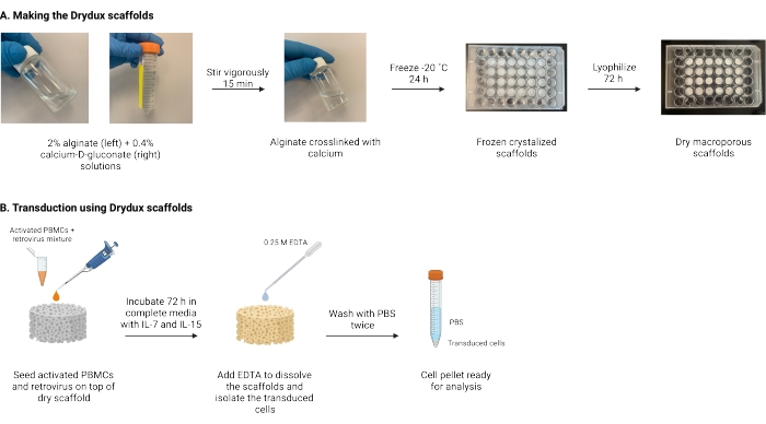
Figure 1: Schematic and timeline of the protocol. (A) Timeline for making the dry macroporous alginate scaffolds. Alginate is cross-linked with calcium-D-gluconate and frozen overnight. The frozen scaffolds are lyophilized for 72 h to create the Drydux scaffolds. (B) Timeline for viral transduction of activated cells. Activated cells and virus (MOI 2) are seeded on top of the scaffold and incubated in complete media supplemented with IL-7 and IL-15. The scaffolds absorb the mixture and promote viral gene transfer. EDTA is used to dissolve the scaffolds and isolate the transduced cells. After washing twice with PBS, the cell pellet can be used for analysis. Abbreviations: PBS = phosphate-buffered saline; PBMCs = peripheral blood mononuclear cells. Please click here to view a larger version of this figure.
These macroporous alginate scaffolds are easy to make and should come out of the lyophilizer as porous, fluffy, and white discs. Although not studied in this experiment, calcium-alginate solution can be cast into different molds to create scaffolds of varying shapes, depending on the needs of the user9,10. The scaffolds are electrostatic and may stick to the lid of the well-plate or to a gloved finger. Figure 2 demonstrates what the scaffolds should look like upon completion. Approximate dimensions of 24- and 48-well scaffolds are also shown in the figure.
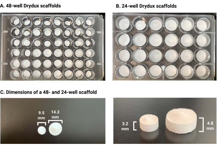
Figure 2: Images of dry macroporous alginate scaffolds. (A) A 48-well plate full of scaffolds. (B) A 24-well plate full of scaffolds. (C) Comparing a 48-well scaffold to a 24-well scaffold. Scaffold dimensions are also shown. Please click here to view a larger version of this figure.
The porosity of the scaffolds is highly important for successful transduction, and we have previously experimented with the porosity of the scaffolds. For more information regarding the porosity, including SEM images of the scaffolds, please refer to papers by Agarwalla et. al. in the references17,18.
Transduction efficiency was analyzed using flow cytometry and the results are shown in Figure 3. Frozen PBMCs from the same donor were activated and used for all experimental groups. A protocol for activating PBMCs can be found in Supplemental File 1, and any standard method to activate T cells and validate activation can be used21,22,23. The activated PBMCs were seeded on either a scaffold with GFP-encoding retrovirus or a scaffold without virus, termed a "blank" scaffold. Activated PBMCs were also seeded on plates spinoculated with retronectin and GFP-encoding retrovirus to compare the transduction efficiency of the alginate scaffolds to the conventional method. Non-transduced (NT) cells-activated PBMCs seeded in uncoated wells of a 24-well plate-were used as the negative control group. As expected, the non-transduced cells and cells isolated from the blank scaffold did not show any transduction. Cells isolated from the scaffold seeded with activated PBMCs and GFP retrovirus showed comparable transduction efficiency to the retronectin-coated plates. The scaffold had an average transduction efficiency of 85%, just below the retronectin group. These results demonstrate that these alginate scaffolds serve as an easier and cheaper alternative to virally transduce T cells without the need for spinoculation of retronectin.
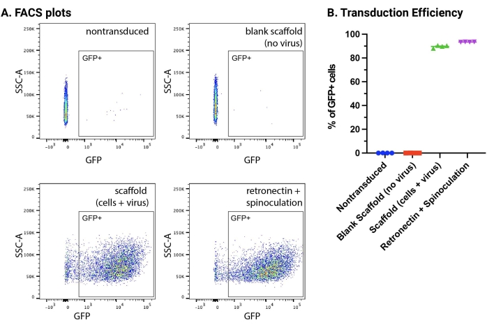
Figure 3: FACS quantification of transduction efficiency. (A) FACS plots showing GFP expression. Cells were gated on viable cells, FSC singlets, and GFP positive cells. (B) Quantification of GFP-positive cells by FACS. Conventional spinoculation of retronectin-coated plates (purple inverted triangle) was used as a positive control. Non-transduced cells (blue circle) and activated cells seeded on the alginate scaffolds without virus (red square) were used as negative controls. Scaffolds seeded with activated cells and GFP retrovirus (green triangle) had a transduction efficiency of 85%, comparable to retronectin. Data represented as mean ± standard deviation with n = 4. Abbreviations: GFP = green fluorescent protein; FACS = fluorescence-activated cell sorting; SSC-A = side scatter-peak area; FSC = forward scatter. Please click here to view a larger version of this figure.
Supplemental File 1: Protocol for activating cells. Please click here to download this File.
Supplemental Figure S1: Images of dry macroporous alginate scaffolds fabricated at -80 ˚C. Freezing scaffolds at -80 ˚C leads to less consistent scaffold appearance and function than when frozen at -20 ˚C. Please click here to download this File.
