Obtaining sterilization is essential for tracheal tissue transplant. Herein, we present a sterilization protocol using low-dose gamma irradiation that is fully tolerated by organs.
One of the main key aspects in ensuring that a transplant evolves correctly is the sterility of the medium. Decellularized tracheal transplantation involves implanting an organ that was originally in contact with the environment, thus not being sterile from the outset. While the decellularization protocol (through detergent exposition [2% sodium dodecyl sulfate], continuous stirring, and osmotic shocks) is conducted in line with aseptic measures, it does not provide sterilization. Therefore, one of the main challenges is ensuring sterility prior to in vivo implantation. Although there are established gamma radiation sterilization protocols for inorganic materials, there are no such measures for organic materials. Additionally, the protocols in place for inorganic materials cannot be applied to organic materials, as the established radiation dose (25 kGy) would completely destroy the implant. This paper studies the effect of an escalated radiation dose in a decellularized rabbit trachea. We maintained the dose range (kGy) and tested escalated doses until finding the minimal dose at which sterilization is achieved. After determining the dose, we studied effects of it on the organ, both histologically and biomechanically. We determined that while 0.5 kGy did not achieve sterility, doses of both 1 kGy and 2 kGy did, with 1 kGy, therefore, being the minimal dose necessary to achieve sterilization. Microscopic studies showed no relevant changes compared to non-sterilized organs. Axial biomechanical characteristics were not altered at all, and only a slight reduction in the force per unit of length that the organ can radially tolerate was observed. We can therefore conclude that 1 kGy achieves complete sterilization of decellularized rabbit trachea with a minimal, if any, effects on the organ.
Sterilization of an implant is a basic requisite for its viability; in fact, prostheses that have proven to be successful are those implanted in sterile areas (blood vessels, heart, bone, etc.)1. The trachea has two surfaces: a surface in contact with the external environment, which is therefore not sterile, and a surface toward the mediastinum, which is sterile. Therefore, from the moment the trachea is extracted, it is not a sterile organ. Despite the subsequent decellularization process being carried out in maximum sterile conditions, it is not a sterilization step2. The implantation of foreign material in itself entails a risk of infection due to the probacterial microenvironment it produces3and an up to 0.014% risk of disease transmission from the donor to the recipient, even if the material has been sterilized4. To ensure correct vascularization of the trachea, in almost all experimental transplant protocols, it first undergoes heterotopic implant5,6,7 to a sterile area (muscle, fascia, omentum, subcutaneous, etc.); this is because implanting a non-sterile element in this medium would lead to infection of the area3.
There are a range of possible strategies to obtain a sterile implant. Using supercritical CO2has achieved terminal sterilization8,9. Other methods, such as ultraviolet radiation or treatment with substances such as peracetic acid, ethanol, oxygen peroxide, and electrolyzed water, have obtained different success rates in sterilization, almost always depending on their dosages, but they have been shown to affect the biomechanical characteristics of implants. Indeed, some substances, such as ethylene oxide, can substantially change the structure of the implanted matrix and can even cause undesirable immunogenic effects. For this reason, many of these strategies cannot be applied to biological models2,10,11,12,13.
The most widely studied and accepted sterilization strategy is that established by the ISO 11737-1:2006 standard for the sterilization of medical devices implanted in humans, with a gamma radiation dose of 25 kGy. However, this regulation focuses only on the sterilization of inert, non-biological elements14,15. Additionally, radiotherapy doses in the radical treatment of carcinoma are three orders of magnitude lower than those used to sterilize medical devices1. With this in mind, we can conclude that said dose would not only kill the microbiota but would also destroy and radically alter the biological structure of the implant. There is also the possibility that it would generate residual lipids upon degradation, which can potentially be cytotoxic and accelerate the enzymatic degradation of the scaffold13,14,15,16,17, even when using doses as low as 1.9 kGy and with damage directly proportional to the radiation dose received17.
Thus, the objective of this paper is to try to identify the radiation dose that allows for a sterile implant to be obtained with minimal harmful effects caused by irradiation2,18,19. The strategy we followed involved the irradiation of decellularized and irradiated tracheas at different escalated doses within a range of kilograys (0.5, 1, 2, 3 kGy, etc.), until achieving a negative culture. Additional tests were carried out for those doses that achieved negative cultures, in order to confirm sterilization. After determining the minimum dose to obtain sterilization, the structural and biomechanical impact of the irradiation on the trachea were checked. All the metrics were compared with the control native rabbit tracheas. The sterilization of the construct was then tested in vivo by implanting the tracheas into New Zealand white rabbits.
The European directive 20170/63/EU for the care and use of laboratory animals was adhered to and the study protocol was approved by the Ethics Committee of the University of Valencia (Law 86/609/EEC and 214/1997 and Code 2018/VSC/PEA/0122 Type 2 of the Government of Valencia, Spain).
1. Tracheal decellularization
NOTE: The decellularization method has been reported elsewhere20.
2. Sterilization
3. Histological analysis
NOTE: Stain the pieces using hematoxylin and eosin21, Masson's trichrome, and orcein22.
4. Biomechanical study
NOTE: Tracheal resistance to longitudinal and transverse forces is measured through axial tensile and radial compression tests23.
5. Surgical technique
NOTE: The surgical technique has been widely reported elsewhere20.
6. Statistical analysis
Decellularization
DAPI staining shows the absence of DNA, and no DNA values higher than 50 ng were detected in any of the tracheae any by electrophoresis, with all fragments being smaller than 200 bp20.
Microbial culture
Two of the eight pieces subjected to 0.5 kGy showed color change in less than 1 week. None of the pieces irradiated at 1 kGy and 2 kGy showed any color change (Figure 1).
Histological analysis
No changes to the collagen or elastic fiber distribution pattern were detected in any of the specimens analyzed (Figure 2).
Determining the radiation dose
Given the results described above, which showed that irradiation at 0.5 kGy did not ensure sterilization of the specimen, whereas doses of 1 kGy and 2 kGy did, we established the minimum possible irradiation dose to achieve sterilization of the tissue as 1 kGy. Therefore, we tested the biomechanical impact of this dose on the tracheas2,17,23.
Biomechanical study
Axial tensile tests
The data obtained in the tensile test on irradiated tracheas are shown in Table 1. Figure 3 shows the corresponding stress-strain curves and breaking points.
Thus, subjecting tracheal pieces to gamma irradiation for sterilization purposes, despite slightly increasing the detected values, does not cause significant effects on the axial biomechanical characteristics of the organs. Hence, both the σmax that the tracheas can tolerate (0.05 MPa; CI [-0.046, 0.144] MPa), as well as εmax (0.096 CI [-0.096, 0.281]), (0.022 MPa; CI [-0.23, 0.274] MPa), and W / Vol (from 0.044 mJ / mm3; CI [-0.018, 0.106] mJ/mm3), are very slightly increased in this sample, but are not in any case applicable to the population estimate.
Radial compression tests
The compression tests performed on both the native tracheas (controls) and on the decellularized, cryopreserved, and irradiated tracheas are shown in Table 2. The corresponding graphs can be seen in Figure 4.
Gamma irradiation causes only a minimal but significant decrease in radial biomechanical characteristics in the variable force per unit of length, which varies by -0.017 N/mm; CI [-0.042, -0.004] N/mm, while the minimal variations detected in W/Vol (0.044 mJ/mm3; CI [-0.018, 0.106] mJ/mm3), R (-0.018 MPa · mm; CI [-0.145, 0.083] MPa · mm), and W/S (-0.081 mJ/mm2; CI [-0.95, 0.74] mJ/mm2), are in no case applicable to the population estimate (Figure 5).
Implant
Macroscopic examination
None of the animals showed inflammatory or infectious symptoms during the postoperative period; their diet was reinstated as planned and antibiotics and analgesics were suspended on day five. Upon euthanasia, integration of the trachea and the flap was observed macroscopically, with no visible signs of inflammation.
Histological examination
The histological examination showed the flap forming highly organized connective tissue – closely linked to the tracheal rings, showing continuity between them and the tissue – in the form of the perichondrium of the native trachea. The cartilage was intact and showed no signs of necrosis. In addition, the presence of macrophages and some isolated giant cells forming sheets were observed. Other than the scarce presence of eosinophils, usual postsurgical mild acute inflammatory cellularity was observed (Figure 6). Incipient neovascularization was also observed around the trachea.
Biomechanical evaluation
After being implanted in the lagomorph, the characteristics of the trachea remained unchanged, except for the force per unit length, which recovered the characteristics of the native trachea only 2 weeks after the transplant (0.006 N/mm, CI [-0.026, 0.04] N/mm) (Figure 7).
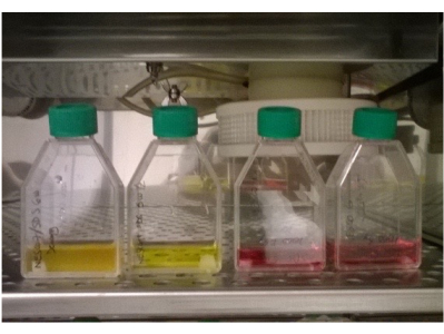
Figure 1: Irradiated tracheas in DMEM without antibiotics or antifungals. The color of the two specimens on the left (0.5 kGy) has changed, indicating a change in pH, and is an indirect sign of bacterial growth. There is also increased turbidity in the first specimen on the left. The two specimens on the right (1 kGy) show no color change. Please click here to view a larger version of this figure.
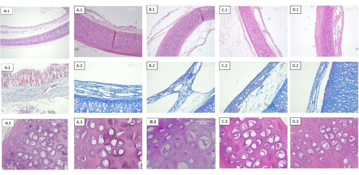
Figure 2: Tracheas decellularized and irradiated at different doses. Each row corresponds to a different staining and each column to different sterilization dosage. 1) Hematoxylin-eosin. Panoramic view of the cartilage, mucosa, submucosa, and serosa. 2) Masson's trichrome stain. Tracheal submucosa. 3) Hematoxylin-eosin. Detailed view of the tracheal cartilage. (A) Non-irradiated tracheas (control). (B) Tracheas irradiated at 0.5 kGy. (C) Tracheas irradiated at 1 kGy. (D) Tracheas irradiated at 2 kGy. The absence of objective histological changes with respect to the radiation dose is observed. Abbreviation: N = native trachea. Please click here to view a larger version of this figure.
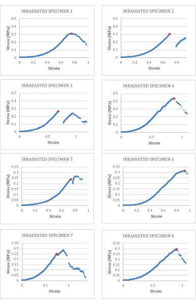
Figure 3: Stress-strain curves for decellularized and irradiated tracheas. The breaking point is marked in orange. Please click here to view a larger version of this figure.
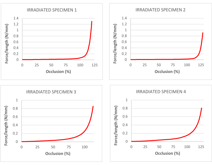
Figure 4: curves for percentage of occlusion corresponding to traction tests in decellularized and irradiated tracheas. Please click here to view a larger version of this figure.
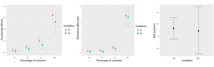
Figure 5: Biomechanical response to irradiation. (A) Graph of marginal effects on the variable force per unit of length, according to the percentage of occlusion of the irradiation interaction. (B) Graph of marginal effects on the variable force per unit of length, according to the percentage of occlusion of the irradiation interaction. (C) Partial dependence plot of the stored energy per unit area model for the irradiation variable. Please click here to view a larger version of this figure.
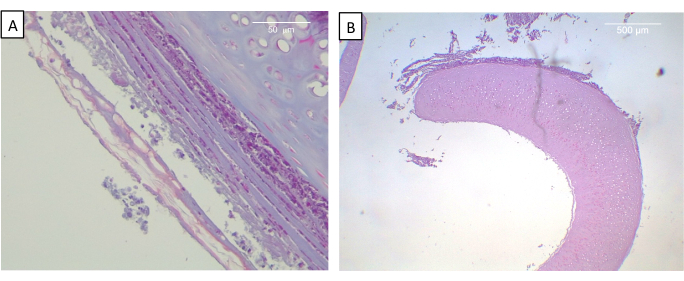
Figure 6: View of implanted trachea at 2 weeks. (A) Masson's trichrome staining. Neoformed connective tissue of the tracheal outer surface organized in concentric layers of fibers and cells is observed. (B) Hematoxylin-eosin. Panoramic view of perfectly preserved cartilage. Please click here to view a larger version of this figure.
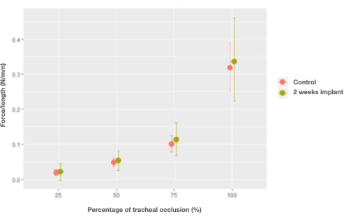
Figure 7: Graph of the marginal effects of the interaction between force per unit length and percentage of occlusion and control (native) tracheas versus trachea implants at 2 weeks. Please click here to view a larger version of this figure.
Table 1: Tensile tests on irradiated tracheas. Controls are native rabbit tracheas. Please click here to download this Table.
Table 2: Compression tests on irradiated, decellularized tracheas. Controls are native rabbit tracheas. Please click here to download this Table.
There are several sterilization strategies that exist. Supercritical CO2fully penetrates tissues, acidifying the medium and deconstructing the cellular phospholipid bilayer with simple elimination by means of depressurization of the implant8,14,25. Ultraviolet radiation has also been used, and its effectiveness in rodent trachea has been published, although there are only a few reports in the literature10. Other methods used include the application of substances such as peracetic acid, ethanol, oxygen peroxide, or electrolyzed water, which have given irregular results and been shown to greatly affect tissue11,12. In contrast to the aforementioned strategies, gamma irradiation has not only been shown to be completely effective in terms of sterilization, but has also been thoroughly and profusely studied, in regards to both its dose and sterilizing effects. In fact, it has been studied so much that there is an ISO standard for the use of gamma radiation in sterilization, in which the dose for the sterilization of inert material to be implanted in humans is established at 25 kGy13,14,15.
On the other hand, in addition to sterilizing material, irradiation has also been shown to cause collateral effects as a limitation of the technique. These include the destruction and alteration of matrices by denaturing protein molecules, including collagen, and generating residual molecules, which can even become toxic. This degradation of organ structure consequently affects both its biological and biomechanical characteristics, with the deleterious effects of irradiation being directly proportional to its dose and being observed at relatively low doses13,14,15,16,17. Here, the objective was therefore twofold: on the one hand, to obtain a sterile construct in order to ensure a viable implant, and on the other to preserve the biological and biomechanical characteristics of the matrix, as the implant would be futile unless both were maintained26. Thus, the challenge was selecting a strategy that allowed for a balance between successful sterilization and preserving tissue structure.
Herein, 1 kGy was established as the minimum dose for sterilization. Histological examination showed that this dose of irradiation has no impact on the tissue. Further, biomechanical characterization of the irradiated tracheas determined that the use of irradiation makes absolutely no difference to traction parameters. There was a slight but statistically significant decrease in the force per unit length that the trachea was able to tolerate in the radial compression tests, however this does not affect its other radial characteristics.
While there are a few papers that discuss the impossibility of sterilization and the destructuration caused by doses as low as 1.5 kGy19, the vast majority are in line with the presented data2,18,19. In this way, authors observe that sterilizing bone at doses of10, 15, 20, and 25 kGy achieves complete sterilization, although in exchange for a reduction in cell incubation capacity and an increase in collagen degradation products at doses higher than 15 kGy18. A dose of 1.5 kGy did not obtain sterilization in decellularized heart valves, but did cause damage to the mechanical qualities of the specimens both in vivo and in vitro; meanwhile, a dose of 3 kGy did achieve sterilization, but caused destructuring and fibrosis19. As regards the trachea, Johnson et al. compared the effects of sterilization at the ISO dose of 25 kGy with a dose of 5 kGy. Both doses obtained terminal sterilization, with the dose of 5 kGy slightly altering the structure of the specimen and the dose of 25 kGy completely destructuring the trachea2.
In addition, effective sterilization is confirmed thanks to an absence of infectious events in regards to the implant after 2 weeks, with sterilization being fully tolerated by the organs. Also, the structure was completely preserved, with no necrosis or denaturation of the organ. Furthermore, as an additional finding, it was observed that the minor alteration in biomechanical characteristics – to the force that the trachea is able to tolerate per unit length – returned to the values of a native trachea only 2 weeks after implantation; therefore, this effect can be disregarded as per the final management of the construct.
Therefore, this paper presents the possibility of obtaining completely sterile organs at much lower doses than the recommended dose of 25 kGy; the proposal troubleshoots the sterilization of New Zealand rabbit tracheas with a dose of 1 kGy. This dose ensures that the histological, ultrastructural, and biomechanical characteristics of these organs are maintained, and shows perfect tolerance to the implantation. A limitation of the study is that it is conducted only on sterilized rabbit tracheas, which generally require a lower dosage due to being smaller in size; however, it can be concluded that the excessively high figures established in the ISO standard for inert implants are not necessary for the sterilization of decellularized tracheas, thus being a huge achievement due to the greatly reduced harm to the tissue. Furthermore, in future studies, depending on the animal, and therefore on the size of its trachea, these doses may be adjusted to much lower doses that are consequently more respectful of the organ's structure and function.
The authors have nothing to disclose.
This paper was supported by the 2018 Spanish Society of Thoracic Surgery Grant to National Multicentric Study [Number 180101 awarded to Néstor J.Martínez-Hernández] and PI16-01315 [awarded to Manuel Mata-Roig] by the Instituto de Salud Carlos III. CIBERER is funded by the VI National R&D&I Plan 2018-2011, Iniciativa Ingenio 2010, Consolider Program, CIBER Actions, and the Instituto de Salud Carlos III, with assistance from the European Regional Development Fund.
6-0 nylon monofilament suture | Monosoft. Covidien; Mansfield, MA, USA | SN-5698G | |
Amphotericin B 5% | Gibco Thermo Fisher Scientific; Waltham, MA USA | 15290018 | |
Bioanalyzer | Agilent, Santa Clara, CA, USA | G2939BA | |
Buprenorphine | Buprex. Reckitt Benckiser Healthcare; Hull, Reino Unido | N02AE01 | |
Compression desktop UTM | Microtest, Madrid, Spain | EM1/10/FR | |
Cryostate | Leyca CM3059, Leyca Biosystems, Wetzlar, Alemania | CM3059 | |
DAPI (4',6-diamino-2-phenylindole) | DAPI. Sigma-Aldrich, Missouri, USA | D9542 | |
Dimethyl sulfoxide (DMSO) | Sigma-Aldrich; MO, USA | D2650 | |
DMEM | Thermo Fisher Scientific; Waltham, MA, USA | 11965084 | |
DNA extraction kit | DNeasy extraction kit Quiagen, Hilden, Germany | 4368814 | |
Enrofloxacin, 2.5% | Boehringer Ingelheim, Ingelheim am Rhein, Germany | 0035-0002 | |
Fetal bovine serum (FBS) | GE Healthcare Hyclone; Madrid, Spain | SH20898.03IR | |
Fluorescence microscope | Leyca DM2500 (Leica, Wetzlar, Germany) | DM2500?? | |
Freezing Container | Mr Frosty. Thermo Fisher; Madrid, Spain | 5100-0001 | |
Isofluorane | Isoflo; Proyma Ganadera; Ciudad Real, Spain | 8.43603E+12 | |
Ketamin | Imalgene. Merial; Toulouse, Francia | BOE127823 | |
Linear accelerator | "True Beam". Varian, Palo Alto, California, USA | H191001 | |
Magnetic stirrer | Orbital Shaker PSU-10i. Biosan; Riga, Letonia | BS-010144-AAN | |
Meloxicam 5 mg/ml | Boehringer Ingelheim, Ingelheim am Rhein, Germany | 6283-MV | |
OCT (Optimal Cutting Temperature Compound) | Fischer Scientific, Madrid, Spain | 12678646 | |
Penicillin-streptomycin 5% | Gibco Thermo Fisher Scientific; Waltham, MA USA | 15140122 | |
Pentobarbital sodium | Dolethal. Vetoquinol; Madrid, España | 3.60587E+12 | |
Phosphate buffered saline (PBS) | Sigma-Aldrich; MO, USA | P2272 | |
Propofol | Propofol Lipuro. B. Braun Melsungen AG; Melsungen, Alemania | G 151030 | |
Proteinase K | Gibco Thermo Fisher Scientific; Waltham, Massachussetts, USA | S3020 | |
PVC hollow tubes | Cristallo Extra; FITT, Sandrigo, Italy | hhdddyyZ | |
PVC stent | ArgyleTM Medtronic; Istanbul, Turkey | 019 5305 1 | |
R software, Version 3.5.3 R Core | R Foundation for Statistical Computing | R 3.5.3 | |
Sodium dodecyl sulfate (SDS) | Sigma-Aldrich; MO, USA | 8,17,034 | |
Spectrophotometer | Nanodrop, Life Technologies; Isogen Life Science. Utrech, Netherlands | ND-ONEC-W | |
Spreadsheet | Microsoft Excel for Mac, Version 16.23, Redmond, WA, USA | 2864993241 | |
Traction Universal Testing Machine | Testing Machines, Veenendaal, Netherlands | 84-01 | |
UTM Software | TestWorks 4, MTS Systems Corporation, Eden Prairie, MN, USA | 100-093-627 F | |
VECTASHIELD Mounting Medium | Vector Labs, Burlingame; CA; USA | H-1000-10 | |
Xylacine | Xilagesic. Calier; Barcelona, España | 20102-003 |