Luminescent identification of functional elements in 3’ untranslated regions (3’UTRs) (3’LIFE) is a technique to identify functional regulation in 3’UTRs by miRNAs or other regulatory factors. This protocol utilizes high-throughput methodology such as 96-well transfection and luciferase assays to screen hundreds of putative interactions for functional repression.
Luminescent Identification of Functional Elements in 3’UTRs (3’LIFE) allows the rapid identification of targets of specific miRNAs within an array of hundreds of queried 3’UTRs. Target identification is based on the dual-luciferase assay, which detects binding at the mRNA level by measuring translational output, giving a functional readout of miRNA targeting. 3’LIFE uses non-proprietary buffers and reagents, and publically available reporter libraries, making genome-wide screens feasible and cost-effective. 3’LIFE can be performed either in a standard lab setting or scaled up using liquid handling robots and other high-throughput instrumentation. We illustrate the approach using a dataset of human 3’UTRs cloned in 96-well plates, and two test miRNAs, let-7c and miR-10b. We demonstrate how to perform DNA preparation, transfection, cell culture and luciferase assays in 96-well format, and provide tools for data analysis. In conclusion 3'LIFE is highly reproducible, rapid, systematic, and identifies high confidence targets.
The overall goal of this method is to detect and precisely map microRNA (miRNA) targets in high-throughput. MiRNAs are endogenous non-coding RNAs ~22 nucleotides in length. Following transcription and processing, mature miRNAs are incorporated in a protein complex called the RNA induced silencing complex (RISC). Each miRNA guides the RISC to target elements located primarily in the 3’untranslated regions (3’UTRs) of messenger RNAs (mRNAs), resulting in either translation repression or mRNA cleavage 1. MiRNA recognize target sites based on standard Watson-Crick and G:U wobble base pairing, and are degenerate in nature, containing multiple mismatched base pairs and bulged regions. Many miRNAs are broadly conserved from plants to humans 2,3, where they play a diverse range of biological roles. In metazoans miRNAs can influence multiple biological processes including cell fate decisions 4, developmental timing 5, and frequently exhibit tissue specific expression patterns 6,7. MiRNA misexpression can also result in aberrant gene regulation, which can have substantial influence on cell behaviour based solely on the function of target genes. As such, miRNAs are linked to a wide range of diseases, including neurodegeneration 8,9, diabetes 10 and cancer 11. Bioinformatic and wet-bench approaches suggest that each miRNA may be capable of targeting hundreds to thousands of distinct mRNAs 12-14, indicating that high-throughput or genome wide approaches are required to probe this large pool of potential interactions.
Identifying target genes is a critical component of mechanistically defining miRNA function, and to do so researchers must be able to reveal targets on a large scale. Several approaches have been developed to identify miRNA targets, including bioinformatic prediction algorithms, high-throughput sequencing of targeted mRNAs, and reporter based assays. Each of these approaches has inherent strengths and weaknesses. Given that miRNA targeting is guided by sequence specificity, most notably of nucleotides 2-6 of the miRNA (termed the seed region), several algorithms have been developed to predict miRNA targets throughout the genome of many organisms. These algorithms are trained using the observed base-pairing motifs of validated miRNA targets, and frequently utilize parameters such as stringent seed pairing, site conservation, and/or thermodynamic stability 15. While these filters refine the large number of putative targets with sufficient complementarity to only high confidence targets, they may exclude species specific and non-canonical miRNA target sites, which recent evidence suggests are widespread 16-24. Furthermore, these predictions do not take into account mechanisms of mRNA processing that exclude miRNA target sites, such as alternative polyadenylation 25, RNA editing 26, RNA methylation 27, and cooperative binding. As such, high false positive and false negative rates have been reported for many algorithms 22,24,28. While these algorithms are useful to identify candidate miRNA targets for subsequent experimental validation, these high error rates limit the efficacy of bioinformatic approaches for systematic miRNA target detection.
To systematically probe for interactions between a given miRNA and potentially targeted 3’UTRs we have developed a high-throughput assay called Luminescent Identification of Functional Elements in 3’UTRs (3’LIFE) 24. This assay measures direct interactions and translational repression of the test 3’UTR by a query miRNA using a dual luciferase reporter system. In this system, the 3’UTR of a gene of interest is cloned downstream of the firefly luciferase (fluc) reporter reading frame. The reporter construct is cotransfected with a query miRNA in HEK293T cells. MiRNA targeting is determined by measuring the relative change between the test fluc::3’UTR reporter and a second non-specific Renilla luciferase reporter. Importantly, luciferase assays detect functional miRNA/mRNA interactions that influence the translational output of the reporter. This is a key advantage over traditional methods to detect miRNA regulation, such as RT-qPCR and Western blots, in that this bypasses differences in mRNA degradation and translational repression, as well as changes in protein abundance independent of 3’UTR based regulation.
Luciferase assays are widely utilized to validate direct miRNA targets because of their relative simplicity and sensitivity, yet their use in high-throughput screens is limited by high costs associated with consumable reagents, the lack of 3’UTR libraries from public sources, and the absence of standardized luciferase protocols, leading to difficulties in comparing functional repression across multiple datasets. To facilitate the use of the 3’LIFE assay, we have placed emphasis on simplification of experimental design, utilization of non-commercial transfection 24 and luciferase reagents 29, creating a 3’UTR library which is regularly updated and expanded, and is available through a public plasmid repository 30.
The scalability of the 3’LIFE assay allows screening of a large 3’UTR library for targeting by a given miRNA without biasing the screen towards bioinformatically identified genes. In addition to testing canonical and predicted interactions, this systematic approach allows the identification of novel targets driven via non-canonical and/or species-specific interactions. Importantly, the effect of miRNA targeting on protein production is generally understood to result in modest translational repression 15,31, suggesting that a primary role of miRNA regulation is to fine-tune protein output, protect against aberrant levels of gene expression, and provide robustness to cell specific programs 32,33. The sensitivity of the luciferase assay combined with the inherently large number of negative miRNA/mRNA interactions in the 3’LIFE screen allows the detection of subtle effects of miRNA targeting on a large number of genes, and the identification of multiple components of gene networks that are regulated by a given miRNA 24.
Here we describe the 3'LIFE protocol, and demonstrate it’s feasibility by screening two well characterized miRNAs, miR-10b and let-7c against a panel of 275 human 3'UTRs (Figure 1).
1. Cell Culture (24-48 hr prior to transfection)
2. Preparation Prior to Transfection
NOTE: The preparation of the buffers and plasmid DNA in step 2.0-2.2 should be performed in the days prior to transfection since the preparation of these reagents may be time consuming.
3. Prepare Following Items Immediately Prior to Transfection
4. Preparation of Plasmid DNA and Cell Mixture
NOTE: The following protocol assumes transfecting three 96-well plates in one experiment for a screen with two miRNAs (miRNA-#1 and miRNA-#2). Each plate will correspond to the same 96-well plate of pLIFE-3’UTR plasmids, and be treated three times with pLIFE-miRNA-blank, pLIFE-miRNA-#1, or pLIFE-miRNA-#2.
5. Transfection
6. Cell Lysate Preparation for Luciferase Assay
7. Dual Luciferase Assay
NOTE: If multiple plates are being measured sequentially on one luminometer, create buffer master mixes with everything except ATP and substrates, adding these reagents followed by pH adjustment immediately before use with each plate. ATP and substrates may degrade over time; consistency in the amount of time these reagents are in the buffer will improve consistency across multiple plates.
8. Data Analysis
The luminometer output file contains raw measurements for both firefly and Renilla luciferase proteins. This raw format is compatible with the “3’LIFE – single plate analysis” and “3’LIFE – multiplate analysis” spreadsheets available from the Mangone lab website (www.mangonelab.com). The single plate analysis spreadsheet automatically calculates firefly/Renilla ratio, normalizes each miRNA to the appropriate negative control, and normalizes repression values across each plate. This spreadsheet automatically identifies wells with low Renilla luciferase signal, highlights wells that exhibit repression compared to the negative control, and provides measures of repression across the entire plate (Figure 2). See 24 for detailed explanation of statistical analysis.
Multiple replicates can be analysed using the “multiplate analysis” spreadsheet. Each replicate is compared side by side, and statistical measures of the data are automatically calculated (Figure 3). In addition to comparing replicates with the “Normalized Repression” columns, the user can compare repression between the two miRNAs under the “miRNA#1/miRNA#2” columns. This measure divides the repression index for each miRNA for each replicate. This measure can indicate erroneous values from the luciferase assay (for example abnormally high or low readings with the negative control, see Figure 3, row A9, Rep #3), and wells where the repression index may not indicate substantial repression, but that do exhibit significant differences between the miRNAs. While this measure may not be used directly to indicate a miRNA target, it is useful for identifying outliers, problematic wells, or patterns in the data that are not solely attributed to direct miRNA regulation.
The Repression Index (RI) is used to call a putative miRNA target, with lower values corresponding to higher relative repression. The threshold for calling putative targets is based on the level of stringency required by the researcher, but combining the RI with 3’UTRs that display statistically significant p-values (p <0.05) will indicate high confidence targets (see Figure 2 Rows B8 and B12).
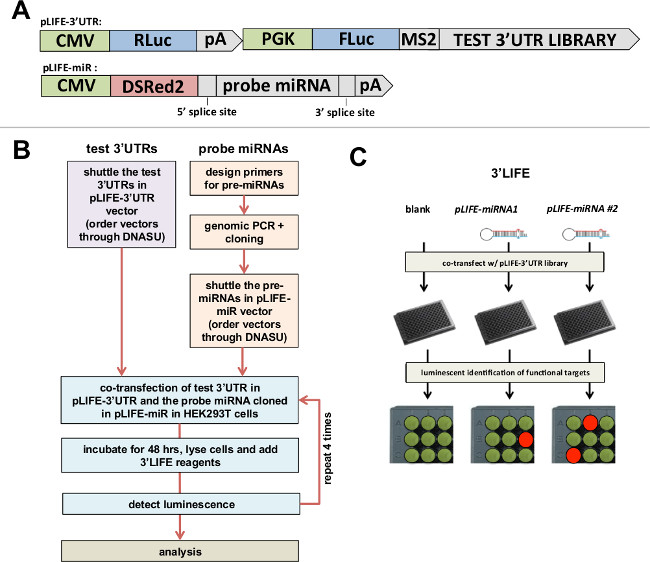
Figure 1: 3’LIFE Assay. (A) Gateway-compatible vectors used in the 3’LIFE assay. Top: The luciferase gene (FLuc) is fused to the test 3’UTR, while the Renilla luciferase gene (RLuc) is fused to the unspecific SV40 pA 3’UTR as control. Bottom: The RFP- miRNA-intron vector – The probe pre-miRNA, plus ~400 nucleotides within its genomic locus (to recapitulate endogenous miRNA processing), is cloned within an intron to allow its co-expression with DSRed2 fluorochrome. Both vectors are publically available (Seiler et al., 2013). (B) Flow chart of the 3’LIFE Assay. (C) 3’LIFE Pipeline: The dual-luciferase vector containing the test 3’UTR with or without the miRNA vectors are co-transfected into HEK293 cells in 96-well plates. The interaction between the miRNA and a bona fide 3’UTR target will lower the relative luminescence in specific wells (exemplified by the orange spot in the experimental plate).
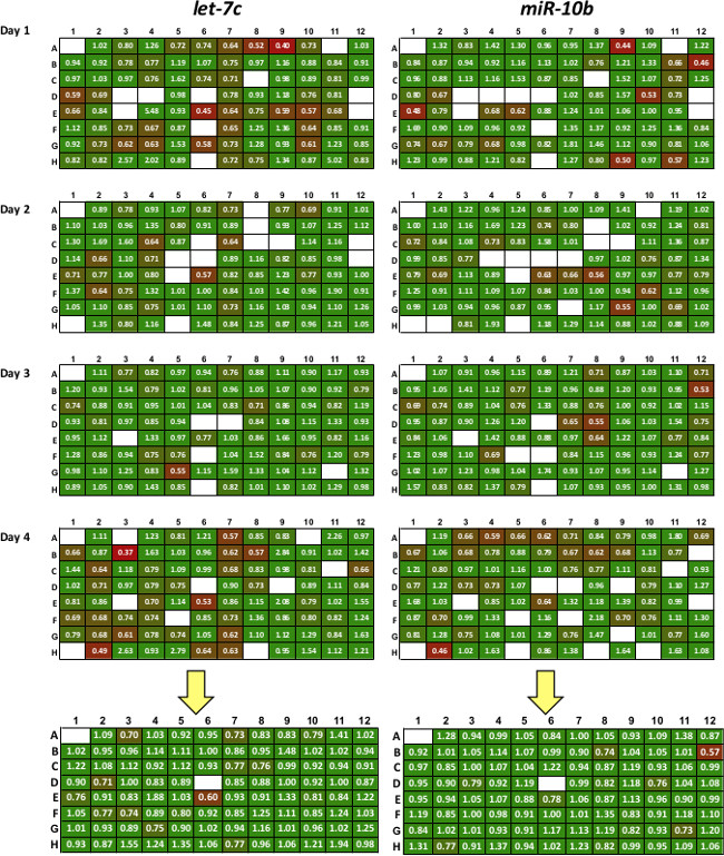
Figure 2: Sample of data produced with 3’LIFE assay. Each probe miRNA is tested in quadruplicate (replicates 1-4). Colors represent repression levels, with red colors indicating strong miRNA/3’UTR interaction. All replicates are averaged to produce high-quality putative targets shown in the summary plate below the yellow arrow. White box represent controls, failed transfections or wells with low transfection efficiency.
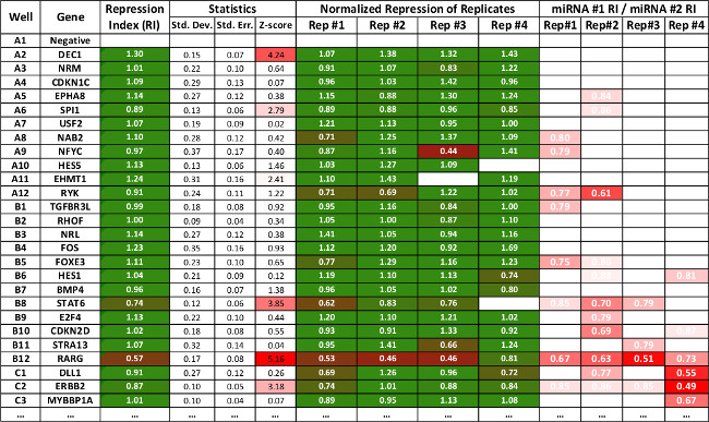
Figure 3: Table representing summary data of a subset of interactions produced using the 3’LIFE spreadsheet. The repression values are as in Figure 2. The software calculates standard deviation, standard error and z-score for each interaction. Statistically significant interactions are marked in red. The last four rows show relative repression of one miRNA to the other, and is used as secondary indicator to compare repression between two different miRNAs. The spreadsheet can be downloaded from www.mangonelab.com
Firefly luciferase buffer reagents | Final concentration (1x) |
Glycylglycine | 25 mM |
KxPO4 (pH 7.8) | 15 mM |
MgSO4 | 15 mM |
DTT (store at 4º) | 1 mM |
EGTA | 4 mM |
ATP* | 2 mM |
Beetle luciferin* | 250 μM |
Table 1: Stock firefly luciferase reagents: 10x Stock solutions of Glycylglycine, KxPO4, MgSO4, DTT and EGTA can be prepared separately and stored prior to buffer reconstitution. 100x Beetle Luciferin (firefly luciferase substrate) can be stored by dissolving 50 mg luciferin in 7.134 ml H20 (25 mM). Aliquot 105 μl/plate of dissolved Beetle luciferin into tubes and store at -80 ºC. Per Promega technical support, this should be stable for >6 months, but may be light sensitive. NOTE: EGTA will not go into solution at neutral pH. Slowly add NaOH to EGTA until it dissolves completely.*Reagents added to final buffer immediately prior to the luciferase assay
Renilla luciferase buffer reagents | Final concentration (1x) |
NaCl | 1.1 M |
Na2EDTA | 2.2 mM |
KH2PO4 | .22 M |
NaN3 | 1.3 mM |
BSA* | .44 mg/ml |
Coelenterazine* | 2.5 μM |
Table 2: Stock Renilla luciferase buffer reagents All the reagents except BSA and Coelenterazine can be mixed at a 1x concentration and stored at room temperature. Coelenterazine can be dissolved in acidified methanol and aliquoted per plate. Acidify methanol by adding HCl to final concentration of 5 mM (<3 pH). Dissolve 250 μg coelenterazine in 2.36 ml acidified methanol (250 μM) aliquot 105 ul/plate. The mix is stable for at least 6 months but may be light sensitive. *Reagents added to buffer immediately before luciferase assay.
The 3’LIFE assay identifies functional miRNA targets in 3’UTRs in high-throughput. This assay is useful for researchers who wish to experimentally identify a large number of putative targets for their miRNA of interest. The 3’LIFE assay is a powerful approach to query for 3’UTR driven regulation, in that the assay provides a functional measure of miRNA targeting, and the binary testing of a single reporter::3’UTR against a single miRNA can confidently address the targeting status of individual genes. To validate this approach, we screened a panel of 275 3’UTRs and against two miRNAs, let-7c and miR-10b, and included 10 previously validated target genes in this library. Eight of these ten genes exhibited repression 24. We also observed a significant enrichment of unvalidated bioinformatically predicted targets, and unpredicted 3’UTRs that contain canonical seed elements among our top hits, suggesting that 3’LIFE is capable of identifying bona fide miRNA targets.
A key indicator of the sensitivity of high-throughput screens is the false positive and false negative rates. While the false positive rate of this assay needs to be evaluated using additional alternative approaches to validate hits, eight of the ten positive controls included in our proof-of-principle screen exhibited repression, suggesting a false negative rate of 20%. However, many techniques are used to identify miRNA targets in different cellular contexts, and 3’UTR processing and regulation by trans-acting factors is known to be highly tissue specific. For example, the majority of 3’UTRs contain multiple polyadenylation sites, which control the length of the 3’UTR in the mature mRNA. In many cases the use of proximal polyadenylation sites is tissue specific, and may exclude miRNA target sites. Additionally, cooperative miRNA targeting, competition with RNA-binding proteins, and mRNA secondary structure may all impact the ability of 3’LIFE to detect miRNA targets in specific tissues. Because of this, the detection of targets by the 3’LIFE assay may vary based on the cellular context in which the assay is performed, complicating the evaluation of absolute error rates. This protocol is optimized for HEK293T cells, but alternative cell lines can be used if the researcher wishes to perform the assay in a specific biological context. However, the optimization of transfection efficiency and cell survival with each cell line will have to be optimized using multiple buffer conditions, pulse codes, and number of cells. An example of an optimization scheme can be found at Wolter et al. 24.
This protocol has been optimized in 96-well format and specifies the use of certain high-throughput instrumentation. In the case that the institution does not possess the equipment required for 96-well Nucleofection, alternative transfection reagents could be used to perform the 3’LIFE assay, as long as the transfection efficiency remains high. Additionally, the luciferase assay is the most time consuming aspect of the 3’LIFE assay. As such, the use of multiple luminometers is recommended for high-throughput screens.
The authors have nothing to disclose.
We thank Stephen Blazie, Karen Anderson, Josh LaBaer for advice and discussion. Karen Anderson, John Chaput, and Josh LaBaer for sharing reagents and instrumentation, Michael Gaskin and Andrea Throop for technical advise and protocols. Justin Wolter is a Maher scholar and thanks the Maher family for their generous support.
Reagents | ||
glycylglycine | Sigma | G1127-25G |
Kx PO4 | Sigma | P2222 |
EGTA | Sigma | E3889 |
ATP | Sigma | FLAAS |
DTT | Sigma | D0632 |
MgSO4 | Sigma | M7506 |
CoA | Sigma | C4282 |
luciferin | Sigma | L9504 |
NaCl | Sigma | S7653 |
Na2EDTA | Sigma | E0399 |
K H2 P O4 | Sigma | 1551139 |
BSA | Sigma | A2153 |
NaN3 | Sigma | S2002 |
Coelenterazine | Sigma | C3230 |
PBS/HEPES | Corning | 21-040-CV |
DMEM | Sigma | D5546 |
FBS | Sigma | F2442 |
Pennicilin/Streptomicin | Sigma | P4333 |
Trypsin | T2600000 | |
Consumables | ||
MaxiPrep Kit | Promega | A2392 |
96-well miniprep plate | Pall | 8032 |
96-well shuttle plates | Lonza | V4SP-2096 |
5x Lysis Buffer | Promega | E1941 |
Instruments | ||
96-well GloMax Plate Reader | Promega | E9032 |
Biomech FX Liquid Handler Robot | Beckmann | A31842 |
4D-Nucleofector Core Unit | Lonza | AAF-1001 |
96-well Shuttle System | Lonza | AAM-1001 |
Cell Counter Countess | Invitrogen | C10227 |