Here, we present a protocol that uses JC-1 dye to assess the mitochondrial membrane potential of cells after being exposed to hypoxia/reoxygenation with or without a protective agent.
Timely and efficient reperfusion of the occluded coronary artery is the best strategy for decreasing myocardial infarct size in patients with a ST-segment elevated myocardial infarction. However, reperfusion per se can result in further cardiomyocyte death, a phenomenon known as reperfusion injury. The opening of the mitochondrial permeability transition pore (mPTP), with the decrease of the mitochondrial membrane potential (MMP), or mitochondrial depolarization, is universally recognized as the final step of reperfusion injury and is responsible for mitochondrial and cardiomyocyte death. JC-1 is a lipophilic cationic dye that accumulates in mitochondria depending on the value of MMP. The higher the MMP is, the more JC-1 accumulates in the mitochondria. The increasing amounts of JC-1 in mitochondria can be reflected by a fluorescence emission shift from green (~530 nm) to red (~590 nm). Therefore, the reduction of the red/green fluorescence intensity ratio can indicate the depolarization of mitochondria. Here, we take advantage of JC-1 to measure MMP, or the opening of mPTP in human cardiac myocytes after hypoxia/reoxygenation, detected by flow cytometry.
Coronary heart disease is the leading cause of death worldwide. The treatment of choice for reducing ischemic injury and limiting infarct size in patients with ST-segment elevated myocardial infarction is timely and effective myocardial reperfusion via primary percutaneous coronary intervention (PCI)1,2. However, reperfusion causes additional damage, which can account for up to 30 percent of the final infarct size3. It is universally acknowledged that the mitochondrial permeability transition pore (mPTP) is not only central in mitochondrial damage and cell death during ischemia/reperfusion (I/R), but is also a converging target of cardioprotective signaling4,5. As the mPTP opening would bring about depolarization of the inner mitochondrial membrane potential (MMP)4, we detected mPTP opening using the 5,5′,6,6′-Tetrachloro-1,1′,3,3′-tetraethyl-imidacarbocyanineiodide (JC-1) assay.
The JC-1 assay is a cytofluorimetric method that is both qualitative and quantitative, and it has been further validated by analyzing the MMP at the level of a single mitochondria6. JC-1 exists as an aggregated form, yielding a red to orange colored emission (590 ± 17.5 nm) in the matrix of mitochondria with normal MMP; with the loss of MMP, JC-1 is converted to the monomeric form that yields green fluorescence with an emission of 530 ± 15 nm. Therefore, a decrease in the red/green fluorescence intensity ratio could indicate the reduction of MMP in conditions such as ischemia/reperfusion (I/R).
In addition to JC-1, MMP has also been studied with membrane-permeable lipophilic cations such as Rhodamine 123 and 3,3′-dihexyloxadicarbocyanine iodide [DiOC6(3)]. However, compared to these two probes, JC-1 is more reliable for analyzing MMP. Rhodamine 123 has relatively poor sensitivity (especially in quenching mode7,8) and poor specificity. The shift in rhodamine 123 sometimes is so small that it is hard for researchers or equipment to observe/detect. Besides, in a single cell, there are different mitochondrion binding sites for rhodamine 123 and so that it may have different fluorescence emissions9. DiOC6(3) is not recommended for detecting MMP either as it reacts sensitively to the depolarization of plasma membrane10.
Therefore, here we use the JC-1 assay to assess the MMP of HCMs after being exposed to hypoxia/reoxygenation with or without a protective agent.
1. Preparation of Reagents and Solutions
2. Establishment of Hypoxia/Reoxygenation Model
3. Preparation of HCMs for Flow Cytometry
4. Flow Cytometer Setup
5. Flow Cytometry Measurement and Data Analysis
Before performing the JC-1 assay to evaluate the changes of MMP, it is highly recommended that experiments be carried out to confirm the conditions successfully set by the researchers. As shown by the flow cytometry results (Figure 2), compared with the normal group, hypoxia/reoxygenation (H/R) significantly induced the apoptosis of HCMs (Annexin V + /PI±), indicating that we had established a cell-based model of I/R (45.00 ± 2.13% vs. 11.50 ± 0.18% in the normal group, p <0.05). Tongxinluo, a Chinese traditional medicine with cardioprotective effects, prevented the apoptosis of HCMs in condition of H/R (24.50 ± 1.13% vs. 45.00 ± 2.13% in the H/R group, p <0.05).
CCCP is a protonophore that can inhibit oxidative phosphorylation in mitochondria by uncoupling the proton gradient (loss of MMP) established during the normal activity of electron carriers in the electron transport chain. Consequently, HCMs treated with CCCP was set as positive control. As shown in Figure 4, the red/green ratio in the CCCP-treated group was almost zero. Consistent with the results from the apoptosis assay, the H/R-treated group displayed a significantly lower red/green ratio compared with the normal group (0.69 ± 0.07 vs. 5.64 ± 0.21 in the normal group, p <0.05) and Tongxinluo reversed such a ratio change (2.92 ± 0.22 vs. 0.69 ± 0.07 in the H/R group, p<0.05).
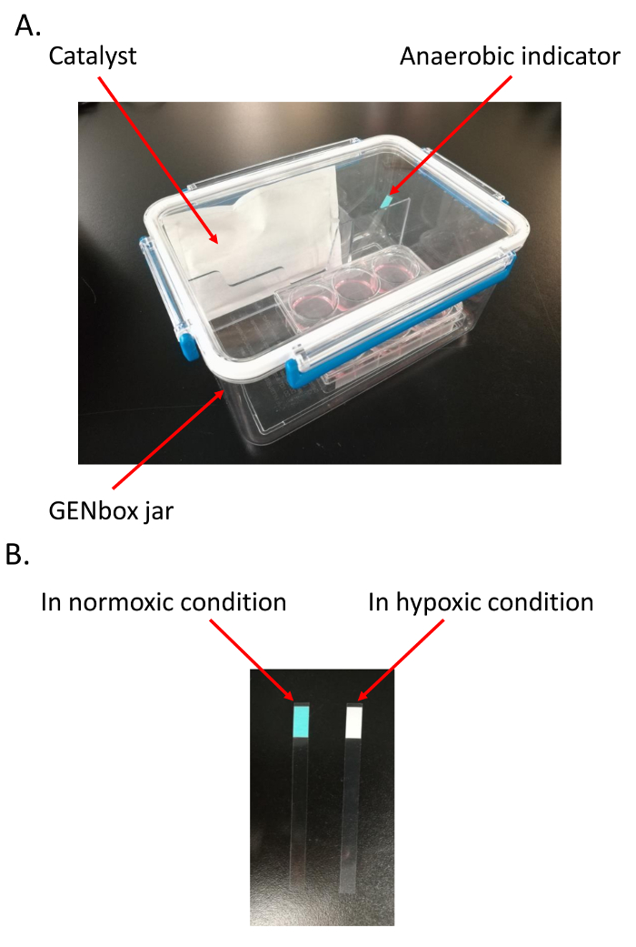
Figure 1. Establishment of hypoxia/reoxygenation (H/R) model. A. Materials needed to establish the H/R model; B. Confirmation of the hypoxic environment in the jar. The color of anaerobic indicator changes from light blue in normoxic environment to pale white in hypoxic condition. Please click here to view a larger version of this figure.
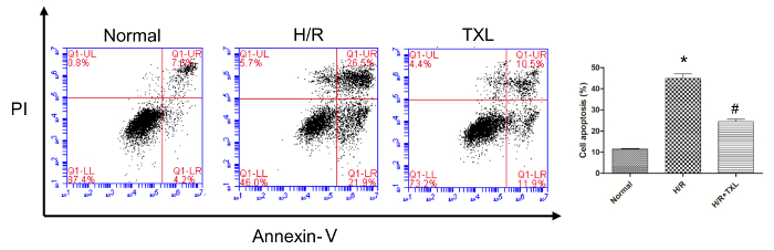
Figure 2. Validation of the establishment of hypoxia/reoxygenation (H/R) model by flow cytometry. Compared with the normal group, the apoptotic rate of human cardiac myocytes was significantly higher in the H/R group, while Tongxinluo reversed this change (*p <0.05 vs. Normal; # p <0.05 vs. H/R; n = 3)11. Please click here to view a larger version of this figure.
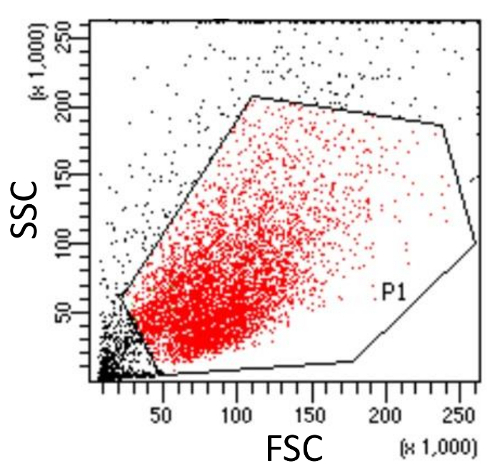
Figure 3. Gating cell population (P1, Red) in the SSC versus FSC dot plot for further analysis. The integrity of the cell population is assessed using side scatter (SSC) and forward scatter (FSC). Cell debris (Outside the gate P1, Black), which have reduced light scattering, are excluded. Please click here to view a larger version of this figure.
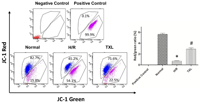
Figure 4. Evaluation of mitochondrial membrane potential in human cardiac myocytes. Compared with the normal group, the H/R group displayed a significantly lower red/green ratio, while Tongxinluo reversed this trend (Red: cells with no fluorescence; Blue, cells with red fluorescence; Purple, cells with green fluorescence. Negative Control, blank and no staining; Positive Control, cells treated with CCCP; *p <0.05 vs. Normal; #p <0.05 vs. H/R; n = 3)11. Please click here to view a larger version of this figure.
Here, we present a protocol that uses JC-1 dye to assess the MMP of cells after being exposed to H/R. Detected by JC-1 assay, the MMP of cells is independent of factors such as mitochondrial size, shape, and density that may influence single-component fluorescence signals14. Consequently, the results of JC-1 assay are relatively reliable. Besides, it is convenient and time-saving to perform the JC-1 assay. This assay has a low requirement for materials and reagents, and generally, it takes less than 1.5 h to do the staining, from cell detachment to MMP measurement.
A few critical steps cannot be overstated when performing the JC-1 assay if researchers want to obtain reliable and satisfactory results. First of all, the cells loaded with JC-1 should be stored in ice-cold staining buffer until they are collected by the flow cytometer. Temperature has a considerable effect on the stability of MMP and the value of one sample varies a lot after a brief time in room temperature. In addition, preliminary experiments are recommended to determine the pretreating time and concentration of CCCP, ensuring that the red/green ratio of the positive control is ~0. It is noteworthy that carbonyl cyanide 4-(trifluoromethoxy) phenylhydrazone (FCCP), which can also eliminate MMP and inhibit oxidative phosphorylation, is an alternative to CCCP and can also be used to set up the positive control10.
The JC-1 assay has a major limitation in that other fluorochromes cannot be combined with JC-1, for its fluorescence emission spans the green, yellow, and part of the red wavelengths of the spectrum. Additionally, JC-1 assay must be combined with one or more methods, such as using calcein-acetoxymethylester15,16 or even genetic manipulation17,18, to determine if the opening of mPTP accounts for the decrease of MMP. This is because the opening of other mitochondrial pores like the ATP-sensitive K+ channels19,20 can also lead to the depolarization of mitochondria.
The JC-1 assay is best suited to more coarse "yes/no" assessments with the aim of evaluating whether mitochondria are largely polarized (e.g., apoptosis-related experiments)8. It has also been applied to measure MMP in a variety of cell types other than cardiomyocytes, including neurons21, hepatocytes22, renal cells23 and even isolated mitochondria24. In summary, we describe a quick, simple and sensitive method for the detection of MMP in HCMs after H/R.
The authors have nothing to disclose.
This study was supported by grants from The National Key Research and Development Program of China (No. 2017YFC1700503), the National Basic Research Program (973 Program) of China (No.2012CB518602), the National Natural Science Foundation of China (No. 81370223 and No. 81573957), and the Postgraduates' Innovative Research Foundation of Peking Union Medical College (2016-1002-01-02).
Mitochondrial membrane potential assay kit with JC-1 | Beyodtime, China | C2006 | In the kit there are JC-1 stock solution (200×), stock staining buffer (5×) and CCCP(10mM) |
Tongxinluo ultrafine powder | Shijiazhuang Yiling Pharmaceutical Co., China | 071201 | |
Annexin V-FITC/PI Kit | Becton-Dickinson, USA | 556547 | |
DMEM | Life Technologies, Grand Island Biological Company, USA | 11966-025 | |
Human cardiac myocyte | Promocell, Germany | C-12810 | |
Myocyte Growth Medium (SupplementMix) |
Promocell, Germany | C-39275 | |
Myocyte Growth Medium (Ready-to-use) | Promocell, Germany | C-22070 | used with Myocyte Growth Medium SupplementMix |
GENbox | BioMérieux, Marcy l’Etoile, France | 96127 | 2.5L |
Catalyst (AnaeroPack) | MITSUBISHI GAS CHEMICAL COMPANY, INC. , Japan | C-1 | |
Anaerobic indicator | BioMérieux, Marcy l’Etoile, France | 96118 | |
Flow cytometer | Becton-Dickinson, USA | FACSAria 2 | |
BD FACSDiva Software | Becton-Dickinson, USA | Version8.0.1 | |
Sample tube | Corning science, USA | 352054 | 12*75mm |
PBS | Hyclone, USA | SH30256.01 |