Fibroblast-Derived 3D Matrix System Applicable to Endothelial Tube Formation Assay
Summary
The aim of this method is to obtain fibroblast-derived 3D matrices as a natural scaffold for subsequent cellular assays. Fibroblasts are seeded in a pre-treated culture plate and stimulated with ascorbic acid for matrix generation. Matrices are decellularized and blocked to culture relevant cells (e.g., endothelial cells).
Abstract
The extracellular matrix (ECM) is a three-dimensional scaffold that acts as the main support for cells in tissues. Besides its structural function, the ECM also participates in cell migration, proliferation, and differentiation. Fibroblasts are the main type of cells modifying ECM fiber arrangement and production. In cancer, CAFs (cancer associated fibroblasts) are in permanent activation status, participating in ECM remodeling, facilitating tumor cell migration, and stimulating tumor-associated angiogenesis, among other pro-tumorigenic roles. The objective of this method is to create a three-dimensional matrix with a fiber composition that is similar to in vivo matrices, using immortalized fibroblasts or human primary CAFs. Fibroblasts are cultured in pre-treated cell culture plates and grown under ascorbic acid stimulation. Then, fibroblasts are removed and matrices are blocked for further cell seeding. In this ECM model, fibroblasts can be activated or modified to generate different kinds of matrix, whose effects can be studied in cell culture. 3D matrices are also shaped by cell signals, like degradation or cross-linking enzymes that might modify fiber distribution. In this context, angiogenesis can be studied, along with other cell types such as epithelial tumor cells.
Introduction
The extracellular matrix (ECM) is a dynamic structure present in all types of tissue. It consists of proteins and polysaccharides that create a net of fibers crucial for cell adhesion, migration, and communication1. ECM composition varies depending on the tissue. While type-I collagen is the most prevalent structural protein, collagen types II, III, V and XI can also be found in various tissues2. Fibronectin generated by fibroblasts is needed for cell adhesion2. Moreover, there are other structural molecules like elastin, laminin and surface receptors called integrins that mediate fiber assembly and are specific to the different ECM tissues2. The ECM plays an important role as a cell scaffold and can also be involved in both physiological and pathological processes1. Abnormalities in the ECM are observed in pathologies such as cancer, which alters ECM composition and/or its organization. In tumors, the ECM represents the non-cellular component of the tumor microenvironment (TME), a complex milieu of cell components such as fibroblasts, immune cells, endothelial cells, pericytes and a variety of soluble factors. It is known that the TME promotes cancer progression and metastasis; cancer-associated fibroblasts, as a predominant cell type in the tumor stroma, take part in this process3. Unlike normal fibroblasts, CAFs are in permanent activation, showing increased secretion of ECM proteins and growth factors (e.g., Transforming Growth Factor-β, TGF-β), as well as a higher expression of some markers, such as α-smooth muscle actin (α-SMA) and fibroblast activation protein (FAP)4. However, CAFs are a heterogeneous cell population, showing different levels of activation or marker expression5. It can be assumed then that the composition and structure of fibroblast-derived matrices will depend on fibroblast status and characteristics.
In this context, the goal of this methodology is to establish an appropriate in vitro model for ECM generation by fibroblasts equivalent to the in vivo ECM setting. We propose this approach as an in vitro translational methodology for further studies of tumor cell functions, like chemoresistance or migration, mediated by ECM. As our group has published elsewhere, CAFs can be obtained from fresh tissue samples, but it has to be noted that the CAFs' survival in culture is limited and their cell passage number is reduced6. In addition, CAF primary cultures established from patients' samples can be used for matrix generation. Manipulation of gene expression in fibroblasts is also an interesting way to produce varied in vitro matrices to assess the possible effects on matrix composition, fiber orientation, etc. Along these lines, our group has recently reported the role of Snail-expressing fibroblasts in the composition and fiber orientation of various derived matrices7.
Furthermore, CAFs and ECM are involved in the vascular system, both in vessel generation and as part of the vase outer layer8. ECM remodeling induces angiogenesis; matrix metalloproteinases (MMPs) seem to be the most important enzyme type contributing to this process9,10. The tissue vascularization of primary cells that generate the ECMs, ECM macromolecules, residual growth factors included in the ECM, matrix elasticity, and matrix thickness are described as factors involved in endothelial cell activation11. In tumors, hypoxia increases ECM stiffness and endothelial sprout generation12. Moreover, CAFs secrete vascular endothelial growth factor (VEGF) and platelet-derived growth factor (PDGF) that stimulate angiogenesis in tumor stroma13. In this field, in vitro matrix generation could be used to study angiogenesis processes or MMP action under different experimental conditions. Thus, the in vitro reproduction of the most analogous in vivo matrix could be a valuable tool to investigate ECM's role in angiogenesis or micro-environmental cell interactions.
The stimulation of cultured fibroblasts with ascorbic acid to enhance matrix deposition and generate an ECM is an accepted way of producing analogous in vivo matrices. Immortalized fibroblast cell lines are easily cultured and are activated by diverse growth factors, like PDGF-BB, Tumor Necrosis Factor-α (TNF-α) or TGF-β14. Within the TME, CAFs synthesize type-I collagen and fibronectin as the main components of ECM4. Similarly, these components are found as major components of in vitro-generated fibroblast-derived matrices (Figure 1).
There are different in vitro methodologies to simulate in vivo ECM. The use of coated culture dishes with mixtures of ECM fibers was extended in past years, but this 2D approach needed improvement to 3D structures, such as cross-linked gels (e.g., Matrigel)1. The Matrigel-like setup has become the standard method for simulating a 3D matrix. Fibrin is also an alternative when generating matrices but fails in terms of strength and durability of the ECM1. Collagen used in combination with other ECM components amends some of the abovementioned issues. However, these collagen gels form a strong network with fibers that can be oriented, but are highly heterogeneous, which can be a problem in experiment repetitions1. Nevertheless, it must be assumed that, depending on the objective of the experiments, the use of Matrigel or other hydrogels is more appropriate (e.g., in matrix contraction studies in which gel contraction can be easily detected).
The potential immunogenicity of the generated matrices could be an issue in experiments with some cell types. Therefore, to reduce the possibility of immune responses due to ECM-generating cells when using our method, matrices are decellularized and washed, although cell fragment removal could not be total15. The ideal ECM needs to be compatible with cell culture and able to communicate and react to cell signals. Our procedure allows the introduction of changes without difficulty during ECM production (e.g., adding fibroblast-stimulating growth factors).
Protocol
Human tissue samples were obtained with the approval of the Research Ethics Board of the Hospital Ramón y Cajal, Madrid.
1. Preparation of Solutions
- Prepare 0.2% gelatin solution: add 1 g of gelatin to 500 mL of PBS. Autoclave the solution and keep at 4 °C. Filter with a 0.22 µm filter before use.
- Prepare 1% glutaraldehyde: Add 1 mL of 25% glutaraldehyde stock solution to 24 mL of PBS. Filter with a 0.22 µm filter before use.
- Prepare 1 M ethanolamine: prepare ethanolamine solution with sterile H2O. Filter with a 0.22 µm filter before use.
NOTE: As ethanolamine is provided with a security cap, a needle and syringe will be needed. - Prepare ascorbic acid: add 0.1 mL of 50 mg/mL stock solution ascorbic acid (light-sensitive) to 100 mL of medium.
- Prepare lysis buffer: prepare PBS 0.5% Triton 100X with 20 nM of NH4OH. Add NH4OH right before use.
- Prepare PBS Pen/Strep: dilute Pen/Strep stock solution to 100 U/mL Pen and 100 µg/mL Strep.
- Prepare DMEM with 10% FBS: Add 10% FBS to 500 mL of DMEM. Supplement with 100 U/mL penicillin, 100 µg/mL streptomycin, 0.1 mg/mL Normocin and 0.25 µg/mL amphotericin B.
- Prepare 2% BSA heat denatured: add 2 g of BSA to 100 mL of sterile water. Warm in boiling water for 7 min.
- Prepare FBS with antibiotics: supplement FBS with 200 U/mL penicillin, 200 µg/mL streptomycin, 100 µg/mL gentamicin and 2.5 g/mL amphotericin B.
2. Cell Culture Preparation
- Immortalized fibroblasts cell line
- Culture recombinant telomerase transfected immortalized human foreskin fibroblasts (BJ-hTERT, ATCC CRL-4001) in DMEM with 10% FBS and maintain at 37 °C and 5% CO2.
- Endothelial cells
- Culture human umbilical vein endothelial cells (HUVECs, ATCC PCS-100-013) in EBM-2 medium containing 2% FBS and maintain at 37 °C and 5% CO2.
- Fibroblast primary culture
NOTE: For CAF establishment and culture, the protocol previously published by our group was followed6.- Briefly, cut tissue samples into small pieces of approximately 2-3 mm3 and seed in FBS with high concentration of antibiotics.
- When the first fibroblasts appeared, replace medium with FBM medium for cell maintenance.
NOTE: Depending on tissue origin, cell cultures can be contaminated easily. Wash samples in PBS supplemented with antibiotics, with highly contaminated tissue (e.g., colon6) and shake for 30-45 min.
3. Fibroblast-derived 3D Matrices (Adapted from Castelló-Cros and Cukierman16)
- Add 2 mL of 0.2% gelatin solution to each well of a 6-well plate and incubate for 1 h at 37 °C or overnight at 4 °C.
- Aspirate gelatin and wash it in 2 mL of PBS.
- Add 2 mL of 1% glutaraldehyde and incubate for 30 min at RT. Glutaraldehyde will cross-link gelatin.
- Aspirate glutaraldehyde and wash wells in 2 mL of PBS for 5 min. Repeat 3 times.
- Add 2 mL of 1 M ethanolamine and incubate for 30 min at RT. Ethanolamine will act to block the remaining glutaraldehyde.
- Aspirate ethanolamine and wash wells in 2 mL of PBS for 5 min. Repeat 3 times.
- Add 1 mL of DMEM with 10% FBS. If the medium immediately turns pink, remove the medium, wash in 2 mL of PBS and add again 1 mL of DMEM with 10% FBS.
- Seed 1 mL of fibroblast suspension, with 5 x 10⁵ cells in each well. Total volume in each well will be 2 mL.
- Culture cells until 100% confluence is reached. Then remove the medium and replace with DMEM with 10% FBS with 50 µg/mL ascorbic acid and additional treatment if used.
- Replace medium with fresh DMEM with 10% FBS and 50 µg/mL ascorbic acid every 2 days for 6 days.
NOTE: If additional treatment is used (e.g., PDGF-BB, TGF-β and/or other grow factors), add them the same days as ascorbic acid treatment is added. - Two days after the last ascorbic acid treatment, remove the medium and wash in 2 mL of PBS.
- Slowly add 1 mL of lysis buffer, pre-heated at 37 °C, to each well. Incubate for 5-10 min at RT until fibroblasts are lysed (observable under the microscope).
- Carefully and without removing lysis buffer, add 2 mL of PBS. Then aspirate approximately 2.5 mL of PBS. Repeat twice for a total of three washes.
- Eventually, remove 2.5 mL of PBS and add 2 mL of PBS with Pen/Strep (100 U/mL and 100 µg/mL, respectively). Seal with film and keep at 4 °C for up to 3 months.
NOTE: Depending on the experiment, long-term storage of the generated matrices may affect results. We recommend using matrices as soon as possible, and even more so if the experiment involves cell seeding, due to possible protein degradation. If matrices are used for structural assays, such as collagen observation, matrices can be fixed and stored for longer periods. This protocol is indicated for a 6-well culture plate. Other plates can be used, but reactive volumes and cell suspension concentration need to be recalculated according to well area.
4. Tube Formation Assay
- Grow HUVEC cells in EBM-2 2% FBS until maximum confluence.
- Replace medium with EBM-2 without FBS for 8 h.
- Prepare matrices before seeding cells.
- Remove matrices from the refrigerator and place for 1 h at RT.
- Block matrices by adding 2 mL of heat-denatured 2% BSA. Incubate 1 h at 37 °C.
- Aspirate BSA and wash in 2 mL of PBS.
- Seed 2 x 10⁵ HUVEC cells in FBS-depleted medium on each well of a 6-well plate previously coated with 3D fibroblast-derived matrices.
- Incubate endothelial cells at 37 °C for 16 h.
- Examine tube-like structure formation under a standard bright field microscope at 20-40x magnification.
Representative Results
PDGF-BB stimulated fibroblasts create a thicker ECM. Herrera et al. showed how PDGF-stimulated fibroblasts generated a thicker matrix as well as higher fiber orientation7. BJ-hTERT fibroblasts were incubated with or without PDGF and representative areas observed showed a more aligned cell distribution in matrices produced by PDGF-stimulated fibroblast (Figure 1a). Collagen I and fibronectin protein expression was increased in matrices derived from PDGF-stimulated fibroblasts (Figure 1b) and, consequently, matrix thickness was increased (Figure 1c). Moreover, collagen I and fibronectin show parallel patterns, as shown in directionality histograms (Figure 1d).
3D matrices derived from PDGF-BB-stimulated fibroblasts induce tubulogenesis in endothelial cells. HUVEC cells were seeded onto decellularized matrices derived from PDGF-stimulated or non-stimulated BJ-hTERT fibroblasts. Endothelial cells seeded on matrices generated by PDGF-stimulated fibroblasts showed more capillary-like structures than under non-stimulated conditions (Figure 2).
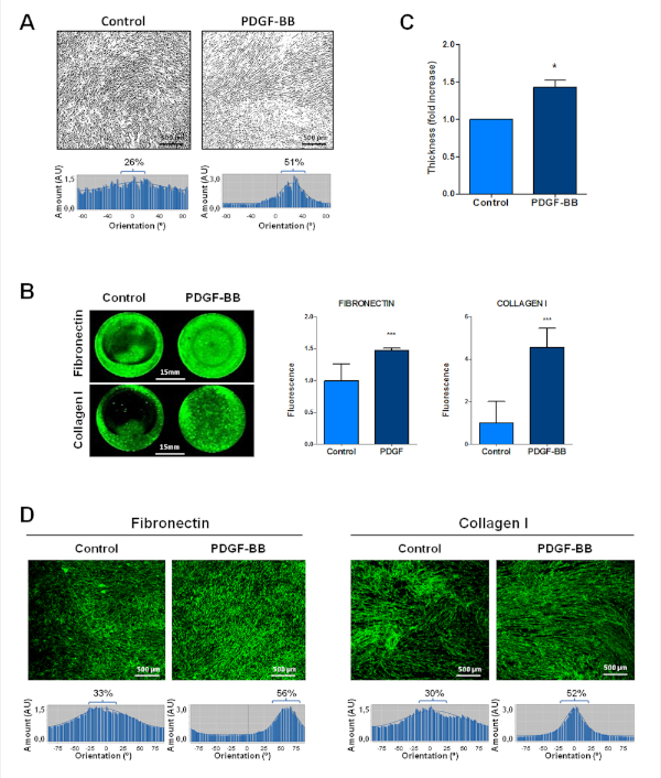
Figure 1: PDGF-stimulated fibroblasts enhance thicker and anisotropic ECM. (A) Binary images representative of cellular orientation of the BJ-hTERT fibroblasts treated or not with PDGF-BB (above) and directionality histograms (below), which represent the frequency of distribution of cell angles (centering on the 0° angle). (B) Increase in protein expression of the extracellular proteins fibronectin and collagen I in PDGF-stimulated fibroblasts derived from ECM. (C) Increase in ECM thickness in those ECMs derived from PDGF-stimulated fibroblasts. (D) Increase in protein and organization of extracellular proteins such as fibronectin and collagen I in PDGF-stimulated fibroblasts derived from ECM. Below, directionality histograms. *p < 0.05; ***p < 0.001. Adapted from Herrera et al.7 Please click here to view a larger version of this figure.
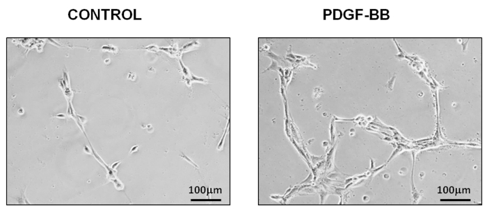
Figure 2: PDGF-stimulated fibroblasts promote endothelial cell activation, which was observed by the formation of capillary-like structures on matrices derived from PDGF-stimulated fibroblasts. The "Particle analysis" tool from ImageJ showed an increase rate of 1.81 in HUVECs seeded on matrices from PDGF-stimulated fibroblasts regarding those seeded on matrices from non-stimulated fibroblasts. Adapted from Herrera et al.7 Please click here to view a larger version of this figure.
Discussion
Matrices can be generated with immortalized fibroblasts or primary fibroblast cultures. Fibroblasts are easy to maintain in in vitro culture with a high growth rate and stress resistance. They can even be isolated from post-mortem tissue3, although contamination could be a restriction, depending on the tissue origin.
The 3D-matrix model here offers a novel and effective system to successfully study ECM composition and fiber orientation. It is also suitable for the analyses of fibroblast activation status, gene/protein expression, interactions with other cell types, etc. It is important to highlight that matrices generated with fibroblasts from patients make personalized study of stroma-related mechanisms in tumors possible. For instance, this approach can be applied to studies of chemo-resistance, in which the ECM has an important role. The translation of this type of study could aid in decision making for the treatment of patients in clinical practice, implementing personalized medicine.
Even though the 3D-matrix protocol can be tedious, interesting results can be achieved by following instructions. It must be noted that multiple washing steps are needed, so avoiding matrix damage is essential. In addition, we recommend that when different cell culture conditions are tested with this protocol, matrices should be generated at the same time due to minimal differences that may occur during experiments (e.g., using the same fibroblast suspension when seeding pre-treated plates).
Other tissue-engineering methods are being developed, but additional steps like sterilization are needed to create compatible ECM and tissues for human use. Although new technologies are arising, they can be costly and may be difficult to adapt to standard laboratory facilities. The use of primary CAFs obtained from patients makes it possible to generate "personalized matrices" to test different treatments. Along this line, our group has been using this methodology to demonstrate that matrices generated by CAFs are different to the ones generated by NFs, showing thicker and more organized matrices7. We have reported that capillary-like structures derived from endothelial cells are dependent on fibroblast Snail1 expression, and there are future studies to observe that effect on CAFs-derived matrices.
ECM requires further study before we can understand the underlying mechanisms responsible for chemotherapy resistance or tumor relapse. Therefore, we suggest the use of CAF-derived matrices from cancer patient tissues as a procedure to investigate microenvironmental behavior in cancer and ECM communication with cancer cells or other stromal cells.
Disclosures
The authors have nothing to disclose.
Acknowledgements
The development of this protocol was supported by PI12/02037, PI15/02101, PI17/01847, PI18/01034 and RD12/0036/0041 from the Instituto de Salud Carlos III; by the Fondo Europeo de Desarrollo Regional (FEDER); by "CIBER de Cáncer", CB16/12/00273 and CB16/12/00446, from the Instituto de Salud Carlos III-FEDER; and by the Fundación Científica AECC (a multifaceted approach to target pancreatic cancer). Cristina Peña is a recipient of a Miguel Servet Contract from the Instituto de Salud Carlos III. M. Eaude helped with the English text. We thank lab members for help and advice throughout this research.
Materials
| Ammonium hydroxide (NH4OH) | Roth | A990.1 | |
| Amphotericin-B | Corning | 30-003-CF | |
| Bovine Serum Albumin (BSA) | Sigma | A7906-50G | |
| Dulbecco's Modified Eagle Medium (DMEM) | Corning | 10-014-CVR | |
| Endothelial Basal Medium (EBM) | Lonza | CC-3121 | add supplements before use |
| Endothelial cell Basal Medium Supplements (EGM-2) | Lonza | CC-4176 | |
| Ethanolamine | Sigma | 411000-100ML | |
| Fetal Bovine Serum (FBS) | Biowest | S181B-500 | heat-inactivated before use |
| Fibroblast Growth Basal Medium (FBM) | Lonza | CC-3131 | add supplements before use |
| Fibroblast Growth Medium Supplements and Growth Factors (FGM-2) | Lonza | CC-4126 | |
| Gelatin from bovine skin | Sigma | G9391-100G | |
| Glutaraldehyde | Sigma | g5882 | |
| L-Ascorbic Acid | Sigma | A92902-100G | light sensitive |
| L-Glutamine | Lonza | BE17-605E | |
| Normocin | Invivogen | 3ANT-NR-2 | |
| Pencillin/Streptomycin (Pen/Strep) | Gibco | 15140122 | |
| Phosphate Buffered Saline (PBS) | Corning | 21-040-CVR | |
| Triton X100 | Roth | 3051 | |
| Recombinant telomerase transfected immortalized human foreskin fibroblasts (BJ-hTERT) | ATCC | ATCC CRL-4001 | |
| Human umbilical vein endothelial cells (HUVECs) | ATCC | PCS-100-013 |
References
- Frantz, C., Stewart, K. M., Weaver, V. M. The extracellular matrix at a glance. Journal of Cell Science. 123 (24), 4195-4200 (2010).
- Kular, J. K., Basu, S., Sharma, R. I. The extracellular matrix: Structure, composition, age-related differences, tools for analysis and applications for tissue engineering. Journal of Tissue Engineering. 5, (2014).
- Kalluri, R. The biology and function of fibroblasts in cancer. Nature Reviews Cancer. 16 (9), 582-598 (2016).
- Erdogan, B., Webb, D. J. Cancer-associated fibroblasts modulate growth factor signaling and extracellular matrix remodeling to regulate tumor metastasis. Biochemical Society Transactions. 45 (1), 229-236 (2017).
- Erez, N., Truitt, M., Olson, P., Hanahan, D. Cancer-Associated Fibroblasts Are Activated in Incipient Neoplasia to Orchestrate Tumor-Promoting Inflammation in an NF-kB-Dependent Manner. Cancer Cell. 17 (2), 135-147 (2010).
- Herrera, M., et al. Colon Cancer-associated Fibroblast Establishment and Culture Growth. Bio-protocol. 6 (7), e1773 (2016).
- Herrera, A., et al. Endothelial cell activation on 3D-matrices derived from PDGF-BB-stimulated fibroblasts is mediated by Snail1. Oncogenesis. 7 (9), 76 (2018).
- MacColl, E., Khalil, R. A. Matrix Metalloproteinases as Regulators of Vein Structure and Function: Implications in Chronic Venous Disease. The Journal of Pharmacology and Experimental Therapeutics. 355 (3), 410-428 (2015).
- Shian, S. G., Kao, Y. R., Wu, F. Y. H., Wu, C. W. Inhibition of Invasion and Angiogenesis by Zinc-Chelating Agent Disulfiram. Molecular Pharmacology. 64 (5), 1076-1084 (2003).
- Neri, S., et al. Cancer cell invasion driven by extracellular matrix remodeling is dependent on the properties of cancer-associated fibroblasts. Journal of Cancer Research and Clinical Oncology. 142 (2), 437-446 (2016).
- Du, P., Subbiah, R., Park, J. H., Park, K. Vascular morphogenesis of human umbilical vein endothelial cells on cell-derived macromolecular matrix microenvironment. Tissue Engineering. (Part A), (2014).
- Bignon, M., et al. Lysyl oxidase-like protein 2 regulates sprouting angiogenesis and type IV collagen assembly in the endothelial basement membrane. Blood. 118 (14), 3979-3989 (2011).
- Huang, L., Xu, A. M., Liu, S., Liu, W., Li, T. J. Cancer-associated fibroblasts in digestive tumors. World Journal of Gastroenterology. 20 (47), 17804-17818 (2014).
- Herrera, A., Herrera, M., Peña, C. The emerging role of Snail1 in the tumor stroma. Clinical and Translational Oncology. 18 (9), 872-877 (2016).
- Sheng, Y., Fei, D., Leiiei, G., Xiaosong, G. Extracellular Matrix Scaffolds for Tissue Engineering and Regenerative Medicine. Current Stem Cell Research & Therapy. 12 (3), 233-246 (2017).
- Castelló-Cros, R., Cukierman, E. Stromagenesis during tumorigenesis: characterization of tumor-associated fibroblasts and stroma-derived 3D matrices. Methods in Molecular Biology. 522, 275-305 (2009).
