This protocol describes the method, materials, equipment and steps for bottom-up preparation of RNA and protein producing synthetic cells. The inner aqueous compartment of the synthetic cells contained the S30 bacterial lysate encapsulated within a lipid bilayer (i.e., stable liposomes), using a water-in-oil emulsion transfer method.
The bottom-up assembly approach for construction of synthetic cells is an effective tool for isolating and investigating cellular processes in a cell mimicking environment. Furthermore, the development of cell-free expression systems has demonstrated the ability to reconstitute the protein production, transcription and translation processes (DNA→RNA→protein) in a controlled manner, harnessing synthetic biology. Here we describe a protocol for preparing a cell-free expression system, including the production of a potent bacterial lysate and encapsulating this lysate inside cholesterol-rich lipid-based giant unilamellar vesicles (GUVs) (i.e., stable liposomes), to form synthetic cells. The protocol describes the methods for preparing the components of the synthetic cells including the production of active bacterial lysates, followed by a detailed step-by-step preparation of the synthetic cells based on a water-in-oil emulsion transfer method. These facilitate the production of millions of synthetic cells in a simple and affordable manner with a high versatility for producing different types of proteins. The obtained synthetic cells can be used to investigate protein/RNA production and activity in an isolated environment, in directed evolution, and also as a controlled drug delivery platform for on-demand production of therapeutic proteins inside the body.
Synthetic cells are artificial cell-like particles, mimicking one or multiple functions of a living cell, such as the ability to divide, form membrane interactions, and synthesize proteins based on a genetic code1,2,3. Synthetic cells that enclose cell-free protein synthesis (CFPS) systems possess high modularity due to their ability to produce various proteins and RNA sequences following alterations in the DNA template. Presenting an attractive alternative to the current approaches of protein production, CFPS systems are based on cell lysate, purified components, or synthetic components and include all the transcription and translation machinery required for protein synthesis such as ribosomes, RNA polymerase, amino acids and energy sources (e.g., 3-phosphoglycerate and adenine triphosphate)4,5,6,7,8,9. The encapsulation of a CFPS system inside lipid vesicles enables the simple and efficient production of proteins without depending on a living cell10. Moreover, this platform allows synthesis of peptides that may degrade inside natural cells, production of proteins that are toxic to living cells, and modify proteins with non-natural amino acids11,12. Synthetic cells have been used as a model for research purposes investigating the minimal cell components required to enable cellular life from an evolutionary perspective1,13. Synthetic cells have also been used to build and implement genetic circuit and as models for directed evolution14,15,16. Other studies have focused on the ability of synthetic cells to mimic the biological activity of natural cells, aiming to replace damaged natural cells, such as beta cells in patients with diabetes17. Furthermore, the ability of these CFPS encapsulating synthetic cells to produce a variety of therapeutic proteins illustrates its potential to be incorporated into clinical use18.
Here we describe a bottom-up lab-scale protocol (Figure 1) for the production of RNA and protein-producing synthetic cells based on a CFPS system encapsulated in a lipid vesicle. This shows the potential use of synthetic cell platforms as novel drug delivery systems for the onsite production of a therapeutic protein drug in vivo19. Previous studies have investigated the optimization of the CFPS reaction and the cell lysate preparation processes4,8,20. Moreover, several techniques have been applied for cell-sized liposome preparation, such as microfluidic and polymer-based droplet stabilization methods21,22,23, which also differ in the liposomes' lipid composition24,25,26. In the presented protocol, synthetic cells are produced using a water-in-oil emulsion transfer method and the encapsulation process is carried out at low temperatures (<4 °C)5,10,24,27,28. These mild conditions have been found to be favorable for retaining the bio-functional integrity of the molecular machinery, namely ribosomes and proteins27,29,30. The lipid composition of the particles consists of both cholesterol and 1-palmitoyl-2-oleoyl-sn-glycero-3-phosphocholine (POPC). The first is found in all mammalian cell membranes and is essential for the stability, rigidity and permeability reduction of the membrane, and the latter mimics mammalian phospholipid composition11,13. The cellular transcription and translation molecular machinery are extracted from the BL21 (DE3) Escherichia coli (E. coli) strain, which is transformed with pAR1219 plasmid overexpressing T7 RNA polymerase to increase CFPS potency and protein synthesis. This system has been used to produce diagnostic and therapeutic proteins, with molecular weights of up to 66 kDa in vitro and in vivo19,31. The following protocol provides a simple and effective method for the production of the synthetic cell system, which can address a wide range of fundamental questions associated with protein synthesis in nature and can also be utilized for drug delivery applications.
NOTE: Illustration of the complete synthetic cells’ production protocol is presented in Figure 1. According to the user’s needs, the protein expression (section 3.2) and synthetic cell formation (section 4) parts of the protocol can also be carried out independently (with some adaptations).
1. Preparation of S30-T7 lysate
2. Preparation of lipids in oil solution
3. Preparations of outer, pre-inner and feeding solutions
4. Preparation of synthetic cells
NOTE: The following volumes are adjusted for the preparation of 100 µL of synthetic cells.
We present a protocol for the preparation of synthetic cells by encapsulating a S30-T7 CFPS system based on BL21 E. coli inside lipid vesicles. A schematic description of the preparation process that includes an image of each stage is presented in Figure 2. The success of the synthetic cell preparation process is dependent on the appropriate performance of each stage and effected by different parameters. The protocol should be adjusted to accommodate the production of a specific protein.
Plasmids expressing the model protein super-folder Green Fluorescent Protein (sfGFP) and Renilla Luciferase under the T7 promoter were introduced into CFPS bulk reactions and synthetic cells, and protein production was evaluated using different methods, including western blot, flow cytometry, microscopy and spectroscopy (Figure 3). A verification of the sfGFP-His6 tagged protein (~27 kDa32) production by western blot analysis is presented in Figure 3A. A sample of 30 µL of synthetic cells diluted 6 folds was mixed with 10 µL of a common SDS-PAGE sample buffer (containing the detergents sodium dodecyl sulfate (SDS) and β-mercaptoethanol) and boiled for 10 min (95 ᵒC). We found that the combination of heat and detergents in the sample buffer is sufficient to disassemble the vesicles and to enable the running of proteins in the SDS-PAGE gel. Protein detection was performed using anti-His polyclonal primary antibody (diluted 1:12,000). As expected, while protein production is detected in samples containing sfGFP-encoding DNA templates ("+sfGFP DNA"), no protein is observed in negative control samples in which the DNA template was excluded ("-DNA"). Samples of purified sfGFP and purified sfGFP encapsulated within synthetic cells' membrane ('sfGFP (encapsulated)), are used as positive controls for the sfGFP CFPS bulk production and for its synthesis within synthetic cells, respectivley. This method can be applied to different protein types. According to Krinsky et al.19, the sfGFP production yield obtained under the detailed protocol is 380 µg/mL in CFPS solution and 5.3 µg/mL, when the CFPS solution is encapsulated inside lipid vesicles.
When the protein that is produced inside the synthetic cells is fluorescent, its production can be evaluated using microscopy and flow cytometry-based methods. We analyzed the synthetic cells using a fluorescent microscope with a filter for GFP fluorescence (Figure 3B). Since the CFPS components have some autofluorescent, the image acquisition parameters should be adapted according to an appropriate negative control sample (synthetic cells with no DNA template, for example).
In addition, we used flow cytometry to determine the mean fluorescence intensity of sfGFP-producing synthetic cells, and the percentage of active synthetic cells, which can produce proteins within them (Figure 3C I&II). 10,000 events were collected for each analyzed sample. The synthetic cell production, which is described in this protocol, usually yields an active population of 21-25% within a solution with an approximated concentration of 107 synthetic cells/mL. The slight fluorescence intensity presented by “-DNA” samples in Figure 3C II is due to the autofluorescence of different components in the CFPS reaction such as the S30 lysate.
Representative size analysis based on the GFP signal (in diameter) of synthetic cells prepared by the method described above shows a mean value of 2.4±0.5 µm (Figure 3D II). As the formation of water in oil emulsion in this method is an outcome of applying mechanical forces, the size distribution of the particles might be affected by different factors, such as the method and speed of pipetting the emulsion up-and-down, the model of the vortex mixer machine, etc.
To test the versatility of synthetic cells’ protein production, the expression of the reporter protein Renilla luciferase inside synthetic cells was analyzed (Figure 3E). The assay quantified Renilla luciferase activity by measuring the luminescence generated from the enzymatic reaction of luciferase and its substrate, h-coelenterazine. To induce the enzymatic reaction, a final concentration of 1 µM of h-coelenterazine was added to the synthetic cells (25 µL sample volume) just before the measurement. “-DNA” sample, not containing the luciferase-encoding DNA template was used as a negative control for testing luminescence due to non-enzymatic oxidation of the substrate. Luminescence was measured using a plate reader.
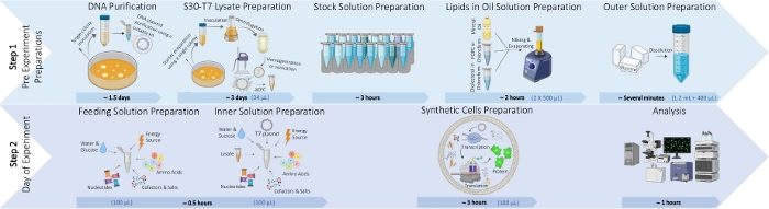
Figure 1: An illustration of the process for typical synthetic cells preparation protocol. The process is divided into two steps. Step 1: Pre-experiment preparations including DNA plasmid purification, S30-T7 lysate preparation, stock solutions preparation required for the inner and feeding solutions, lipids-in-oil solution preparation and outer solution preparation. Step 2: Synthetic cell formation that includes feeding and inner solutions preparation, synthetic cells preparation and analysis. An example of the required volume of each ingredient for preparing 100 µL of synthetic cells solution is presented in blue in brackets. Please click here to view a larger version of this figure.
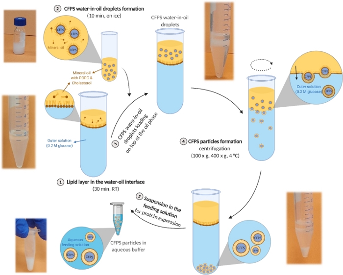
Figure 2: Schematic illustration of the synthetic cell preparation protocol. An image of a well-performed process illustrates each stage of the protocol. This figure has been modified from Krinsky et al.19. Please click here to view a larger version of this figure.
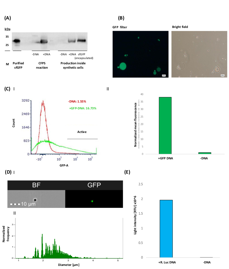
Figure 3: Representative results of the production of sfGFP and Renilla luciferase inside synthetic cells. (A) A western blot analysis of sfGFP-His6 production in both CFPS bulk reactions and inside synthetic cells. Protein detection was carried out using anti-His polyclonal primary antibody (diluted 1:12,000). Purified sfGFP-His6 was used as a positive control (7.8 µg). sfGFP (encapsulated) is positive control of purified sfGFP encapsulated in synthetic cells. Samples without DNA templates were used as a negative control for the production analysis ("-DNA"). (B) Representative images of sfGFP producing synthetic cells taken using a fluorescent microscope with a bright-field and a GFP filter. Scale bars = 50 µm. (C) I&II Flow cytometry analysis of sfGFP producing synthetic cells activity (Data collected using digital, 4-laser analyzer). Samples were measured after 3 h of incubation. 10,000 events were collected for each analyzed sample. FSC (500 V) and SSC (300 V) filters were used to define the total synthetic cell population. Then, a FITC (600 V) filter was used to detect GFP fluorescence.. (I) Calculation of the active synthetic cell population [%] was based on GFP fluorescence intensity threshold, defined by the “-DNA” sample (red histogram), allowing an error of ~1%. (II) Mean fluorescence intensity of sfGFP producing synthetic cells. This value was calculated from the active synthetic cell population and normalized to the “-DNA” sample by dividing the mean fluorescence of the active synthetic cells by the mean of the synthetic cells without sfGFP-DNA. (D) I&II Synthetic cell size analysis. The emission spectrum was detected by 505-560 nm and 642-745 nm for GFP and bright field signals, respectively. (I) A representative image of the analyzed synthetic cells. (II) Synthetic cells’ size distribution. Analysis was performed and the diameter distributions of the active synthetic cells was calculated based on the GFP signal. (E) Production of active Renilla luciferase inside synthetic cells. Luciferase activity was quantified with luminescence measurements after the addition of 1 µM h-coelenterazine using a plate reader and presented as light intensity [RLU] x 106. Please click here to view a larger version of this figure.
Table 1: Buffer and stock solutions preparation. | |
Comments | |
Luria Bertani (LB) agar (1.5%) plate: | Prepare and sterilize. Add Ampicillin at a final concentration of 50 µg/mL only after cooling to room temperature. |
10 g/L Bacto-tryptone | |
10 g/L Sodium chloride (NaCl) | |
5 g/L Bacto-Yeast extract | |
15 g/L Agar agar purified | |
50 µg/mL Ampicillin | |
LB media (20 mL): | Minimal media. Prepare and sterilize in advance. Just prior to inoculating the bacteria, add Ampicillin at a final concentration of 50 µg/mL. |
10 g/L Bacto-tryptone | |
10 g/L Sodium chloride (NaCl) | |
5 g/L Bacto-Yeast extract | |
50 µg/mL Ampicillin | |
Terrific Broth (TB) media (1 L): | Rich media. Prepare and sterilize in advance. Just prior to inoculating the bacteria, add Ampicillin at a final concentration of 50 µg/mL. |
12 g/L Bacto-tryptone | |
24 g/L Bacto-Yeast extract | |
4% (v/v) Glycerol anhydrous | |
2.32 g/L K2HPO4 | |
12.54 g/L KH2PO4 | |
50 µg/mL Ampicillin | |
S30 lysate buffer (1.5 L): | Prepare and sterilize in advance (*excluding DTT and 2-mercaptoethanol). Store at -4 °C. |
10 mM Tris-acetate at pH = 7.4 | Trisma-base. Prepare and adjust pH to 7.4 using Acetic acid. |
14 mM magnesium acetate | |
60 mM potassium acetate | |
*1 mM DTT | Add just prior to use |
*0.5 mL/L 2-mercaptoethanol | Add just prior to use |
1 M HEPES-KOH (pH = 8): | Dissolve HEPES to a final concentration of 1 M. Adjust to pH 8 using KOH solution. |
HEPES | |
Potassium hydroxide (KOH) |
Table 2: Inner solution composition. | ||||||||||
Final requested Inner solution volume | ||||||||||
Number | Reagent | Stock conc. | Final conc. | 25 µL | 50 µL | 100 µL | 200 µL | 300 µL | 400 µL | |
1 | HEPES KOH pH=8 | 1 M | 55 mM | 1.375 | 2.75 | 5.5 | 11 | 16.5 | 22 | Pre-Inner solution |
2 | Magnesium acetate | 1 M | 14 mM | 0.35 | 0.7 | 1.4 | 2.8 | 4.2 | 5.6 | |
3 | Potassium acetate | 1 M | 50 mM | 1.25 | 2.5 | 5 | 10 | 15 | 20 | |
4 | Ammonium acetate | 5.2 M | 155 mM | 0.75 | 1.5 | 3 | 6 | 9 | 12 | |
5 | PEG 6000 | 50% | 3% | 1.5 | 3 | 6 | 12 | 18 | 24 | |
6 | 3-PGA | 0.5 M | 40 mM | 2 | 4 | 8 | 16 | 24 | 32 | |
7 | Amino acids – mixture I | 50 M | 2.5 mM | 1.25 | 2.5 | 5 | 10 | 15 | 20 | |
8 | Amino acids – mixture II | 50 M | 2.5 mM | 1.25 | 2.5 | 5 | 10 | 15 | 20 | |
9 | ATP | 100 mM | 1.2 mM | 0.3 | 0.6 | 1.2 | 2.4 | 3.6 | 4.8 | |
10 | GTP | 50 mM | 1 mM | 0.5 | 1 | 2 | 4 | 6 | 8 | |
11 | UTP | 100 mM | 0.8 mM | 0.2 | 0.4 | 0.8 | 1.6 | 2.4 | 3.2 | |
12 | IPTG | 100 mM | 1 mM | 0.25 | 0.5 | 1 | 2 | 3 | 4 | |
13 | Sucrose | 2 M | 200 mM | 2.5 | 5 | 10 | 20 | 30 | 40 | |
14 ** | H2O UPW | ** | ** | ** | ** | ** | ** | |||
15 | S30-T7 Lysate | 34% | 8.5 | 17 | 34 | 68 | 102 | 136 | ||
16 * | DNA plasmid | 10 µg/mL | * | * | * | * | * | * |
Table 3: Feeding solution composition. | |||||||||
Final requested feeding solution volume | |||||||||
Number | Reagent | stock conc. | final conc. | 25 µL | 50 µL | 100 µL | 200 µL | 300 µL | 400 µL |
1 | HEPES KOH pH=8 | 1 M | 83.3 mM | 2.08 | 4.17 | 8.33 | 16.67 | 25 | 33.33 |
2 | Magnesium acetate | 1 M | 21.2 mM | 0.53 | 1.06 | 2.12 | 4.24 | 6.36 | 8.48 |
3 | Potassium acetate | 1 M | 75.5 mM | 1.89 | 3.78 | 7.55 | 15.1 | 22.66 | 30.2 |
4 | Ammonium acetate | 5.2 M | 236.4 mM | 1.14 | 2.27 | 4.55 | 9.09 | 13.64 | 18.18 |
5 | PEG – 6000 | 50% | 4.54% | 2.27 | 4.54 | 9.08 | 18.16 | 27.24 | 36.32 |
6 | 3-PGA | 0.5 M | 60.1 mM | 3 | 6.01 | 12.01 | 24.02 | 36.04 | 48.04 |
7 | Amino acids – mixture I | 50 mM | 3.8 mM | 1.89 | 3.78 | 7.56 | 15.12 | 22.68 | 30.24 |
8 | Amino acids – mixture II | 50 mM | 3.8 mM | 1.89 | 3.78 | 7.56 | 15.12 | 22.68 | 30.24 |
9 | ATP | 100 mM | 1.8 mM | 0.45 | 0.91 | 1.81 | 3.62 | 5.43 | 7.24 |
10 | GTP | 50 mM | 1.5 mM | 0.76 | 1.51 | 3.02 | 6.04 | 9.06 | 12.08 |
11 | UTP | 100 mM | 1.2 mM | 0.3 | 0.61 | 1.21 | 2.42 | 3.63 | 4.84 |
12 | IPTG | 100 mM | 1.5 mM | 0.38 | 0.76 | 1.51 | 3.02 | 4.53 | 6.04 |
13 | Glucose | 2 M | 200 mM | 2.5 | 5 | 10 | 20 | 30 | 40 |
14 | H2O UPW | 5.92 | 11.83 | 23.7 | 47.38 | 71.06 | 94.76 |
Table 4: Required solutions for synthetic cells preparation. | |
Solution & materials required | Comments |
Lipids in oil | Store at room temperature until use. Prepared according to section 2 |
Outer solution | Prepared according to section 3.1 |
Inner solution | Prepared according to section 3.2. |
Prepare tests + controls samples according to section 4.1. | |
Keep on crushed ice until use. | |
Feeding solution | Prepared according to section 3.3. |
Keep on crushed ice until use. |
This protocol introduces a simple and affordable method for the production of large quantities of protein-producing synthetic cells. The yield of active cells is dependent on careful and accurate execution of the protocol with emphasis on several critical steps. In the lysate preparation section of this method, it is essential to reach the appropriate bacteria density before cell lysis to achieve a sufficient amount of proteins in the bacterial lysate. Second, the lysis process should be performed at 4 °C and the lysate frozen quickly with liquid nitrogen to maintain protein activity. Moreover, in this protocol we used E. coli BL21(DE3) cells transformed with pAR1219 plasmid expressing T7 RNA polymerase. In cases where a different strain of bacteria is used, changes to the lysate concentration may be required to reach a satisfactory amount of RNA polymerase and ribosomes. Even when the same bacterial strain is used, different lysate productions may have some batch-to-batch variability.
The vesicle production section also includes a couple of significant steps. Solubilization of the lipids and removal of the chloroform from the mineral oil is important for establishing the water-in-oil emulsion. The centrifugation of the water-in-oil droplets into the outer aqueous buffer is also a key step in the protocol to generate synthetic cells with a lipid bilayer. Changes in the inner solution of the vesicles and the lipid composition might lead to a layer of droplets at the oil-water interphase without any observable pellet. To overcome this, a quick solution is to increase the centrifugation speed to 1,000 x g in the emulsion transfer step. In case this does not solve the issue, the specific gravity of the outer aqueous solution can be altered ensuring that the inner solution will have a higher specific gravity than the outer solution. Furthermore, the osmolality of the inner solution may also vary between different lysate sources and productions, ranging between 800-1100 mOsm/kg. Variability in the inner solution's osmolality is mostly due to changing concentrations of the lysate during its production process. Usually this does not lead to significant changes in protein production, yet a large dilution might lead to a reduced yield due to low concentrations of the transcription and translation enzymes in the lysate itself. Measuring the total protein concentration in each lysate batch can assist in tuning lysate concentrations in the inner solution to maintain constant values (approximately 22 mg/mL measured by Bradford assay).
Nevertheless, this method has some limitations that should be mentioned. Not all of the generated synthetic cells are active and capable of producing proteins due to incomplete encapsulation of all the required components. We measured approximately 21-25% of active cells when producing sfGFP expressing synthetic cells using flow cytometry (no significant size differences were observed between active and inactive cells). Oil and lipid excess residues occasionally remain in the bilayer lipid membrane after the transfer of the synthetic cells into the aqueous phase. Optimizing the lipid and cholesterol concentrations in the oil phase can improve this issue. It is also important to note that the size distribution of the obtained synthetic cells is quite wide in comparison to alternative microfluidic methods, with vesicles ranging from approximately 1-50 µm.
The high yield of the emulsion transfer method makes it especially suitable for synthetic cells encapsulating cell-free protein synthesis systems for therapeutic protein production. Different variations of the emulsion transfer method have been used in synthetic cell studies and included changes in the oil preparation procedure, membrane composition, sample volume, inner solution to oil ratio and centrifugation speeds5,24,28. Microfluidic and polymer-based droplet stabilization methods have also been used for synthetic cell preparation, however, encapsulation of the whole CFPS system has not been performed yet with these methods21,22,23.
The synthetic cells obtained by this method were shown in vivo to produce proteins and treat cancer in murine models19. The capabilities of these cells can be further expanded in the future beyond protein expression. Integration of other cellular processes such as cellular communication, cytoskeleton modification and cell division in synthetic cells have been recently described33,34,35,36. Substitution of the bacterial lysate with eukaryotic cell lysate will allow expression of proteins with higher complexity and post-translational modifications, and will perhaps be less immunogenic even without a cleaning procedure, therefore opening more therapeutic frontiers37.
The authors have nothing to disclose.
This work was supported by ERC-STG-2015-680242.
The authors also acknowledge the support of the Technion Integrated Cancer Center (TICC); the Russell Berrie Nanotechnology Institute; the Lorry I. Lokey Interdisciplinary Center for Life Sciences & Engineering; the Israel Ministry of Economy for a Kamin Grant (52752); the Israel Ministry of Science Technology and Space – Office of the Chief Scientist (3-11878); the Israel Science Foundation (1778/13, 1421/17); the Israel Cancer Association (2015-0116); the German-Israeli Foundation for Scientific Research and Development for a GIF Young grant (I-2328-1139.10/2012); the European Union FP-7 IRG Program for a Career Integration Grant (908049); the Phospholipid Research Center Grant; a Rosenblatt Foundation for cancer research, a Mallat Family Foundation Grant; and the Unger Family Foundation. A. Schroeder acknowledges Alon and Taub Fellowships. O. Adir acknowledges the Sherman and Gutwirth fellowships. G. Chen acknowledges the Sherman Fellowship. N. Krinsky acknowledges the Baroness Ariane de Rothschild Women Doctoral Program from the Rothschild Caesarea Foundation.
A. Reagents required for step 1 (S30-T7 lysate preparation) | |||
E.coli BL21 (DE3) | NEB | C2527 | E.coli BL21 (DE3). |
pAR1219 | Sigma | T2076 | TargeTron vector for transformation. |
Stock solution of 50 mg/mL Ampicillin | Sigma | A9518 | Stored at -20 °C. |
10 g/L Bacto-tryptone | BD Bioscience | 211705 | For preparation of Luria Bertani (LB) agar (1.5%) plate. |
10 g/L Sodium chloride (NaCl) | Bio-Lab | 19030591 | |
5 g/L Bacto-Yeast extract | BD Bioscience | 212750 | |
15 g/L Agar agar purified | Merck | 1.01614.5007 | |
50 µg/mL Ampicillin | Sigma | A9518 | |
10 g/L Bacto-tryptone | BD Bioscience | 211705 | For preparation of Luria Bertani (LB) media (20 mL). |
10 g/L Sodium chloride (NaCl) | Bio-Lab | 19030591 | |
5 g/L Bacto-Yeast extract | BD Bioscience | 212750 | |
50 µg/mL Ampicillin | Sigma | A9518 | |
12 g/L Bacto-tryptone | BD Bioscience | 211705 | For preparation of Terrific Broth (TB) media (1 L). |
24 g/L Bacto-Yeast extract | BD Bioscience | 212750 | |
4% (v/v) Glycerol anhydrous | Bio-Lab | 7120501 | |
2.32 g/L K2HPO4 | Spectrum chemical | P1383 | |
12.54 g/L KH2PO4 | Spectrum chemical | P1380 | |
50 µg/mL Ampicillin | Sigma | A9518 | |
Stock solution of 100 mM Isopropyl β-D-1-thiogalactopyranoside (IPTG) | INALCO | INA-1758-1400 | Filtered using 0.2 µm hydrophilic PVDF syringe filter. |
Stock solution of 0.1 M dithiothreitol (DTT) | TCI | D1071 | Filtered using 0.2 µm hydrophilic PVDF syringe filter. |
10 mM Tris-acetate at pH = 7.4 | Sigma | T1503 | S30 lysate buffer (1.5 L) |
14 mM magnesium acetate | Merck | 1.05819.0250 | |
60 mM potassium acetate | Carlo Erba | 470147 | |
1 mM DTT | TCI | D1071 | |
0.5 mL/L 2-mercaptoethanol | Sigma | M6250 | |
Equipment required for step 1 | |||
100 mL sterilized Erlenmeyer flasks | Thermo Scientific | 50-154-2846 | 2 flasks |
2 L sterilized Erlenmeyer flasks with baffles | KIMAX-KIMBLE | 25630 | 2 flasks |
Floor incubator shaker | MRC | TOU-120-2 | Laboratory shaker incubator 450x450mm, 400rpm, 70 °C |
Centrifuge | Thermo Scientific | 75004270 | (75003340) – Fiberlite F10-6 x 100 LEX Fixed-Angle Rotor. Should enable at least 13,000 x g. * Pre-cooled to 4 °C. |
High pressure homogenizer | AVESTIN | EmulsiFlex-C3 | Pre-cooled to 4 °C. |
-80oC freezer | SO-LOW | U85-18 | |
Sterilized 1.5 mL plastic tubes | Eppendorf | 30120086 | Preferably pre-cooled to -20 °C. |
Spectrophotometer | TECAN | IN-MNANO | Infinite M200 pro |
96-well transparent plate | Thermo Scientific | 167008 | |
Sterilized graduated cylinder | Corning | ||
Sterilized centrifuge tubes | Eppendorf | 30120086 | Preferably pre-cooled to -20 °C. |
Sterilized pipette tips | Corning | Preferably pre-cooled to -20 °C. | |
Crushed ice bucket | Bel-Art | M18848-4001 | |
Small liquid nitrogen tank | NALGENE | 4150-4000 | |
B. Reagents required for step 3 (lipids in oil solution preparation): | |||
1-palmitoyl-2-oleoyl-sn-glycero-3-phosphocholine (POPC) | Lipoid | 556400 | Powder |
Cholesterol | Sigma | C8667 | Powder |
Chloroform | Bio-Lab | 3082301 | |
Mineral oil | Sigma | M5904 | Light oil |
Equipment required for step 2 | |||
Vortex mixer | Scientific industries | SI-0256 | |
Heating block | TECHNE | FDB03AD | Pre-heated to 80 °C. Should enable controlled temperature. |
2 mL screw neck glass vials | CSI Analytical Innovations | VT009M-1232 | For a larger scale, use 50 mL falcons and evaporate the chloroform using rotary evaporator. |
9mm Screw Cap | CSI Analytical Innovations | C395R-09LC | |
C. Reagents required for step 3 (inner and feeding reaction mixtures): | |||
HEPES | Spectrum | H1089 | 1 M HEPES-KOH (pH = 8) – pH buffer |
Potassium hydroxide (KOH) | Frutarom | 55290 | |
1 M Magnesium acetate | Merck | 1.05819.0250 | Co-factor and negative charge stabilizor. |
1 M Potassium acetate | Carlo Erba | 470147 | Negative charge stabilizor. |
5.2 M Ammonium acetate | Merck | 1.01116.1000 | Stabilizes negative charge. |
50% (w/v) Polyethylene glycol 6000 (PEG) | Merck | 8.07491.1000 | Increases the concentration of the macromolecules. |
0.5 M 3-phosphoglycerate (3-PGA) | Sigma | P8877 | Secondary energy source. |
50 mM Amino acids mixture I | Sigma | LAA21-1KT | Amino acids additive. Contains: 50 mM of each of the following 17 natural amino acids – alanine, arginine, asparagine, aspartic acid, cysteine, glutamine, glutamic acid, glycine, histidine, isoleucine, leucine, lysine, methionine, proline, serine, threonine, and valine. |
50 mM Amino acids mixture II | Sigma | LAA21-1KT | Amino acids additive. Contains: 50 mM of each of the following 3 natural amino acids – tryptophan, phenylalanine, and tyrosine. |
100 mM Adenine triphosphate (ATP) | Sigma | A3377 | Nucleotides & energy source. |
50 mM Guanidine triphosphate (GTP) | Sigma | G8877 | Nucleotides & energy source. |
100 mM Uridine triphosphate (UTP) | ACROS ORGANICS | 226310010 | Nucleotides additive. |
100 mM Isopropyl β-D-1-thiogalactopyranoside (IPTG) | INALCO | INA-1758-1400 | Genes expression induction. |
2 M Sucrose | J.T. Baker | 1933078 | Generating a density gradient. |
2 M Glucose | Sigma | 16301 | Generating a density gradient. |
H2O UltraPure Water (UPW) | Bio-Lab | 2321777500 | DNase & RNase free |
S30-T7 lysate | _ | _ | Prepared at step 1. Source of transcription & translation components. Store at -80 °c, thaw on crashed ice just before usage. |
Stock of DNA plasmid of choice | _ | _ | Contains the sequence for the requested protein. Under T7 promotor |
D. Equipment required for step 4 (synthetic cells preparation) | |||
Floor incubator shaker or Thermomixer | MRC | TOU-120-2 | Laboratory shaker incubator 450x450mm, 400rpm, 70 °C |
PHMT Grant Bio | PSC18 | Thermomixer | |
Centrifuge | Thermo Scientific | 75004270 | (75003629) – TX-400 4 x 400mL Swinging Bucket Rotor. Suited for 15 mL sized tubes. Preferably swinging buckets. Should enable at least 1000 x g. Pre-cooled to 4 °C. |
Table centrifuge | Thermo Scientific | 75002420 | (75003424) – 24 x 1.5/2.0mL rotor with ClickSeal. Suited for Eppendorf vials. Pre-cooled to 4 °C. |
Vortex mixer | Scientific industries | SI-0256 | |
Crushed ice bucket | Bel-Art | M18848-4001 | |
2 mL screw neck glass vials | CSI Analytical Innovations | VT009M-1232 | |
Sterile 15 mL plastic tubes | Thermo Scientific | 339651 | |
Sterilized 1.5 mL plastic tubes | Eppendorf | 30120086 | |
Sterilized pipette tips | Corning | Sterilized by autoclave. |