Transthoracic echocardiography is the first-line diagnostic test for post-resuscitation left ventricular dysfunction and structural changes in a pig model of cardiac arrest.
One of the main causes of out-of-hospital cardiac arrest is acute myocardial infarction (AMI). After successful resuscitation from cardiac arrest, approximately 70% of patients die before hospital discharge due to post-resuscitation myocardial and cerebral dysfunction. In experimental models, myocardial dysfunction after cardiac arrest, characterized by an impairment in both left ventricular (LV) systolic and diastolic function, has been described as reversible but very little data are available in cardiac arrest models associated with AMI in pigs. Transthoracic echocardiography is the first-line diagnostic test for the assessment of myocardial dysfunction, structural changes and/or AMI extension. In this pig model of ischemic cardiac arrest, echocardiography was done at baseline and 2-4 and 96 hours after resuscitation. In the acute phase, the examinations are done in anesthetized, mechanically ventilated pigs (weight 39.8 ± 0.6 kg) and ECG is recorded continuously. Mono- and bi-dimensional, Doppler and tissue Doppler recordings are acquired. Aortic and left atrium diameter, end-systolic and end-diastolic left ventricular wall thicknesses, end-diastolic and end-systolic diameters and shortening fraction (SF) are measured. Apical 2-, 3-, 4-, and 5-chamber views are acquired, LV volumes and ejection fraction are calculated. Segmental wall motion analysis is done to detect the localization and estimate the extent of myocardial infarction. Pulsed Wave Doppler echocardiography is used to record trans-mitral flow velocities from a 4-apical chamber view and trans-aortic flow from a 5-chamber view to calculate LV cardiac output (CO) and stroke volume (SV). Tissue Doppler Imaging (TDI) of LV lateral and septal mitral anulus is recorded (TDI septal and lateral s', e', a' velocities). All the recordings and measurements are done according to the recommendations of the American and European Societies of Echocardiography Guidelines.
Cardiac arrest frequently happens minutes after the onset of typical chest pain, and in some cases it is the first manifestation of coronary artery disease1. In fact, 48% of survivors of out-of-hospital cardiac arrest present occlusion of a coronary artery on angiography2. In addition, for patients who return to spontaneous circulation (ROSC) after cardiac arrest, cardiac dysfunction is one of the most important determinants of morbidity and mortality3.
Transthoracic echocardiography (TTE) is a non-invasive diagnostic and prognostic tool used in patients to assess post-resuscitation myocardial dysfunction, structural changes and/or AMI extension after ROSC and in the days that follow. In experimental ischemic and non-ischemic cardiac arrest models in pigs, TTE is frequently used to noninvasively serially assess cardiac systolic function, hemodynamics, and the response to therapy. In 2008, changes in diastolic dysfunction were described in terms of increase in mitral E velocity and tissue Doppler (TDI) e' velocity ratio (E/e') and decrease in mitral E velocity and A velocity ratio (E/A) shortly after resuscitation in a non-ischemic pig model of cardiac arrest4.
The present study describes the different methodologic steps followed to assess left ventricular (LV) structure and LV systolic and diastolic function by TTE at different time-points in an ischemic pig model of cardiac arrest.
All procedures involving animals and their care conformed with national and international laws and policies. Approval of the study was obtained from the institutional review board of the University of Milan and governmental institution (Ministry of Health approval no. 84/2014-PR). Data that support the findings of this study are available from the corresponding author on reasonable request. The experimental model and echocardiographic protocol diagrams are detailed in Figure 1 and Figure 2.
1. Animal preparation
2. Induction of anesthesia and maintenance, antibiotic prophylaxis
3. Mechanical ventilation electrocardiographic and hemodynamic monitoring
4. Baseline transthoracic echocardiography
NOTE: On average echocardiography takes 20-30 min. For TTE, a phased-array multifrequency 2.5 to 5 MHz probe is used, while ECG is continuously recorded. Sets of frames and cine-loops consisting of at least three consecutive cardiac cycles are stored for off-line analysis.
5. Induction of myocardial infarction
6. Cardiac arrest
7. Cardiopulmonary resuscitation
8. Post-cardiac arrest supportive care
9. Four-hour (h) observation
10. 96-hours observation and euthanasia
11. Echocardiographic measurements
NOTE: Take all recordings and measurements according to the recommendations of the American and European Societies of Echocardiography Guidelines6,7. Send all echocardiographic recordings by a remote desktop connection to be stored in a local database for analysis. A cardiologist blinded to the study groups averages at least three measurements for each variable.
12. Statistical analysis
Twelve pigs underwent coronary artery occlusion followed by 12 min of ventricular fibrillation and 5 min of CPR. Eight pigs were successfully resuscitated, and seven survived at 96 h post AMI-cardiac arrest-ROSC. All echocardiographic variables at different time-points during the study are summarized in Table 1.
Changes in heart rate (HR) and systolic echocardiographic parameters
HR increased significantly at 2 h and 4 h post-AMI-cardiac arrest-ROSC compared with baseline (BL) (mean ± SEM: +64 ± 9 and +56 ± 12 bpm, p < 0.001 and p < 0.01, respectively) together with ESV (+15 ± 3 and +18 ± 4 mL, p < 0.01 for both), while EDV did not change significantly at the different times. The mean differences in LVEF between BL and 2 h and 4 h were -40 ± 4.1 and -39 ± 4.0 absolute points %, respectively (p < 0.001 for both) (Figure 4).
From 2 h to 96 h post AMI-cardiac arrest-ROSC, HR tended to normalize, (mean ± SEM difference -49 ± 9.1 bpm, p < 0.05). LVEF improved, rising 24.9 ± 2.5 points percent (p < 0.05), but it remained below BL. Changes in LV volumes were minimal and not significant; results were similar for changes between 4 h and 96 h post-AMI-cardiac arrest-ROSC (Figure 4 and Figure 5).
Changes in diastolic echocardiographic parameters
DT was the only echocardiographic diastolic variable that changed significantly at the different study time-points (Figure 6). At 2 h, DT decreased 16% from BL and maintained the decrease at 4 h post AMI-cardiac arrest-ROSC. At 96 h post AMI-cardiac arrest-ROSC, DT returned similar to those at BL.
LV regional motility 96 h post AMI-cardiac arrest-ROSC
The mean ± SEM number of akinetic/dyskinetic (A/D) segments was 4.2 ± 0.7 and WMSI was 26 ± 4.4%. The most frequently compromised segments were mid anterolateral, mid-inferoseptal, apical anterior, and apical inferior.
Table 1: Echocardiographic variables at different times after AMI-cardiac arrest-ROSC. BL, baseline; HR, heart rate; AoD, aortic diameter; LAD, left atrium diameter; AWThd, diastolic anterior wall thickness; AWThs, systolic anterior wall thickness; EDD, end-diastolic diameter; ESD, end-systolic diameter; IPWThd, diastolic infero-posterior wall thickness; IPWThs, systolic infero-posterior wall thickness; SF, shortening fraction; EDV, end-diastolic volume; ESV, end-systolic volume; LVEF, left ventricle ejection fraction; E vel, peak mitral inflow E velocity; A vel, peak mitral inflow A velocity; DT, deceleration time; CO, cardiac output; SV, stroke volume; s' sept, TDI-derived mitral annular s' septal velocity; e' vel, TDI-derived mitral annular e' septal velocity; a' vel, TDI-derived mitral annular a' septal velocity; s' lat, TDI-derived mitral annular s' lateral velocity; e' lat, TDI-derived mitral annular e' lateral velocity; a' lat, TDI-derived mitral annular a' lateral velocity; E/e' septal ratio, peak mitral inflow velocity (E vel) to TDI-derived mitral annular e' septal velocity ratio; E/e' lateral ratio, peak mitral inflow velocity (E vel) to TDI-derived mitral annular e' lateral velocity ratio. Data are mean ± SEM. Please click here to download this Table.
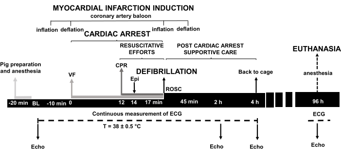
Figure 1: Experimental model of cardiac arrest. VF, ventricular fibrillation; CPR, cardiopulmonary resuscitation; Epi, epinephrine; ROSC, return of spontaneous circulation; BL, baseline; ECG, electrocardiogram; Echo, echocardiography; h, hours; min, minutes. Please click here to view a larger version of this figure.
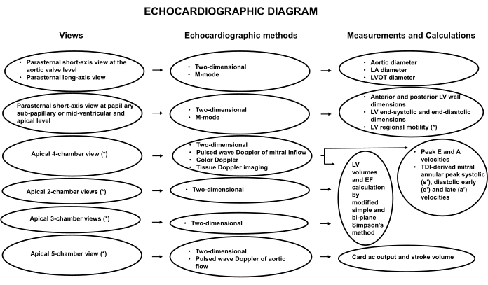
Figure 2: TTE flow-chart in a pig model of ischemic cardiac arrest. LA, left atrium; M-mode, mono-dimensional; LV, left ventricle; LVOT, left ventricle outflow tract; LVEF, left ventricular ejection fraction; PW, pulsed-wave; TDI, tissue Doppler imaging. Please click here to view a larger version of this figure.
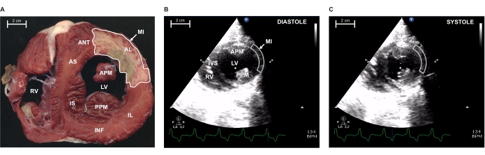
Figure 3: Myocardial infarction (MI) extension at papillary level by morphometry and bi-dimensional echocardiography 96 h after coronary artery occlusion. (A) Representative ex vivo 0.5 cm slice of pig heart at the papillary level, stained with triphenyl tetrazolium chloride (TTC) to display the healthy myocardial zone (red) against the infarcted one (brown). Echocardiographic 2D-parasternal short-axis view at the papillary level in diastole (B) and in systole (C). Arrows indicate the delimited MI areas indicated in A, B, and C. RV, right ventricle; IS, infero-septal wall; AS, antero-septal wall; IVS, intraventricular septum; APM, anterior papillary muscle; PPM, posterior papillary muscle; LV, left ventricle; AL, antero-lateral wall; ANT, anterior wall; INF, inferior wall; IL, infero-lateral wall. Please click here to view a larger version of this figure.
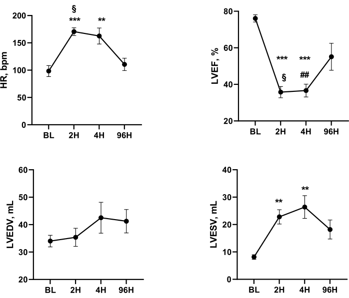
Figure 4: Systolic function parameters with heart rate at BL and after AMI, cardiac arrest, and resuscitation. One-way ANOVA for repeated measurements and Tukey's post-hoc test: *** p < 0.001, ** p < 0.01 vs BL; § p < 0.05 2 h vs 96 h; # p < 0.05, ## p < 0.01 4 h vs 96 h. BL, baseline; 2H, 2 h after AMI-cardiac arrest-ROSC; 4H, 4 h AMI- cardiac arrest-ROSC; 96H, 96 h AMI- cardiac arrest -ROSC; HR, heart rate; LVEF, left ventricular ejection fraction; LVEDV, left ventricular end-diastolic volume; LVESV, left ventricular end-systolic volume. Please click here to view a larger version of this figure.
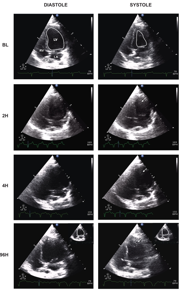
Figure 5: Apical four-chamber view at different times after AMI-cardiac arrest-ROSC. BL, baseline; H, hour; LV, left ventricle; RV, right ventricle; LA, left atrium; RA, right atrium. Arrows indicate apical thrombi near akinetic segments. Baseline and 96 h LV systolic and diastolic internal borders are shown in white. Please click here to view a larger version of this figure.
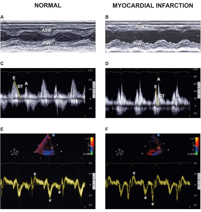
Figure 6: M-mode traces of short axis, MV color Doppler and TDI images in a healthy pig and 96 h after myocardial infarction (MI)-cardiac arrest-ROSC. Representative images of the LV from M-mode echocardiography at baseline (A) and 96 h after AMI-cardiac arrest-ROSC (B). ASW, anteroseptal wall; PIW, posteroinferior wall. * = normo-kinetic; ** = severely hypokinetic. Apical four-chamber view: pulse wave Doppler (PW) of the trans-mitral valve flow at baseline (C) and 96h after AMI-cardiac arrest-ROSC (D). Evel, PW early peak mitral inflow velocity; Avel, PW late peak mitral inflow velocity; DT, deceleration time. Representative images of septal and lateral TDI velocities at baseline (E) and (F) 96 h after MI-cardiac arrest-ROSC. s', TDI systolic velocity; e' TDI early diastolic velocity; a', TDI late diastolic velocity. Please click here to view a larger version of this figure.
A complete echocardiographic examination in a pig experimental model of AMI, cardiac arrest and resuscitation may give different information on the evolution of LV function and LV structural changes, although some amount of data are available in the literature5,8. In "pure" models of experimental cardiac arrest (restricted to induced ventricular fibrillation), myocardial function impairment reverses in the first days after ROSC, but little is known of what happens when AMI is the cause of cardiac arrest.
This study in pigs investigated the short- and mid-term post-AMI-cardiac arrest changes in LV structure, regional motility, and global LV function. At 2 h and 4 h after resuscitation, ESV significantly increased and LVEF decreased compared to baseline. These results are explained by a 26% akinetic/dyskinetic wall motion score index due to the post-AMI injury of mid-anterolateral and apical segments (Figure 3).
Myocardial stunning due to post-ROSC ischemia-reperfusion injury is well known. Yang l et al. found that diastolic parameters in pigs post-ROSC without AMI normalized in 24 h, while LV systolic function normalized in 48 h8. To the best of our knowledge, no data are available regarding a longer follow-up. Vammen et al.9, in a post-ROSC and AMI model in pigs, showed that the lower LVEF in both sham and AMI animals did return to normal at 48 h. In a previous work, the authors pointed out the relationship between smaller infarct, lower high-sensitivity troponin plasma concentration and better left ventricular function recovery 96 h after ROSC5,10.
Cardiac magnetic resonance (CMRI) is the gold standard imaging method to examine cardiac structure and function11, but it is expensive and requires lengthy acquisition and post-processing times. TTE is a less time-consuming, cheaper and more easily available method for experimental in vivo research and can follow repeated examinations in the same animal during experimental studies.
TTE in experimental cardiac arrest models in pigs is very challenging, but the method presents several difficulties obtaining good quality images during the acute phase post-ROSC mechanical ventilation due to: 1) the curtain effect of the left lung, 2) the increased chest resistance, 3) suboptimal animal positioning, and 4) the need for experienced sonographers. In fact, full training in the field is essential, particularly when an assessment of hemodynamics and LV function is required at the same time.
A limitation of our study is the absence of a sham group (cardiac arrest without AMI), in order to assess the level of LV systolic dysfunction ascribable to myocardial necrosis after coronary artery occlusion and that due to the post-ROSC myocardial injury.
In conclusion, TTE is a reliable, non-invasive diagnostic method for investigating the evolution of LV dysfunction in the post-cardiac arrest syndrome after AMI in a pig experimental model.
The authors have nothing to disclose.
We are grateful to Judith Bagott for language editing.
Aquasonic | Parker | – | ultrasound gel |
Adult foam ECG disposable monitoring and stress testing, wet gel, non-invasive patien | Philips | 40493E | ECG electrode |
Bellavista 1000 | Bellavista | MB230000 | ventilator with infrared capnometer |
ComPACS | Medimatic SRL | – | local database and software |
CX50 | Philips | – | Echocardiographic machine |
InTube Tracheal tube | Intersurgical Ltd | 8040080 | cuffed tracheal tube |
LUCAS2 | Phisio-Control Inc | – | mechanical chest compressor |
MRx defibrillator | Philips | – | defibrillator |
S5-1 | Philips | – | Phased array probe |
Swan-Ganz catheter 2 lumen 5fr | Edwards | 110F5 | for the coronary artery occlusion |
Swan-Ganz catheter 2 lumen 7fr | Edwards | 111F7 | for mean arterial pressure measurement |
Swan-Ganz catheter for thermodiluition 7fr | Edwards | 131F7 | to measure right atrial pressure, core temperature and cardiac output |