Presented here is a step-by-step procedure to induce acute lung injury in mice by direct intratracheal lipopolysaccharide instillation and to perform FACS analysis of blood samples, bronchoalveolar lavage fluid, and lung tissue. Minimal invasiveness, simple handling, good reproducibility, and titration of disease severity are advantages of this approach.
Airway administration of lipopolysaccharide (LPS) is a common way to study pulmonary inflammation and acute lung injury (ALI) in small animal models. Various approaches have been described, such as the inhalation of aerosolized LPS as well as nasal or intratracheal instillation. The presented protocol describes a detailed step-by-step procedure to induce ALI in mice by direct intratracheal LPS instillation and perform FACS analysis of blood samples, bronchoalveolar lavage (BAL) fluid, and lung tissue. After intraperitoneal sedation, the trachea is exposed and LPS is administered via a 22 G venous catheter. A robust and reproducible inflammatory reaction with leukocyte invasion, upregulation of proinflammatory cytokines, and disruption of the alveolo-capillary barrier is induced within hours to days, depending on the LPS dosage used. Collection of blood samples, BAL fluid, and lung harvesting, as well as the processing for FACS analysis, are described in detail in the protocol. Although the use of the sterile LPS is not suitable to study pharmacologic interventions in infectious diseases, the described approach offers minimal invasiveness, simple handling, and good reproducibility to answer mechanistic immunological questions. Furthermore, dose titration as well as the use of alternative LPS preparations or mouse strains allow modulation of the clinical effects, which can exhibit different degrees of ALI severity or early vs. late onset of disease symptoms.
Experimental animal models are indispensable in basic immune research. Administration of whole bacteria or microbial components has been frequently used in small animal models to induce local or systemic inflammation1. Lipopolysaccharide (LPS, or bacterial endotoxin) is a cell wall component and surface antigen of gram-negative bacteria (e.g., Enterobacteriaceae, Pseudomonas spp., or Legionella spp.). The thermostable and large molecule (molecular weight 1-4 x 10⁶ kDa) consists of a lipid moiety (Lipid A), core region (oligosaccharide), and an O polysaccharide (or O antigen). Lipid A, with its hydrophobic fatty acid chains, anchors the molecule into a bacterial membrane and mediates (upon degradation of bacteria) the immunological activity and toxicity of LPS. Following binding to the LPS binding protein (LBP), LPS:LBP complexes ligate the CD14/TLR4/MD2 receptor complex located on the surface of many cell types, inducing a strong proinflammatory reaction with NF-κB nuclear translocation and subsequent upregulation of cytokine expression2.
Acute lung injury (ALI) is defined as acute hypoxemic respiratory failure with bilateral pulmonary edema in the absence of heart failure3. Airway administration of LPS is a common way to induce pulmonary inflammation and ALI4,5,6,7. Although the sterile substance is not suitable to study pharmacologic interventions in infectious diseases, mechanistic immunological questions may be answered with adequate precision. Instillation of LPS into the trachea induces a robust inflammatory reaction with leukocyte invasion, upregulation of proinflammatory cytokines, and disruption of the alveolo-capillary barrier within hours to days, depending on the LPS dosage3,6,7.
The presented protocol describes a detailed step-by-step procedure to induce ALI in mice by intratracheal LPS instillation. The model has been validated by assessing cytokine expression, neutrophil granulocyte invasion, and intra-alveolar albumin leakage as previously described8.
This animal protocol was approved by the local committee for animal care (LANUV, Recklinghausen, Germany; protocol no. 84-02.04.2015) and was performed in accordance with the National Institutes of Health guidelines for the use of live animals (NIH publication No. 85-23, revised 1996).
1. ALI induction
2. Blood sampling, bronchoalveolar lavage, organ harvesting
NOTE: Timing of euthanization depends on the scientific issue addressed. Usually, it is performed 12-72 h following LPS instillation3,4,9,10. Severity of ALI can be determined clinically by regular observation of body temperature and respiratory distress symptoms11.
3. Tissue preparation for FACS analysis
The described approach to induce ALI in mice was validated by assessing cytokine expression, neutrophil granulocyte infiltration, and alveolo-capillary barrier disruption 24 h and 72 h after LPS instillation. PBS-injected animals served as control. Intratracheal LPS administration induced a robust pulmonary proinflammatory response. Expression of TNF-α in lung tissue was significantly upregulated, reaching a sustained and more than 50-fold increase compared to the control animals [RQ (TNF-α/18s); 24 h: 53.7 (SD = 11.6); 72 h: 55.0 (SD = 20.6); p < 0.05)] (Figure 3A). Leukocyte invasion into tissue and alveolar space is a hallmark and characteristic for the development of ALI13. FACS analysis revealed a significant infiltration of neutrophil granulocytes (NG) into the lung interstitium, with absolute cell count having increased almost 9-fold compared to the controls after 24 h [65,243 (SD = 15,855) vs. 7,358 (SD = 4,794), p < 0.05] (Figure 3B). Absolute NG count slightly decreased after 72 h; however, the factor increases compared to the controls remained stable [48,946 (SD = 5,223) vs. 5,510 (SD = 654), p < 0.05]. Consistent with interstitial NG infiltration, MMP-9 expression in whole lung tissue was likewise significantly increased over the total observation period [RQ (MMP-9/18s), 24 h: 7.4 (SD = 1.5); 72 h: 10.4 (SD = 2.0); p < 0.05] (Figure 3C).
NG were not only increased in the lung tissue but also in the BAL fluid. The fold increase compared to control animals was more pronounced than in lung tissue, with absolute NG counts 24 h following ALI induction of 52,005 (SD = 21,906) vs. 1,829 (SD = 1,724) (p < 0.05) (Figure 3D). After 72 h, NG were increased to 37,254 (SD = 4,478) vs. 17.0 (SD = 10.8) (p < 0.05). Lung edema due to severe impairment of the alveolo-capillary barrier is pathognomonic for the development of ALI, with LPS rapidly inducing endothelial apoptosis and increased permeability14,15. Analysis of albumin content in BAL fluid by ELISA revealed a significant loss of barrier function. 24 h following LPS instillation, albumin in BAL fluid was 43 ng/mL (SD = 13), compared to 20 ng/mL (SD = 9) under control conditions (p < 0.05) (Figure 3E). After 72 h, in ALI animals, albumin content was 48 ng/mL (SD = 14), compared to 29 ng/mL (SD = 9) (p < 0.05).
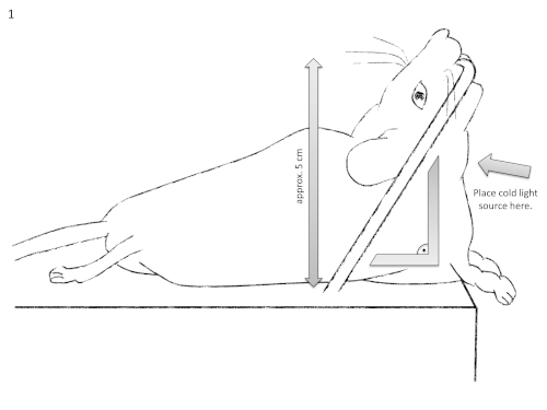
Figure 1: Schematic diagram of the intubation setting. It should be noted that the mouse's neck should be super-extended at a 90° angle relative to the operation table. Please click here to view a larger version of this figure.
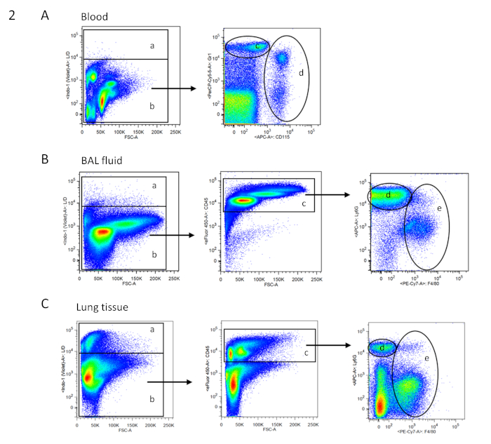
Figure 2: FACS gating strategy for blood, BAL, and tissue cells. Exemplary dot blots of FACS analysis are shown in two-parameter (dual color fluorescence) pseudocolor plots. Gating strategy for the respective samples is based on single cells. (A) Gating tree for blood cells: a = dead cells; b = living cells (according to live/dead cell staining; no CD45 staining necessary as in the blood, high autofluorescence makes the cell populations clearly distinguishable); d = neutrophil granulocytes; e = monocytes (according to Gr1 and CD115 staining). (B) Gating tree for bronchoalveolar lavage (BAL) fluid: a = dead cells; b = living cells (according to live/dead cell staining); c = CD45+ immune cells; d = neutrophil granulocytes; and e = macrophages (according to Ly6G and F4/80 staining). (C) Gating tree for lung tissue: a = dead cells; b = living cells (according to live/dead cell staining); c = CD45+ immune cells; d = neutrophil granulocytes; and e = macrophages (according to Ly6G and F4/80 staining). Please click here to view a larger version of this figure.
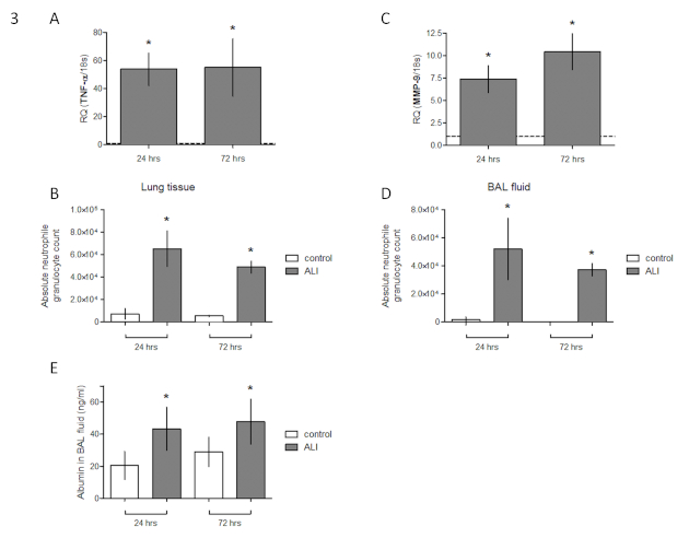
Figure 3: Validation of murine ALI model against control animals. (A) Expression of TNF-α in lung tissue of female C57BL/6 mice 24 h and 72 h following intratracheal LPS instillation (fold change of expression of sham operated animals). (B) FACS analysis of absolute neutrophil granulocyte count in lung tissue. (C) Expression of MMP-9 in lung tissue (fold change of expression of sham operated animals). (D) FACS analysis of absolute neutrophil granulocyte count in bronchoalveolar lavage fluid. (E) Albumin content in BAL fluid [mean ± SD, n = 7, Mann-Whitney U test, *p < 0.05 (vs. PBS control)]. This figure has been modified from Ehrentraut et al.8. Please click here to view a larger version of this figure.
Name of Material/ Equipment | Volume (mL) |
Dulbecco's Phosphate Buffered Saline (PBS), without calcium chloride and magnesium chloride, sterile | 1000 |
Fetal calf serum (FCS) | 1 |
Ethylenediaminetetraacetic acid (EDTA) solution | 1 |
Sodium azide (NaN3) | 0.1 |
Table 1: Composition of FACS buffer.
Name of Material/ Equipment | Suggested dilution | Mastermix for 10 samples: add to 200 µl FACS buffer (= 20 µl per sample): |
Anti-CD115 (c-fms) APC | 0.5 µL/100 µL | 5 µL |
Anti-CD11b (M1/70) – FITC | 0.5 µL/100 µL | 5 µL |
Anti-CD45 (30-F11) – eF450 | 0.5 µL/100 µL | 5 µL |
Anti-F4/80 (BM-8) – PE Cy7 | 0.5 µL/100 µL | 5 µL |
Anti-Gr1 (RB6-8C5) | 0.5 µL/100 µL | 5 µL |
Anti-Ly6C (HK1.4) PerCP-Cy5.5 | 0.5 µL/100 µL | 5 µL |
Anti-Ly6G (1A8) APC/Cy7 | 0.5 µL/100 µL | 5 µL |
Table 2: Preparation of master mix for FACS staining. The table describes master mix preparation for 10 samples.
Minimal invasiveness, simple handling, and good reproducibility are key features of the presented approach to induce ALI in a small rodent model. The use of LPS instead of whole bacteria in animal models has advantages. It is a stable and pure compound and can be stored in lyophilized form until use. It is a potent stimulant for innate immune responses via the TLR4 pathway, and its biological activity may readily be quantified, facilitating the titration of disease severity with good reproducibility. Moreover, the use of LPS has been shown to serve as safe model to induce acute bronchitis in human healthy volunteers and thus allows translation from bench to bedside16. Rittirsch et al. have demonstrated the dose- and time-dependent developments of the characteristic alveolo-capillary leakage in a murine model of intratracheal LPS instillation6. This allows dose titration to achieve certain desired effects, which can illustrate different degrees of ALI severity or early vs. late onset of disease symptoms. However, if distinct infectiological or pharmacological issues are to be addressed (e.g., antibiotic therapy), ALI induced by sterile LPS instillation is not a suitable model.
Moreover, compared to intrapulmonary or intravenous delivery of bacteria, the disruption of the alveolo-capillary barrier was described as being rather mild3, questioning the suitability of this model and whether altered permeability should be investigated particularly. Correct placement of the catheter to deliver the LPS bilaterally into the lower respiratory tract is the critical step of the approach. To ensure proper intratracheal intubation, visualization and identification of the larynx is facilitated by an external cold light source. Changes in respiratory pattern (e.g., coughing or gasping) verify correct intratracheal instillation of the fluid.
Moreover, the choice of mouse strain and LPS are crucial for the induction of ALI and generation of reproducible results in this model and depend on the scientific issue addressed. According to literature, the dosage administered to elicit a maximum effect with no further increase with escalating dosages ranges from 10 µg/mouse (when LPS from Pseudomonas aeruginosa F-D type 1 is injected into female BALB/c mice) to 50 µg/mouse when injecting E. coli (serotype O111:B4) LPS (which was also used in the protocol) into male C57BL/6 mice6,9. In general, BALB/c mice are supposed to react sensitively when challenged with LPS, whereas C57BL/6 mice seem to be more resistant3. Thus, initial dose titration experiments respecting individual conditions are recommended. This also applies to the timing of blood, BAL, and organ sampling. Severity of ALI can be determined clinically by regular observation of body temperature and respiratory distress symptoms. Furthermore, since mice only share approximately 50% homology of the TLR4 receptor with humans, careful interpretation of the results is mandatory3.
Alternatives to the herein presented approach comprise the route of endotoxin administration to the lungs. As described by Szarka et al., LPS may also be administered via intranasal instillation9. Liu et al. compared the direct intratracheal deposition with the inhalation of aerosolized LPS5. Based on their findings, they concluded that the inhalational route induces a more uniform type of ALI. However, their experiments were performed in rats with a directed-flow nose-only inhalation and may therefore not necessarily be transferred to the herein presented approach. In contrast, mice are often exposed to aerosolized LPS in a chamber10. Chamber size, LPS concentration, and the number of mice treated simultaneously are variables that limit the comparability between different studies and make an individual model establishment recommendable. Last, intravenous or intraperitoneal administration of LPS is often used to induce remote ALI17,18. As the data from Szarka et al. suggest, the intratracheal instillation seems to be superior to the i.v. or i.p. route when specific pulmonary inflammatory effects are being addressed9. In conclusion, the protocol represents a simple and reproducible approach to induce sterile ALI in mice to address specific immunological issues.
The authors have nothing to disclose.
The authors wish to thank Jan Kleiner and Susanne Schulz for providing technical support. The authors acknowledge the excellent support of the flow cytometry core facility at the medical faculty of Bonn University. The authors received no funding from any external organization. Part of the data given in the results section and depicted in Figure 3 has already been shown in a previous publication8.
1 ml syringes | BD, Franklin Lakes, NJ, USA | 300013 | |
10 ml syringes | BD, Franklin Lakes, NJ, USA | 309110 | |
Anti-CD115 (c-fms) APC | Thermo Fisher, Waltham, MA, USA | 17-1152-80 | |
Anti-CD11b (M1/70) – FITC | Thermo Fisher, Waltham, MA, USA | 11-0112-81 | |
Anti-CD45 (30-F11) – eF450 | Thermo Fisher, Waltham, MA, USA | 48-0451-82 | |
Anti-F4/80 (BM-8) – PE Cy7 | Thermo Fisher, Waltham, MA, USA | 25-4801-82 | |
Anti-Gr1 (RB6-8C5) | BD Biosciences, Franklin Lakes, NJ, USA | 552093 | |
Anti-Ly6C (HK1.4) PerCP-Cy5.5 | Thermo Fisher, Waltham, MA, USA | 45-5932-82 | |
Anti-Ly6G (1A8) APC/Cy7 | Bio Legend, San Diego, CA | 127623 | |
Buprenorphine hydrochloride | Indivior UK Limited, Berkshire, UK | ||
C57BL/6 mice, female, 10 – 12 weeks old | Charles River, Wilmongton, MA, USA | ||
CaliBRITE APC-beads (6µm) | BD Biosciences, Franklin Lakes, NJ, USA | 340487 | |
Canula 23 gauge 1'' | BD, Franklin Lakes, NJ, USA | 300800 | |
Canula 26 gauge 1/2'' | BD, Franklin Lakes, NJ, USA | 303800 | |
Cell strainer 70 µm | BD Biosciences, Franklin Lakes, NJ, USA | 352350 | |
Collagenase Type I | Sigma-Aldrich, St. Louis, MO, USA | 1148089 | |
Deoxyribonuclease II | Sigma-Aldrich, St. Louis, MO, USA | D8764 | |
Dulbecco's Phosphate Buffered Saline (PBS), sterile | Sigma-Aldrich, St. Louis, MO, USA | D8662 | |
Dulbecco’s Phosphate Buffered Saline (PBS), without calcium chloride and magnesium chloride, sterile | Sigma-Aldrich, St. Louis, MO, USA | D8537 | |
Ethylenediaminetetraacetic acid (EDTA) solution | Sigma-Aldrich, St. Louis, MO, USA | E7889 | |
FACS tubes, 5 ml | Sarstedt, Nümbrecht, Germany | 551579 | |
Fetal calf serum (FCS) | Sigma-Aldrich, St. Louis, MO, USA | F2442 | |
Forceps | Fine Science Tools, Heidelberg, Germany | 11049-10 | |
Isoflurane | Baxter, Unterschleißheim, Germany | ||
Ketamine hydrochloride | Serumwerk Bernburg, Bernburg, Germany | ||
Lipopolysaccharides (LPS) from Escherichia coli O111:B4 | Sigma-Aldrich, St. Louis, MO, USA | L2630 | |
LIVE/DEAD Fixable Dead Cell Green Kit | Thermo Fisher, Waltham, MA, USA | L23101 | |
Purified Rat Anti-Mouse CD16/CD32 (Mouse BD Fc Block™), Clone 2.4G2 | BD, Franklin Lakes, NJ, USA | 553141 | |
Red blood cell lysis buffer | Thermo Fisher, Waltham, MA, USA | 00-4333-57 | |
RPMI-1640, with L-glutamine and sodium bicarbonate | Sigma-Aldrich, St. Louis, MO, USA | R8758 | |
Scissors | Fine Science Tools, Heidelberg, Germany | 14060-09 | |
Sodium azide (NaN3) | Sigma-Aldrich, St. Louis, MO, USA | S2002 | |
Spring scissors | Fine Science Tools, Heidelberg, Germany | 15018-10 | |
Tissue forceps | Fine Science Tools, Heidelberg, Germany | 11021-12 | |
Tubes | Eppendorf, Hamburg, Germany | 30125150 | |
Venous catheter, 22 gauge | B.Braun, Melsungen, Germany | 4268091B | |
Xylazine hydrochloride | Serumwerk Bernburg, Bernburg, Germany |