Summary

기능적 근적외선 분광법과 하지의 로봇 보조 다관절 포인팅 운동을 이용한 실험

Published: June 07, 2024
doi:

Summary

전 세계 6명 중 1명은 일생 동안 뇌졸중을 앓아 장기적인 장애를 유발하는 것으로 추정되며, 이러한 재활 메커니즘은 아직 잘 알려져 있지 않습니다. 이 연구는 하지 로봇 치료 세션 동안 기능적 근적외선 분광법(fNIRS)으로 뇌 활성화를 평가하는 프로토콜을 제안합니다.

Abstract

뇌졸중은 매년 전 세계적으로 약 1,700만 명에게 영향을 미치며 장기 장애의 주요 원인입니다. 로봇 치료는 뇌졸중 환자가 잃어버린 운동 기능을 회복하는 데 도움이 될 수 있는 가능성을 보여주었습니다. 운동 회복이 어떻게 일어나는지에 대한 이해를 높일 수 있는 한 가지 방법은 건강한 사람의 치료 대상이 되는 운동 중 뇌 활성화를 연구하는 것입니다. 기능적 근적외선 분광법(fNIRS)은 운동 기능의 신경 토대를 검사하기 위한 유망한 신경 영상 기술로 부상했습니다. 이 연구는 건강한 피험자의 복잡한 하지 움직임의 fNIRS 신경 상관 관계를 조사하는 것을 목표로 했습니다. 참가자들은 운동 재활을 위한 로봇 장치를 사용하여 6분 동안 휴식과 움직임의 주기를 수행하도록 요청받았습니다. 이 작업에는 컴퓨터 화면에 표시된 표적을 가리키기 위해 무릎과 발목 관절의 조정된 움직임이 필요했습니다. 로봇이 제공하는 다양한 수준의 이동 지원을 사용하는 두 가지 실험 조건을 탐색했습니다. 그 결과, fNIRS 프로토콜은 작업 중 운동 제어와 관련된 뇌 영역을 효과적으로 감지하는 것으로 나타났습니다. 특히, 모든 피험자는 도움을 받은 조건에 비해 도움이 없는 상태에서 반대쪽 전운동 영역에서 더 큰 활성화를 보였습니다. 결론적으로, fNIRS는 하지의 다관절 포인팅 운동과 관련된 옥시헤모글로빈 농도의 변화를 감지하는 데 유용한 접근법인 것으로 보입니다. 이 연구는 뇌졸중 운동 회복 메커니즘에 대한 이해에 기여할 수 있으며 뇌졸중 환자를 위한 개선된 재활 치료를 위한 길을 열 수 있습니다. 그러나 운동 기능 연구와 임상 환경에서의 응용에 대한 fNIRS의 잠재력을 완전히 설명하기 위해서는 추가 연구가 필요합니다.

Introduction

역학 데이터에 따르면 전 세계적으로 매년 ~1,700만 건의 새로운 뇌졸중 사례가 발생하며 저소득 및 중간 소득 국가에서 발병률이 증가하고 있습니다1. 신규 확진자 수는 2030년까지 7,700만 명으로 증가할 것으로 추정2. 뇌졸중으로 인한 운동 장애는 종종 환자의 이동성과 일상 생활 활동 참여에 영향을 미쳐 삶의 질을 저하시킵니다. 전통적인 운동 재활에는 도수 치료가 포함되지만 지난 수십 년 동안 재활을 위한 로봇 시스템이 개발되었습니다. 이러한 시스템은 고강도, 용량, 정량화성, 신뢰성, 반복성및 유연성으로 치료를 제공할 수 있으며3 급성 및 만성 뇌졸중 환자 모두에게 효과적인 재활 치료로서의 잠재력을 보여주었습니다 4,5,6. 재활을 위한 로봇 시스템은 치료를 제공하는 것 외에도 환자의 움직임 운동학적/운동학적 데이터를 측정할 수 있는 센서를 장착할 수 있으므로 평가 도구로 사용할 수 있습니다 7,8. 상지 운동 재활의 경우, 이러한 데이터는 로봇 치료에 의해 유도된 환자의 운동 회복 수준을 평가하는 데 유용한 것으로 입증되었을 뿐만 아니라 전통적인 임상 평가9,10에 대한 보충 도구로 사용될 뿐만 아니라 뇌졸중11에서 운동 회복 과정에 대한 이해를 증진하는 데 기여했다. 12 건강한 피험자의 운동 및 운동 학습의 신경 제어 3,13,14. 결과적으로, 이러한 발견은 재활 치료를 강화하기 위한 기초를 제공하였다15.

지난 20년 동안 하지 신경 재활을 위한 많은 로봇 장치가 제안되었는데, 이는 걷는 동안 환자의 체중을 지탱하는 외골격(예: Lokomat16과 같은 러닝머신 위)에서 환자가 걷지 않고 발목, 무릎 또는 발을 운동할 수 있도록 하는 고정식 로봇 시스템(예: Rutgers Ankle17, 고성능 발목 재활 로봇(High-Performance Ankle Rehabilitation Robot)(18), 광주과학기술원(GIST) 발목/발 재활 로봇(19) 또는 환자가 지상 또는 러닝머신 위를 걸을 때 착용하는 작동식 외골격인 능동 발 보조기(예: Powered Gait Orthosis20 및 MIT Anklebot21). 하지 재활을 위한 로봇에 대한 리뷰는 22,23,4를 참조하십시오.

뇌졸중 환자에 대한 하지 재활을 위한 로봇 장치의 임상 연구 결과는 고무적이었으며, 이러한 시스템은 특정 장치 및 임상 프로토콜에 따라 관절의 운동 범위(ROM), 근력 또는 보행을 개선할 수 있음을 보여주었습니다(재활을 위한 하지 로봇의 효능에 대한 검토는 24,25 참조). 로봇 보조 치료가 신경가소성 변화를 촉진하여 궁극적으로 운동 능력을 향상시킨다는 가정이 있지만26 뇌졸중으로부터 운동 회복 과정이 정확히 어떻게 발생하는지, 그리고 어떤 로봇 훈련 프로토콜이 하지 운동 능력의 회복 과정을 최적화하는지는 대부분 불분명하다. 실제로, 재활 로봇의 개발이 증가하는 것(학술 연구자 또는 상업 단체에 의한)과 운동 회복의 기초가 되는 신경생리학적 메커니즘에 대한 제한된 이해 사이에는 상당한 격차가 커지고 있다4. 내장된 센서로 측정한 운동 운동학 또는 관절 토크의 측정은 환자가 하지 운동 능력27,28,29을 회복할 때 발생하는 운동 행동 변화를 정량적으로 설명하는 데 기여하여 이 격차를 부분적으로 메우는 데 기여했습니다. 그러나 이러한 변화의 기저에 있는 신경 상관 관계는 덜 연구되었습니다. 이것은 몇 가지 이유 때문입니다.

뇌 기능 영상은 시간이 많이 걸리고 때로는 임상시험의 맥락에서 완료하기 어려우며, 환자의 연구 순응 가능성을 극대화하기 위해 환자의 부담을 최소화해야 하는 경우가 많습니다. 뇌졸중 후 피로와 근육 약화가 자주 관찰된다는 사실을 감안할 때 뇌졸중을 앓은 적이 있는 개인에게 특히 해당된다30. 또한 기능적 자기공명영상(fMRI)과 같이 자기장을 기반으로 하는 영상 기법은 환자와 로봇 하드웨어 모두 자석으로부터 안전해야 합니다.

비침습적 이미징 방식 중에서 기능적 근적외선 분광법(fNIRS)은 로봇 치료를 받는 피험자의 뇌 활성화 영역을 평가하는 데 특히 적합한 이미징 기술입니다. fMRI와 유사하게 fNIRS는 뇌의 혈액 산소화/탈산소화를 측정합니다. 그러나 fMRI와 달리 fNIRS는 로봇 하드웨어와 완벽하게 호환되며 종종 휴대가 가능하며 침대 옆에서도 사용할 수 있습니다. 또한, fNIRS는 비용이 저렴하고 모션 아티팩트 31,32,33에 대한 민감도가 낮다.

70년대 후반에 처음 도입된 이래 많은 임상 환경에서 fNIRS의 분명한 장점과 광범위한 사용에도 불구하고34, 하지 운동 및 뇌졸중 운동 회복과 관련된 뇌 활성화를 정량화하기 위해 fNIRS를 사용한 연구는 소수에 불과합니다. 운동의 신경 조절 기전을 규명하거나 뇌졸중으로부터 운동 회복을 평가하기 위한 FNIRS 연구는 주로 단일관절 운동(예: 배측굴곡, 발바닥 굴곡 또는 무릎 신전 운동35,36,37), 걷기38,39,40,41,42,43 또는 사이클링44을 조사하였다. 리뷰는45를 참조하십시오. 마찬가지로, 하지를 위한 로봇 보조 치료에 대한 fNIRS 연구는 대부분 로봇 보조 보행 재활에 중점을 두었습니다. 리뷰는46을 참조하십시오. 몇몇 연구들은 fNIRS를 뇌-컴퓨터 인터페이스(Brain-Computer Interface, BCI) 시스템의 일부로 사용하여 로봇 장치에 대한 제어 신호를 유도하는 데 초점을 맞추었다(47,48); 이 연구 분야는 fNIRS 신호 처리에도 의존하지만, 그 목표는 다르며 주로 환자의 의도(예: 중증 운동 장애 환자)를 해독하는 데 중점을 둡니다.

여기에 제시된 파일럿 연구는 하지 재활을 위한 로봇 시스템의 효과를 조사하기 위한 초기 노력의 일부입니다. 이 로봇은 일상적인 다관절 움직임에 대한 훈련을 포함하는 목표 지향적인 하지 재활을 제공할 수 있을 뿐만 아니라 하지의 단일 관절(예: 무릎 또는 발목)에 대한 치료를 제공할 수 있습니다(즉, 상향식 재활 프로그램 구현).

이 연구는 하지, 다관절 포인팅 운동을 수행하는 동안 fNIRS 데이터 수집이 필요한 실험 프로토콜의 타당성을 조사하는 것을 목표로 했습니다. 6분으로 제한된 이 연구의 데이터 수집 기간은 일반적인 fNIRS 프로토콜보다 짧습니다. 이는 특히 이동성이나 근력이 제한된 환자에서 이 연구의 실용성과 임상적 적용 가능성을 높이기 위한 목적으로 이루어진 신중한 선택이었습니다. 이러한 복잡한 다관절 운동의 fNIRS 상관 관계를 식별하고 로봇 지원에 의해 뇌 활성화가 어떻게 조절되는지에 대한 통찰력을 얻는 것도 관심 지점이었습니다. 이를 위해 동일한 참가자를 대상으로 두 세션의 실험을 수행했는데, 하나는 로봇의 도움을 받지 않았고 다른 하나는 로봇의 도움을 받았습니다. 마지막으로, 본 연구는 로봇 치료가 목표로 하는 움직임 중 뇌 활성화의 평가 및 기록 프로토콜 타당성 측면에서 향후 연구의 토대를 마련하기 위해 건강한 피험자에 초점을 맞췄다는 점에 주목하는 것이 중요합니다.

기구
하지 재활을 제공하도록 설계된 휴대용 로봇( 그림 1 참조)이 실험을 수행하는 데 사용되었습니다. 이 로봇은 3D 접근 가능한 작업 공간을 가지고 있으며 무게가 약 35파운드로 작고 가벼워 운반 및 설치가 쉽습니다.

Figure 1
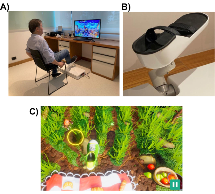
그림 1: 실험 설정. (A) 하지용으로 설계된 로봇 시스템(바닥에 설치됨). 자원봉사자가 오른발로 인터페이스를 활용하는 모습이 보입니다. (B) 로봇 시스템에 부착할 수 있는 피험자의 발을 지지하는 구조. (C) 피크닉 게임의 스크린샷. 게임의 목표는 발(녹색과 흰색 신발)을 목표물(노란색 원)로 이동하는 것입니다. 이 그림의 더 큰 버전을 보려면 여기를 클릭하십시오.

로봇 시스템은 환자가 가리키기나 발차기와 같은 일상 업무에서 수행하는 것과 유사한 하지 움직임을 수행하는 데 도움을 주도록 설계되었습니다. 로봇 장치 앞에 놓인 컴퓨터 모니터 또는 텔레비전 화면에 표시되는 대화형 가상 현실 게임을 사용합니다( 그림 1 참조). 로봇 엔드 이펙터는 환자의 하지(예: 발목)에 부착되고, 그 위치는 화면 상의 커서 위치에 매핑됩니다. 일반적인 게임은 환자의 움직임 목표(예: 가리킬 물체 또는 공을 찰 위치)를 보여줍니다.

이동 작업을 완료하기 위해 로봇은 완전한 지원에서 전혀 지원하지 않는 수준까지 다양한 수준의 지원으로 환자를 도울 수 있습니다. 로봇 지원 수준은 환자의 운동 장애 수준에 따라 각 재활 세션이 시작될 때 선택됩니다. 피험자가 수행한 움직임은 게임에서 환자의 수행 능력에 점수를 매기고 수행에 대한 피드백(예: ROM, 움직임 횟수 및 로봇 지원 수준)을 제공하는 데 사용됩니다. 게임은 환자의 관심과 관심을 유지하기 위해 상호 작용하고 재미있게 설계되었습니다. 이 연구에서 참가자들은 “피크닉 게임”을 했는데, 플레이어는 곤충이 수건에 손을 뻗어 음식을 훔치는 것을 막아야 했습니다(스크린샷은 그림 1, 하단 패널 참조).

데이터 수집은 2개의 서로 다른 연속파 옵토드(760nm 및 850nm), 8개의 듀얼 팁 LED 소스 및 8개의 듀얼 팁 활성 검출기가 있는 휴대용 fNIRS 수집 시스템으로 수행되었습니다. 신호는 10.17Hz의 샘플링 속도를 사용하여 수집되었습니다. fNIRS 시스템에서 생성된 Wi-Fi 네트워크를 사용하여 교정 최적화 및 신호 기록에 노트북을 사용했습니다.

캡은 미리 결정된 위치에 옵토드를 고정하는 데 사용되었습니다. 소스와 검출기는 10-10 국제 EEG 시스템에 따라 격자 공간 분포에 배치되었습니다. 각 fNIRS 채널은 약 30mm의 옵토드 간 거리를 갖는 소스-검출기 쌍에 의해 정의되었습니다. 옵토드는 그림 2에 표시된 위치의 보조 모터, 프리모터 및 모터 영역 위에 배치되었습니다. 총 채널 수는 28개였으며, 여기서 8개는 광섬유 어댑터를 사용하여 단일 검출기에 각 소스에 연결된 단거리 채널이었습니다. 하드웨어의 멀티플렉싱 설정을 감안할 때 하나의 검출기만 사용하여 모든 소스에서 근거리 정보를 수집할 수 있습니다.

Figure 2
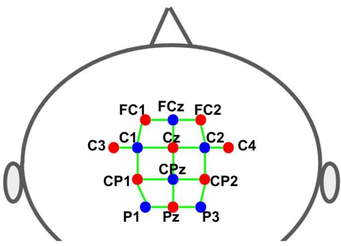
그림 2: 10-10 EEG 시스템을 사용한 몽타주 레이아웃. 문자와 숫자는 소스/감지기 위치를 나타냅니다. 빨간색과 파란색 점은 각각 소스 및 검출기 옵토드를 나타냅니다. 녹색 선은 소스 쌍과 검출기 쌍으로 구성된 fNIRS 채널을 나타냅니다. 이 그림의 더 큰 버전을 보려면 여기를 클릭하십시오.

실험 설계
실험은 피험자의 움직임에 대해 로봇이 제공하는 지원 수준이 다른 두 가지 뚜렷한 실험 조건에서 수행되었습니다. 첫 번째 조건에서 로봇은 피험자의 움직임에 어떤 도움도 주지 않도록 프로그래밍된 반면, 두 번째 조건에서는 로봇이 피험자의 발과 다리 움직임을 제어했다(로봇 보조 움직임).

각 실험은 그림 3과 같이 운동 작업(게임 플레이 – 30초)과 휴식(30초)의 교대 주기를 포함하는 블록 설계 패러다임을 따랐습니다. 각 단계(플레이/게임 또는 휴식)의 시작과 종료는 컴퓨터 화면을 통해 피험자에게 시각적으로 신호를 보냈다. 휴식 단계에서 일시 중지를 나타내는 메시지가 표시되었습니다. 각 주기(플레이/게임 + 휴식)의 지속 시간은 60초이고 6번 반복되어 총 실행 시간은 360초(6분)입니다.

참가자들은 곤충이 수건에 닿아 음식을 훔치는 것을 막는 것이 목표인 “피크닉 게임”을 했습니다. 이 게임은 지정된 홈 타겟(초기 위치)에서 시작하여 홈 타겟으로 돌아오기 전에 세 개의 바깥쪽 타겟 중 하나를 향해 뻗는 일련의 하지 움직임을 포함했습니다. 화면에서 바깥쪽 표적은 움직이는 움직이는 곤충으로 시각적으로 표현되었으며, 참가자들은 이 곤충에 손을 뻗어 밟아야 했습니다. 바깥쪽에 도달하는 표적은 3개였으며, 각 표적은 무작위로 동일한 횟수로 제시되었으며, 모든 움직임에 대해 공통된 홈 표적이 있었습니다. 발이 홈 타겟에서 외부 타겟의 위치까지 이동하는 데 필요한 거리는 약 26cm의 호를 형성합니다. 운동 작업은 다관절 운동의 실행이 필요했기 때문에 무릎 굴곡/신전, 발바닥 굴곡 및 배측 굴곡 운동 간의 조정이 필요했습니다.

fNIRS 데이터 기록은 로봇에 의해 생성된 TTL(Transistor-Transistor-Logic) 펄스를 통해 게임이 피험자에게 제공하는 시각적 자극과 동기화되었습니다. 펄스는 각 단계(플레이/게임 및 휴식)의 시작 시 생성되었습니다. 따라서 모든 타이밍 제어는 게임에 의해 수행되었으며, 이는 참가자에게 각 동작을 시작하도록 시각적 단서(대상)를 제공하고, 뇌 활동 기록을 표시하기 위해 fNIRS 시스템에 TTL 신호를 보내고, 실험에 필요한 경우 로봇 제어 시스템에 신호를 보내 움직임 지원을 시작합니다.

Protocol

이 연구는 UNICEP(Centro Universitario Paulista)의 지역 윤리 검토 위원회의 승인을 받았습니다. 모든 참가자는 인간과 관련된 과학 연구에 관한 모든 기관 지침 및 연방 규범에 따라 정보에 입각한 동의를 제공했습니다. 그들은 브라질 연방 규정에서 요구하는 금전적 보상을 받지 못했다. 1. fNIRS 시스템 8개의 광원(760nm 및 850nm)과 8개의 광 검출기( 그…

Representative Results

6명의 피험자 모두 두 실험을 모두 완료했습니다. 지원이 없는 조건에서 각 피험자는 평균 76.67건의 시도(표준 10.73)를 완료했습니다(참고로, 각 피험자에 대해 이전 목표에 도달한 경우에만 새로운 목표가 표시되었기 때문에 시도 횟수는 성공한 도달 횟수에 따라 달라졌습니다). 로봇이 피험자의 움직임을 완전히 보조하는 보조 조건에서 모든 피험자는 70번의 시도를 완료했습니다. 모든 피험자로?…

Discussion

이 개념 증명 연구에서는 하지 재활을 위해 로봇을 사용하여 다양한 유형의 움직임으로 운동하는 동안 건강한 피험자의 fNIRS 데이터를 사용하여 뇌 활성화 매핑에 대한 추론을 할 수 있는 가능성을 조사했습니다. 성인의 일반적인 fNIRS 기록 세션은 6분54보다 깁니다. 그러나 재활 환경의 맥락에서 녹음을 가능하게 하려면 피험자의 불필요한 피로와 노력을 피하기 위해 실험의 전?…

Disclosures

The authors have nothing to disclose.

Acknowledgements

본 연구의 의견, 가설, 결론 및 권고는 저자의 것이며 반드시 연구비 지원 기관의 의견을 나타내는 것은 아닙니다. JRS는 기술 지원을 해준 상파울루 연구 재단(FAPESP, 보조금 번호 2021/05332-8, 2018/04654-9, 2018/21934-5 및 2023/02538-0)과 Jackson Cionek에 감사드립니다. AMM과 Vivax Ltda는 FAPESP(상파울루 연구 재단)와 FINET(브라질 혁신청)에 감사의 뜻을 전합니다. 이 프로젝트는 FAPESP(보조금 번호 2018/09559-4) 및 FINEP(보조금 번호 2019/09933-6)의 보조금으로 자금을 지원받았습니다.

Materials

32 inch Smart TV Samsung N/A TV connected to robot via HDMI cable
8-detector silicon photodiode (SiPD) optodes for optical detection with dual tip NIRx Medical Technologies (Glen Head, NY, USA) https://nirx.net/nirsport
8-source optodes bundle for optical illumination with dual tip NIRx Medical Technologies (Glen Head, NY, USA) https://nirx.net/nirsport
Aurora acquisition software NIRx Medical Technologies (Glen Head, NY, USA) https://nirx.net/nirsport
Laptop Precision XPS 13 Dell Technologies (Round Rock, TX, USA)
nirsLAB fNIRS Analysis software NIRx Medical Technologies (Glen Head, NY, USA) https://nirx.net/nirsport
NIRSports2 fNIRS acquisition system NIRx Medical Technologies (Glen Head, NY, USA) https://nirx.net/nirsport It has two different continuous wave optics (760 and 850 nm), 8 dual-ended LED sources and 8 dual-ended active detectors.
R R-project.org (open source software) https://www.r-project.org/
Standard cut cap, black color for up to 128 holders. Easycap GmbH (Wörthsee, Germany) https://www.easycap.de/
Vivax Assistive Rehabilitation Machine (ARM)  Vivax Ltda (São Paulo, Brazil) https://vivaxbr.com/home/ It is a portable robot designed to deliver lower limb rehabilitation. It has a 3D reachable workspace and is compact and light, weighing about 35 lb., which makes it easy to transport and to install. 

References

  1. GBD 2016 Stroke Collaborators. Global, regional, and national burden of stroke, 1990-2016: a systematic analysis for the Global Burden of Disease Study 2016. The Lancet. Neurology. 18 (5), 439-458 (2019).
  2. GBD 2019 Stroke Collaborators. Global, regional, and national burden of stroke and its risk factors, 1990-2019: a systematic analysis for the Global Burden of Disease Study 2019. The Lancet. Neurology. 20 (10), 795-820 (2021).
  3. Huang, V. S., Krakauer, J. W. Robotic neurorehabilitation: a computational motor learning perspective. Journal of NeuroEngineering and Rehabilitation. 6, 5 (2009).
  4. Hobbs, B., Artemiadis, P. A Review of Robot-Assisted Lower-Limb Stroke Therapy: Unexplored Paths and Future Directions in Gait Rehabilitation. Frontiers in neurorobotics. 14, 19 (2020).
  5. Bertani, R., Melegari, C., De Cola, M. C., Bramanti, A., Bramanti, P., Calabrò, R. S. Effects of robot-assisted upper limb rehabilitation in stroke patients: a systematic review with meta-analysis. Neurological Sciences. 38 (9), 1561-1569 (2017).
  6. Warutkar, V., Dadgal, R., Mangulkar, U. R. Use of robotics in gait rehabilitation following stroke: A review. Cureus. 14 (11), e31075 (2022).
  7. Dipietro, L., et al. Changing motor synergies in chronic stroke. Journal of Neurophysiology. 98 (2), 757-768 (2007).
  8. Dipietro, L., et al. Learning, not adaptation, characterizes stroke motor recovery: evidence from kinematic changes induced by robot-assisted therapy in trained and untrained task in the same workspace. IEEE Trans Neural Syst Rehabil Eng. 20 (1), 48-57 (2012).
  9. Bosecker, C., Dipietro, L., Volpe, B., Krebs, H. I. Kinematic robot-based evaluation scales and clinical counterparts to measure upper limb motor performance in patients with chronic stroke. Neurorehabilitation and Neural Repair. 24 (1), 62-69 (2010).
  10. Krebs, H. I., et al. Robotic measurement of arm movements after stroke establishes biomarkers of motor recovery. Stroke. 45 (1), 200-204 (2014).
  11. Volpe, B. T., et al. Robotic devices as therapeutic and diagnostic tools for stroke recovery. Archives of Neurology. 66 (9), 1086-1090 (2009).
  12. Hogan, N., et al. Motions or muscles? Some behavioral factors underlying robotic assistance of motor recovery. Journal of Rehabilitation Research and Development. 43 (5), 605-618 (2006).
  13. Shadmehr, R., Wise, S. P. . The Computational Neurobiology of Reaching and Pointing: A Foundation for Motor Learning. , (2005).
  14. Dipietro, L., Poizner, H., Krebs, H. I. Spatiotemporal dynamics of online motor correction processing revealed by high-density electroencephalography. J Cogn Neurosci. 26 (9), 1966-1980 (2014).
  15. Krebs, H., et al. Rehabilitation robotics: Performance-based progressive robot-assisted therapy. Autonomous Robots. 15, 7-20 (2003).
  16. Colombo, G., Joerg, M., Schreier, R., Dietz, V. Treadmill training of paraplegic patients using a robotic orthosis. Journal of Rehabilitation Research and Development. 37 (6), 693-700 (2000).
  17. Girone, M., et al. A Stewart platform-based system for ankle telerehabilitation. Autonomous Robots. 10, 203-212 (2001).
  18. Saglia, J. A., Tsagarakis, N. G., Dai, J. S., Caldwell, D. G. A high-performance redundantly actuated parallel mechanism for ankle rehabilitation. The International Journal of Robotics Research. 28 (9), 1216-1227 (2009).
  19. Yoon, J., Ryu, J. A novel reconfigurable ankle/foot rehabilitation robot. , 2290-2295 (2005).
  20. Ruthenberg, B. J., Wasylewski, N. A., Beard, J. E. An experimental device for investigating the force and power requirements of a powered gait orthosis. Journal of Rehabilitation Research and Development. 34 (2), 203-213 (1997).
  21. Forrester, L. W., et al. Clinical application of a modular ankle robot for stroke rehabilitation. NeuroRehabilitation. 33 (1), 85-97 (2013).
  22. Díaz, I., Gil, J. J., Sánchez, E. Lower-limb robotic rehabilitation: Literature review and challenges. Journal of Robotics. 2011, 759764 (2011).
  23. Zhang, X., Yue, Z., Wang, J. Robotics in lower-limb rehabilitation after stroke. Behavioural Neurology. 2017, 3731802 (2017).
  24. Zhang, M., Davies, T. C., Xie, S. Effectiveness of robot-assisted therapy on ankle rehabilitation – a systematic review. Journal of NeuroEngineering and Rehabilitation. 10, 30 (2013).
  25. Lo, K., Stephenson, M., Lockwood, C. Effectiveness of robotic assisted rehabilitation for mobility and functional ability in adult stroke patients: a systematic review protocol. JBI Database of Systematic Reviews and Implementation Reports. 15 (1), 39-48 (2017).
  26. Belda-Lois, J. M., et al. Rehabilitation of gait after stroke: a review towards a top-down approach. Journal of NeuroEngineering and Rehabilitation. 8, 66 (2011).
  27. Bortole, M., et al. The H2 robotic exoskeleton for gait rehabilitation after stroke: early findings from a clinical study. Journal of NeuroEngineering and Rehabilitation. 12, 54 (2015).
  28. Banala, S. K., Kim, S. H., Agrawal, S. K., Scholz, J. P. Robot assisted gait training with active leg exoskeleton (ALEX). IEEE Transactions on Neural Systems and Rehabilitation Engineering. 17 (1), 2-8 (2009).
  29. Bartenbach, V., Wyss, D., Seuret, D., Riener, R. A lower limb exoskeleton research platform to investigate human-robot interaction. 2015 IEEE International Conference on Rehabilitation Robotics (ICORR). 2015, 600-605 (2015).
  30. Hinkle, J. L., et al. Poststroke fatigue: Emerging evidence and approaches to management: A scientific statement for healthcare professionals from the American heart association. Stroke. 48 (7), e159-e170 (2017).
  31. Balardin, J. B., Zimeo Morais, G. A., Furucho, R. A., Trambaiolli, L. R., Sato, J. R. Impact of communicative head movements on the quality of functional near-infrared spectroscopy signals: negligible effects for affirmative and negative gestures and consistent artifacts related to raising eyebrows. Journal of Biomedical Optics. 22 (4), 4601 (2017).
  32. Nazeer, H., Naseer, N., Mehboob, A., Khan, M. J., Khan, R. A., Khan, U. S., Ayaz, Y. Enhancing classification performance of fNIRS-BCI by identifying cortically active channels using the z-score method. Sensors. 20 (23), 6995 (2020).
  33. Ayaz, H., et al. Optical imaging and spectroscopy for the study of the human brain: status report. Neurophotonics. 9, S24001 (2022).
  34. Chen, W. L., et al. Functional near-infrared spectroscopy and its clinical application in the field of neuroscience: Advances and future directions. Frontiers in Neuroscience. 14, 724 (2020).
  35. Yamamoto, K., Miyata, T., Onozuka, A., Koyama, H., Ohtsu, H., Nagawa, H. Plantar flexion as an alternative to treadmill exercise for evaluating patients with intermittent claudication. European Journal of Vascular and Endovascular Surgery. 33 (3), 325-329 (2007).
  36. Formenti, D., et al. Effects of knee extension with different speeds of movement on muscle and cerebral oxygenation. PeerJ. 6, 5704 (2018).
  37. Miyai, I., et al. Cortical mapping of gait in humans: a near-infrared spectroscopic topography study. NeuroImage. 14 (5), 1186-1192 (2001).
  38. Miyai, I., et al. Premotor cortex is involved in restoration of gait in stroke. Annals of Neurology. 52 (2), 188-194 (2002).
  39. Mihara, M., et al. Sustained prefrontal activation during ataxic gait: a compensatory mechanism for ataxic stroke?. NeuroImage. 37 (4), 1338-1345 (2007).
  40. Rea, M., et al. Lower limb movement preparation in chronic stroke: A pilot study toward an fNIRS-BCI for gait rehabilitation. Neurorehabilitation and Neural Repair. 28 (6), 564-575 (2014).
  41. Holtzer, R., Verghese, J., Allali, G., Izzetoglu, M., Wang, C., Mahoney, J. R. Neurological gait abnormalities moderate the functional brain signature of the posture first hypothesis. Brain Topography. 29 (2), 334-343 (2016).
  42. Kim, H. Y., Yang, S. P., Park, G. L., Kim, E. J., You, J. S. Best facilitated cortical activation during different stepping, treadmill, and robot-assisted walking training paradigms and speeds: A functional near-infrared spectroscopy neuroimaging study. NeuroRehabilitation. 38 (2), 171-178 (2016).
  43. Khan, H., Nazeer, H., Engell, H., Naseer, N., Korostynska, O., Mirtaheri, P. Prefrontal cortex activation measured during different footwear and ground conditions using fNIRS-A case study. 2021 IEEE International Conference on Artificial Intelligence and Mechatronics Systems (AIMS). , 1-6 (2021).
  44. Lin, P. Y., Chen, J. J., Lin, S. I. The cortical control of cycling exercise in stroke patients: an fNIRS study). Human Brain Mapping. 34 (10), 2381-2390 (2013).
  45. Yang, M., Yang, Z., Yuan, T., Feng, W., Wang, P. A systemic review of functional near-infrared spectroscopy for stroke: Current application and future directions. Frontiers in Neurology. 10, 58 (2019).
  46. Berger, A., Horst, F., Müller, S., Steinberg, F., Doppelmayr, M. Current state and future prospects of EEG and fNIRS in robot-assisted gait rehabilitation: A brief review. Frontiers in Human Neuroscience. 13, 172 (2019).
  47. Khan, R. A., Naseer, N., Qureshi, N. K., et al. fNIRS-based Neurorobotic Interface for gait rehabilitation. J NeuroEngineering Rehabil. 15 (1), 7 (2018).
  48. Khan, H., Naseer, N., Yazidi, A., Eide, P. K., Hassan, H. W., Mirtaheri, P. Analysis of Human Gait Using Hybrid EEG-fNIRS-Based BCI System: A Review. Front. Hum. Neurosci. 14, (2020).
  49. Delpy, D. T., Cope, M. Quantification in tissue near-infrared spectroscopy. Philosophical Transactions of the Royal Society B: Biological Sciences. 352 (1354), 649-659 (1997).
  50. Huppert, T. J. Commentary on the statistical properties of noise and its implication on general linear models in functional near-infrared spectroscopy. Neurophotonics. 3, 010401 (2016).
  51. Alexandre, F., Heraud, N., Oliver, N., Varray, A. Cortical implication in lower voluntary muscle force production in non-hypoxemic COPD patients. PLoS One. 9 (6), 100961 (2014).
  52. Yoon, T., Vanden Noven, M. L., Nielson, K. A., Hunter, S. K. Brain areas associated with force steadiness and intensity during isometric ankle dorsiflexion in men and women. Experimental Brain Research. 232 (10), 3133-3145 (2014).
  53. Ciccarelli, O., et al. Identifying brain regions for integrative sensorimotor processing with ankle movements. Experimental Brain Research. 166 (1), 31-42 (2005).
  54. Udina, C., et al. Functional near-infrared spectroscopy to study cerebral hemodynamics in older adults during cognitive and motor tasks: A review. Frontiers in Aging Neuroscience. 11, 367 (2020).
  55. Thickbroom, G. W., Phillips, B. A., Morris, I., Byrnes, M. L., Mastaglia, F. L. Isometric force-related activity in sensorimotor cortex measured with functional MRI. Experimental Brain Research. 121 (1), 59-64 (1998).
  56. Derosière, G., Alexandre, F., Bourdillon, N., Mandrick, K., Ward, T. E., Perrey, S. Similar scaling of contralateral and ipsilateral cortical responses during graded unimanual force generation. NeuroImage. 85 (1), 471-477 (2014).
  57. Shi, P., Li, A., Yu, H. Response of the cerebral cortex to resistance and non-resistance exercise under different trajectories: A functional near-infrared spectroscopy study. Frontiers in Neuroscience. 15, 685920 (2021).
  58. Dettmers, C., et al. Relation between cerebral activity and force in the motor areas of the human brain. Journal of Neurophysiology. 74 (2), 802-815 (1995).
  59. Keisker, B., Hepp-Reymond, M. C., Blickenstorfer, A., Kollias, S. S. Differential representation of dynamic and static power grip force in the sensorimotor network. The European Journal of Neuroscience. 31 (8), 1483-1491 (2010).
  60. Harada, T., Miyai, I., Suzuki, M., Kubota, K. Gait capacity affects cortical activation patterns related to speed control in the elderly. Experimental Brain Research. 193 (3), 445-454 (2009).
  61. Saleh, S., et al. The role of premotor areas in dual tasking in healthy controls and persons with multiple sclerosis: An fNIRS imaging study. Frontiers in Behavioral Neuroscience. 12, 296 (2018).
  62. Bonnal, J., et al. Relation between cortical activation and effort during robot-mediated walking in healthy people: A functional near-infrared spectroscopy neuroimaging study (fNIRS). Sensors. 22 (15), 5542 (2022).
  63. Shibuya, K., Sadamoto, T., Sato, K., Moriyama, M., Iwadate, M. Quantification of delayed oxygenation in ipsilateral primary motor cortex compared with contralateral side during a unimanual dominant-hand motor task using near-infrared spectroscopy. Brain Research. 1210, 142-147 (2008).
  64. Dai, T. H., Liu, J. Z., Sahgal, V., Brown, R. W., Yue, G. W. Relationship between muscle output and functional MRI-measured brain activation. Experimental brain research. 140 (3), 290-300 (2001).
  65. Cabibel, V., Hordacre, B., Perrey, S. Implication of the ipsilateral motor network in unilateral voluntary muscle contraction: the cross-activation phenomenon. Journal of Neurophysiology. 123 (5), 2090-2098 (2020).
  66. Akselrod, M., Martuzzi, R., Serino, A., vander Zwaag, W., Gassert, R., Blanke, O. Anatomical and functional properties of the foot and leg representation in areas 3b, 1 and 2 of primary somatosensory cortex in humans: A 7T fMRI study. NeuroImage. 159, 473-487 (2017).
  67. Brigadoi, S., Cooper, R. J. How short is short? Optimum source-detector distance for short-separation channels in functional near-infrared spectroscopy. Neurophotonics. 2 (2), 025005 (2015).
  68. Funahashi, S. Prefrontal contribution to decision-making under free-choice conditions. Frontiers in Neuroscience. 11, 431 (2017).
  69. Simon, S. R., Meunier, M., Piettre, L., Berardi, A. M., Segebarth, C. M., Boussaoud, D. Spatial attention and memory versus motor preparation: premotor cortex involvement as revealed by fMRI. Journal of Neurophysiology. 88 (4), 2047-2057 (2002).
  70. Desmurget, M., Sirigu, A. A parietal-premotor network for movement intention and motor awareness. Trends in Cognitive Sciences. 13 (10), 411-419 (2009).
  71. Nachev, P., Kennard, C., Husain, M. Functional role of the supplementary and pre-supplementary motor areas. Nature reviews. Neuroscience. 9 (11), 856-869 (2008).
  72. Thoenissen, D., Zilles, K., Toni, I. Differential involvement of parietal and precentral regions in movement preparation and motor intention. The Journal of neuroscience: the official journal of the Society for Neuroscience. 22 (20), 9024-9034 (2002).
  73. Al-Quraishi, M. S., Elamvazuthi, I., Tang, T. B., Al-Qurishi, M., Adil, S. H., Ebrahim, M. Bimodal data fusion of simultaneous measurements of EEG and fNIRS during lower limb movements. Brain Sciences. 11 (6), 713 (2021).
  74. Bishnoi, A., Holtzer, R., Hernandez, M. E. Brain Activation Changes While Walking in Adults with and without Neurological Disease: Systematic Review and Meta-Analysis of Functional Near-Infrared Spectroscopy Studies. Brain sciences. 11 (3), 291 (2021).
  75. Oh, S., Song, M., Kim, J. Validating attentive locomotion training using interactive treadmill: an fNIRS study. Journal of NeuroEngineering and Rehabilitation. 15 (1), 122 (2018).
check_url/cn/66004?article_type=t

Play Video

Cite This Article
Ricardo Sato, J., Carolyna Gianlorenço, A., Borges Fernandes, E., Frigo da Rocha, T., Massato Makiyama, A., Dipietro, L. An Experiment Using Functional Near-Infrared Spectroscopy and Robot-Assisted Multi-Joint Pointing Movements of the Lower Limb. J. Vis. Exp. (208), e66004, doi:10.3791/66004 (2024).

View Video