Guided endodontics describes a template-aided approach for access cavity preparation. The procedure requires cone-beam computed tomography and a surface scan to produce a template. An incorporated sleeve guides the drill to the target point. This allows the preparation of minimally invasive endodontic access cavities in calcified teeth.
Pulp canal obliterations (PCO) are often a consequence of dental trauma, such as luxation injuries. Even though dentin apposition is a sign of vital pulp, pulpitis or apical periodontitis may develop in the long term. Root canal treatment of teeth with severe PCO and pulpal or periapical pathosis is challenging for general practitioners and even for well-equipped endodontic specialists. To ensure detection of the calcified root canal and avoid excessive loss of tooth structure or root perforation, static navigation using templates (“Guided Endodontics”) was introduced a few years ago. The general workflow includes three-dimensional imaging using cone-beam computed tomography (CBCT), a digital surface scan, and superimposition of both in a planning software. This is followed by virtual planning of the access cavity and the design of a template that will guide the drill to the desired target point. To do this, a true-to-scale virtual image of the drill must be placed in a way that the tip of the drill reaches the orifice of the calcified root canal. Once the template has been fabricated using computer-aided design and computer-aided manufacturing (CAD/CAM) or a 3D printer, guided preparation of the access cavity can be performed clinically. For research purposes, a postoperative CBCT image can be used to quantify the accuracy of the access cavity performed. This work aims to present the technique of static guided endodontics from imaging to clinical implementation.
Pulp canal obliterations (PCO) are signs of vital pulp, and are often observed after dental trauma1 or as a response to stimuli like caries, restorative procedures2, or vital pulp therapy3. When no clinical or radiographic signs of pathology are present, root canal treatment is not indicated. In the long term, however, the remaining pulp tissue can develop a pathosis4. In cases where clinical or radiographic signs of pulpal or apical pathology are present, non-surgical root canal treatment would be the treatment of choice for tooth preservation.
For a successful outcome of the root canal treatment, the preparation of an adequate access cavity is crucial. Teeth with PCO needing a root canal treatment are difficult to treat, even for dentists that are specialized in the field of endodontics5. Attempting to locate a calcified root canal may result in a high loss of tooth structure and thus weakening or even perforation of the root. This reduces the prognosis of the tooth, and extraction may be indicated6.
As template-based (static) navigation is already successfully used in oral implantology, its application in endodontics was first described in the literature a few years ago7. Since then, numerous case reports and studies have demonstrated the benefits of template-aided endodontic access cavity preparation in cases with PCO8,9.
The aim of this work is to present the technique of guided access cavity preparation using guided endodontics. For research purposes, a treatment evaluation (determination of angular and spatial deviation between planned and performed access cavity) is possible after a postoperative CBCT scan, which is also presented in this article.
Approval or consent to perform this study was not required since the use of patients' data is not applicable. In this study, DICOM data from a maxillary model consisting of extracted, de-identified human teeth are used. Teeth were extracted due to reasons not related to this study.
1. Virtual access cavity planning
2. Access cavity preparation
3. Treatment evaluation
Figure 10A shows the occlusal view of a prepared endodontic access cavity in a first maxillary molar after template-aided access cavity preparation of the mesio-buccal canal. Figure 10B shows the insertion of three endodontic handfiles to confirm successful root canal detection after preparation of the palatal and disto-buccal access cavities. After matching the postoperative CBCT data to the preoperative planning data, virtual bur placement generates information about the deviation (Figure 11A). Here, the angular deviation is 0.7°, 0.74 mm 3D deviation at the base of the bur, and 0.87 mm 3D deviation at the tip of the bur. For better visualization, the deviation can be shown in different planes or a 3D-rendered view (Figure 11B).
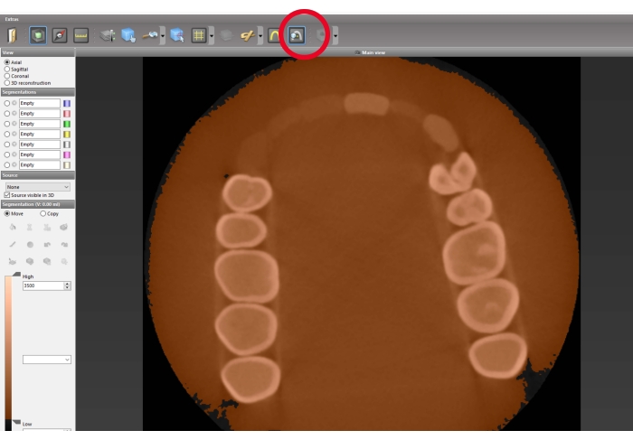
Figure 1: Segmentation preparation. Measurement of the HU density for the tooth enamel and the surrounding material. Calculate the mean value. Red circle: button for the density measuring tool. Left-click to activate, which allows density measurements in the axial view by left-clicking and holding in the desired area. Please click here to view a larger version of this figure.
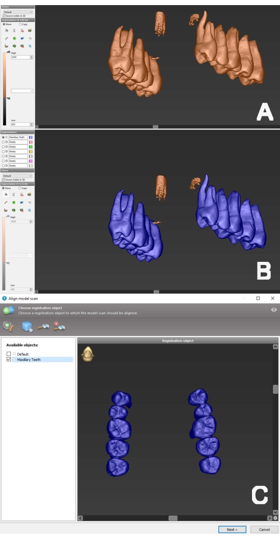
Figure 2: Segmentation process and preparation for alignment with surface data. (A) 3D view of preoperative CBCT data. The lower threshold has been adjusted to the determined mean value. (B) The flood fill tool was utilized to perform a segmentation of the tooth structure (color blue) and was named "Maxillary Teeth". (C) The performed segmentation can be selected (here: "Maxillary Teeth") for the registration step. Please click here to view a larger version of this figure.
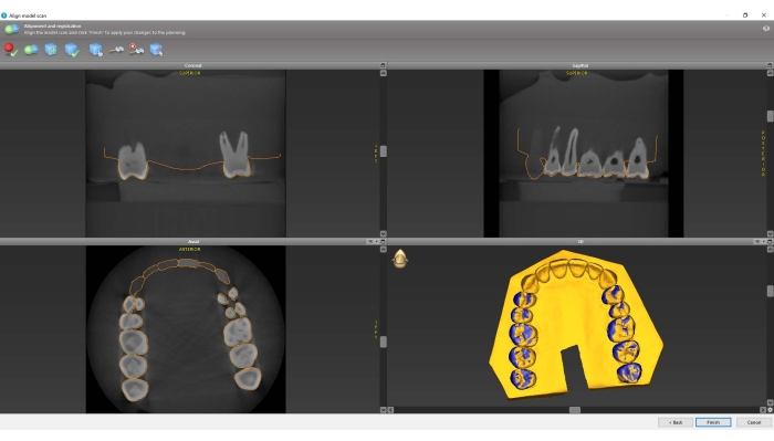
Figure 3: Alignment of CBCT and surface scan datasets. Verify in all planes that matching is accurate and complete the registration step. Note the "camouflage pattern" between segmentation and surface scan data in the 3D reconstruction, which indicates a highly precise matching of the data. Please click here to view a larger version of this figure.
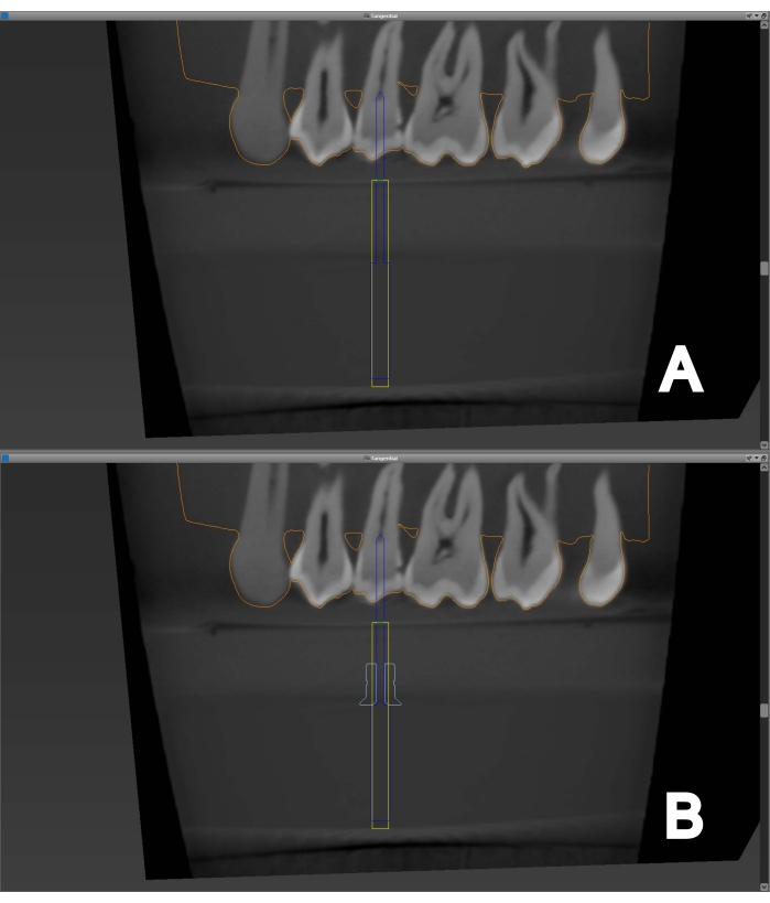
Figure 4: Access cavity planning. (A) An endodontic bur is virtually placed to the root canal orifice of a maxillary second premolar, providing straight-line access. (B) A suitable sleeve can be added to the endodontic bur. There must be enough space between the sleeve and coronal tooth structure to avoid interference when later placing the template on the dental arch. Please click here to view a larger version of this figure.
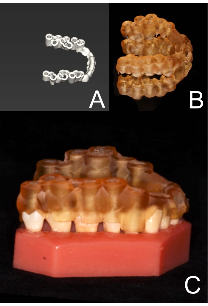
Figure 5: Template for static navigation. (A) The entire template has been designed (here, a maxillary study model with multiple planned access cavities in the posterior tooth area). It is now ready to be exported and 3D printed. (B) The template has been 3D printed. (C) The sufficient fit of the template on the dental arch is checked. Please click here to view a larger version of this figure.
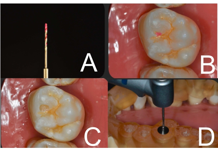
Figure 6: Access cavity preparation. (A) Dye (here: caries detector) at the bur's tip is used to mark enamel at the access cavity site. (B) Enamel has been marked through the template and sleeve. (C) Enamel at the access cavity site has been removed using a diamond bur in a contra-angle handpiece. (D) After sleeve insertion, the template is placed on the dental arch, and the guided endodontic access cavity can be performed with the endodontic bur in a contra-angle handpiece. Please click here to view a larger version of this figure.
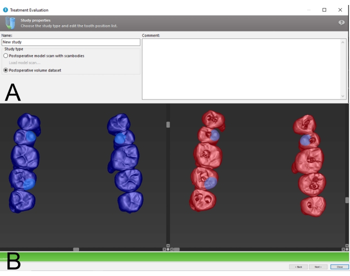
Figure 7: Preparation for treatment evaluation. (A) Choose Postoperative Volume Dataset as a data source for treatment evaluation. (B) Landmark registration between pre- and postoperative CBCT data. Choosing anatomically prominent regions (cusp tips, marginal ridges) as landmarks and their spatial distribution can facilitate semi-automatic registration. Please click here to view a larger version of this figure.
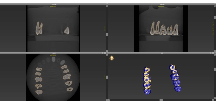
Figure 8: Postoperative CBCT alignment. Matched pre- and postoperative data is shown in all planes and in 3D reconstruction. Note the "camouflage pattern" between the datasets in the 3D Reconstruction, which indicates a highly precise matching of the data. Please click here to view a larger version of this figure.
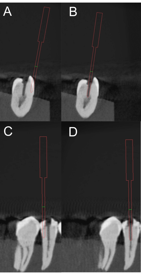
Figure 9: Marking of the access cavity. For treatment evaluation, a virtual bur is placed in the direction of access cavity preparation, which can be withdrawn from the postoperative CBCT data ((A) coronal plane, (C) sagittal plane). Confirm adequate bur positioning in both planes ((B) coronal plane, (D) sagittal plane). Please click here to view a larger version of this figure.
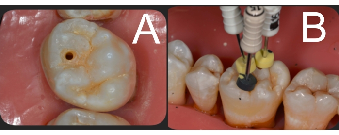
Figure 10: Clinical view after access cavity preparation. (A) Template-aided endodontic access cavity preparation of a maxillary first molar of the mesio-buccal canal. (B) After disto-buccal and palatal root canals are accessed in the same manner, handfiles are inserted to confirm successful root canal detection. Please click here to view a larger version of this figure.
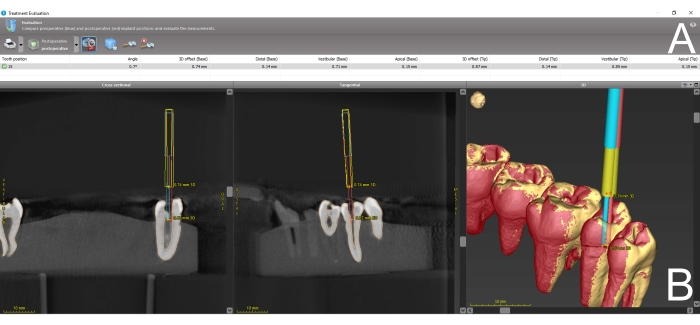
Figure 11: Treatment evaluation. (A) After correct matching of pre- and postoperative CBCT data and correct bur placement, the software calculates the angular and spatial deviation between planned and performed access cavity preparation. The results are presented in a table. (B) Visualization of the deviation is also provided in sagittal or coronal view, or in 3D reconstruction. Please click here to view a larger version of this figure.
Supplementary File 1: A sample stl file of the template. Please click here to download this File.
The introduction of template-aided access cavity preparations in endodontics has brought immense progress to non-surgical endodontic treatment in teeth with PCO. Conventional access cavity preparation can be very time consuming5 and is prone to error in cases with severe PCO. In vitro studies and clinical case reports demonstrate the feasibility of the guided endodontics approach, generating satisfying results in terms of root canal detection and an overall low deviation between the planned and performed access cavities8. However, the implementation of guided endodontics should be limited to cases where the conventional freehand access cavity preparation is accompanied by a higher risk of iatrogenic errors, since the use of ionizing radiation (CBCT) is required10.
To minimize the deviation between the planned and finally performed access cavity, a few factors need to be considered. When performing full-arch surface scans, local deviations and inaccuracies may occur11. This can lead to a certain degree of error in the CBCT data matching process, thus leading to deviations in access cavity preparation. Hence, highly precise surface scanners would also provide more accurate results for the guided endodontics approach. Different planning software and types of template manufacturing (additive versus subtractive) were investigated and found to have an influence on the outcome as well12.
Furthermore, the quality and accuracy of the 3D printing process also play a role in minimizing deviations in access cavity preparation. In addition to the various processes in 3D printing13, the alignment of the printed object14 also plays a decisive role in manufacturing precision. Since additive manufacturing processes are subject to constant further development, the manufacturing process should be critically examined on a regular basis in order to achieve the highest possible precision. Also, the fitting precision between the bur and sleeve plays an important role in the accuracy of the entire procedure. To avoid heat development and allow the bur to slide smoothly, a certain amount of “loose fit” is necessary. Particularly when the distance from the sleeve to the apical target point is large, a small deviation at the bur’s base might result in a larger deviation at the bur’s tip. To avoid a possible disadvantage from a sleeve-based system due to reduced vertical space in the patient’s mouth, a sleeveless guide system has been successfully described in a recent case report15. A further investigation comparing the accuracy of sleeve-containing versus sleeveless systems would be desirable for research in the field of guided endodontics in the future. Besides reduced vertical space, another limitation for template-aided preparation of endodontic access cavities is the presence of mobile teeth. To enable accurate planning and precise treatment, teeth with increased mobility can be splinted beforehand.
When the evaluation of the accuracy is performed utilizing postoperative CBCT data, it is important to assure that CBCT machine settings and the setting of the HU thresholds in the software are the exact same as in the preoperative data. It has been shown that different CBCT and threshold settings result in differing segmentation volumes16, therefore hindering the exact alignment of the imaging data and leading to incorrect results. Yet, even in an ideally matched dataset, error is unavoidable since the virtual bur is placed manually and underlies a subjective error. For the accuracy validation of oral implants, different methods were compared, and an automatic evaluation method was found to be superior to the manual matching method17. Hence, an automatic method should be considered to improve the quality of evaluation itself, and to create comparability between future research findings in the field of guided endodontics.
To the best of our knowledge, no commercially available software exists to date that automates the accuracy evaluation of access cavities. A difficulty that arises in comparison to the evaluation of implant positions is that access cavities are not radiopaque, and therefore, an automatic evaluation is difficult to implement.
Besides static navigation, dynamic navigation systems (DNS) were also described for endodontic purposes. DNS can circumvent the disadvantages of template-guided access preparation18, but require more equipment and are therefore still associated with high costs.
The authors have nothing to disclose.
None.
Accuitomo 170 | Morita Manufacturing | NA | CBCT machine |
coDiagnostiX | Dental Wings Inc | Version 10.4 | Planning software, which is mainly intended for implant surgery. Endodontic access cavities can be planned by adding the utlized bur to the implant database |
Endoseal drill | Atec Dental GmbH | NA | Carbide bur, which is used for the guided access cavity preparation |
StecoGuide Endo-Sleeve | steco-system-technik | REF M.27.28.D100L5 | Sleeves, which are inserted into the fabricated template |
TRIOS 3 | 3Shape A/S | NA | Surface scanner |
P30 | Straumann | NA | 3D Printer |
P pro Surgical Guide Clear | Straumann | NA | Light-curing resin for the additive manufacturing |