All methods described here have been approved by the Institutional Animal Care and Use Committee (IACUC) of the University of Georgia.
1. Washing using Traditional Centrifugation
- Prepare phosphate buffer solution (PBS). Add 8.0 g of NaCl, 0.2 g of KCl, 1.44 g of Na2HPO4 and 0.24 g of KH2PO4 to 800 mL of distilled water (dH2O). Adjust the pH to 7.4 using 0.1 N HCl and bring the solution to 1 L using dH2O.
- Prepare motility buffer. Add 6.5 g of NaCl, 4.5 g of glucose, 0.444 g of CaCl2 and 11.5 g of N-tris-[hydroxymethyl] methyl-2-amino-ethanesulfonic acid (TES) to 800 mL of dH2O. Adjust the pH to 7.4 using 1 M NaOH and bring the solution to 1 L using dH2O.
- Pipette 0.5 mL of semen into a polypropylene microcentrifuge tube. Add 1.0 mL of PBS and mix gently.
- Centrifuge at 1,500 x g for 10 min at room temperature (RT) and discard the supernatant. Resuspend the sperm pellet with PBS up to 1.5 mL.
- Centrifuge at 1,500 x g for 10 min at RT. Resuspend the sperm pellet with motility buffer up to 0.5 mL.
2. Performing the PDGC technique
NOTE: Perform the entire process of PDGC at room temperature.
- Make 3.0 mL of 1.08 g/mL and 1.07 g/mL Percoll solutions in two separate tubes.
- In a clean test tube, add 1.712 mL of the 1.13 g/mL original Percoll to 0.3 mL of 1.5 M NaCl solution. Add 0.988 mL of dH2O and mix by gentle inversion to make 3.0 mL of a 1.08 g/mL density solution.
- In a clean test tube, add 1.482 mL of the 1.13 g/mL original Percoll to 0.3 mL of 1.5 M NaCl solution. Add 1.218 mL of dH2O and mix by gentle inversion to make 3.0 mL of a 1.07 g/mL density solution.
- In a clean test tube, dilute 1.0 mL of semen sample 1:2 with 2.0 mL of PBS. Mix gently by pipetting.
- Pipet 3.0 mL of the 1.07 g/mL density solution into a sterile 15 mL conical tube. Carefully pipet 3.0 mL of the 1.08 g/mL density solution beneath the 1.07 g/mL density solution. Ensure that the two layers do not mix. A long-form (9 in) Pasteur pipette can make this step easier.
- Pipet 3.0 mL of diluted semen sample overtop the PDG. To ensure that the semen sample does not mix with the PDG, gently tilt the conical tube containing the PDG at a 45° angle. Pipet the sample along the wall of the tube and allow it to flow down the tube and over the PDG.
- Prepare a blank tube to match the mass of the PDG with overlaid sample. Centrifuge both tubes at 1500 x g for 20 min. Be careful to maintain the discontinuous gradient while transferring the tubes from the bench to the balance and then to the centrifuge.
NOTE: Do not use the brake at the end of centrifugation. - Observe the results. Ensure that three distinct semen layers have formed in the tube, as seen in Figure 1.
- Aspirate isolated semen layers with a pipette. Collect the top layer of semen first, the middle layer second, and last the hard pellet at the bottom of the tube. Transfer each to a clean and sterile polypropylenemicrocentrifuge tube.
- Dilute each sample to 1.5 mL with PBS. Centrifuge at 1500 x g for 10 min.
- Pour off the supernatant. Reconstitute sperm pellet with motility buffer by gentle pipetting.
NOTE: Alternative densities may be used to suit investigator needs. Determine amounts of ingredients used with the following equation, where v0 is volume of stock density solution used, v is final volume of solution desired, p is density of final density solution desired and p0 is the density of the stock density solution:
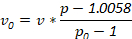
Always use 0.3 mL of 1.5 M NaCl in preparation of density solutions to match the NaCl concentration of physiological saline.
3. Determining Sperm Quality
- Calculate the sperm concentration as previously described22.
- Perform eosin-nigrosin vital staining as previously described12 with the following modifications:
- Prepare 100 µL of sperm solution at a concentration of 1 x 10⁸ cells/mL.
- Pipet 50 µL of sperm solution in a polypropylene microcentrifuge tube containing an equal volume of eosin-nigrosin stain. Incubate the mixture for 5 min at room temperature.
- Place a 20 µL drop of stained sperm sample at one end of a glass slide and smear uniformly in a manner similar to that used for blood smears. Air-dry the smeared slides at room temperature for 3-5 min.
- Observe the smear under microscope. Count the number of live sperm (no stain) and dead sperm (stained pink) and calculate the percentage of live sperm.
- Perform the Accudenz assay, as previously validated for the chicken sperm, to objectively assess the sperm mobility8 with the following modifications:
- Pipet 1.0 mL of 6% assay solution into polystyrene cuvettes, as illustrated in Figure 2. Incubate to 41 °C.
NOTE: 41 °C is used to match the internal temperature of a hen. The incubation temperature should match that of the female reproductive tract of the species being investigated. - Overlay the preheated assay solution with 100 µL of semen sample at a concentration of 5 x 10⁸ cells/mL.
- Place the cuvette containing overlaid sperm sample in the spectrophotometer. Record the absorbance value at 550 nm.
- Pipet 1.0 mL of 6% assay solution into polystyrene cuvettes, as illustrated in Figure 2. Incubate to 41 °C.
- Perform IPVL-penetration assay as previously described11 with the following modifications:
- Cut a piece (0.5 cm x 0.5 cm) of non-germinal disc region of intact IPVL.
- Adjust sperm concentration to 4 x 10⁶ cells/mL
- Incubate sperm in motility buffer with IPVL in a small glass vial for 15 min at 37 °C, as illustrated in Figure 3.
- Immerse the IPVL piece in 3% NaCl to stop the interaction between the IPVL and sperm.
- Mount the IPVL piece on a microscope slide and stain with Schiff's reagent for 10 min following fixation with 10% formalin for 20 s.
- Observe the IPVL under a microscope for successful sperm penetration holes and count the number of all visible holes per 0.25 mm2 at 40X magnification.
The PDGC technique resulted in distinct separation of three layers of sperm by degree of quality across all parameters. Sperm separates into a high-quality layer below the higher density solution, a medium-quality layer between the higher and lower density solution and a low-quality layer above the lower density solution. These differences in quality are evidenced by clear differences in viability (Figure 4), mobility (Figure 5) and penetrability (Figure 6). Sperm isolated from the high-quality layer of the PDG exhibited increases in all three parameters relative to those of the traditionally washed sample. Those layers noted as medium- and low-quality upon separation by PDGC display moderate and dramatic, respectively, decreases across all test parameters.
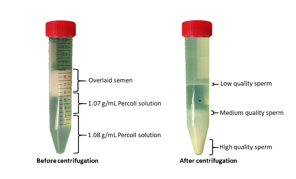
Figure 1: Isolation of sperm with differential quality using PDGC. Overlay 3 mL of sperm solution on prepared PDG solution. Centrifuge at 1500 x g for 20 min. Collect three distinct groups of sperm with low-, medium- and high-quality. Please click here to view a larger version of this figure.
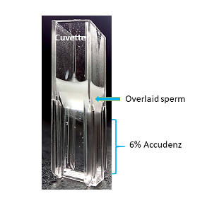
Figure 2: Determination of sperm mobility using Accudenz assay. Overlay 100 µL of sperm sample on a 6% assay solution in a cuvette. Incubate at 41 °C for 5 min. Record the mobility of sperm as a function of absorbance at 550 nm. Please click here to view a larger version of this figure.

Figure 3: A model of formation of sperm penetration holes in IPVL. Incubate sperm with a small section of IPVL at 37 °C for 15 min. Upon contact with IPVL, sperm cells bind with the IPVL, undergo an acrosome reaction and penetrate the IPVL, creating penetration holes. Count the number of penetration holes and measure the penetrability of sperm. Please click here to view a larger version of this figure.
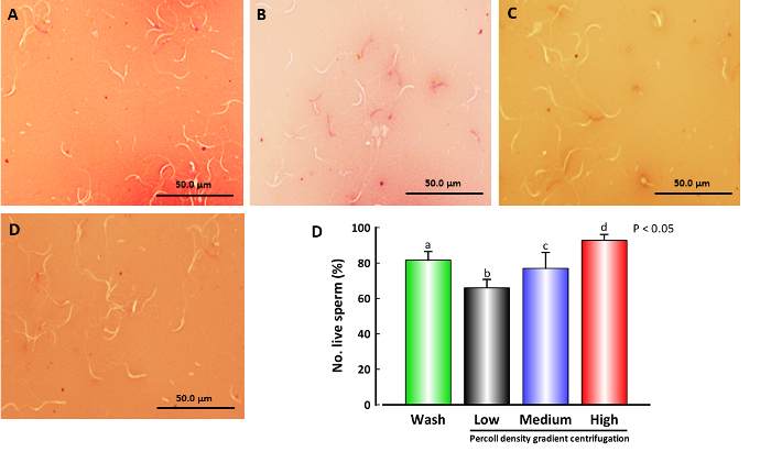
Figure 4: Viability of sperm as assessed by eosin-nigrosin vital staining. Digital micrographs showing eosin-nigrosin vital staining of sperm obtained through a process of traditional wash (A) and PDGC (B, low motile sperm; C, medium motile sperm; D, high motile sperm). Scale bar = 50 µm at 40 x magnification. Pink staining indicates dead sperm which have taken up eosin stain. Data were subjected to one-way ANOVA, followed by the Tukey-Kramer test. Error bars indicate the standard error of the mean (SEM). Values are presented as the mean ± SEM (n = 5). a-d Values without a common superscript differed (P < 0.05). Sperm isolated by PDGC into low-, medium- and high-quality groups (E) reveal significant differences in percentage viability, 66.0 ± 4.7, 77.0 ± 8.9 and 92.8 ± 3.4, respectively. Sperm washed by traditional methods displayed an 81.6 ± 4.9% viability, lower than the high-quality group but higher than the medium- and low-quality groups. Please click here to view a larger version of this figure.
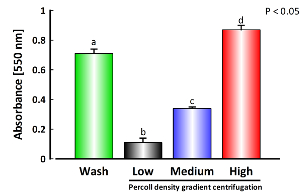
Figure 5: Mobility of sperm as assessed by Accudenz assay. Absorbance values act as a proxy measure for sperm mobility in assessment by the Accudenz assay. Data were subjected to one-way ANOVA, followed by the Tukey-Kramer test. Error bars indicate the standard error of the mean (SEM). Values are presented as the mean ± SEM (n = 5). a-d Values without a common superscript differed (P < 0.05). Sperm isolated into low-, medium- and high-quality groups by the PDGC technique exhibited markedly different low (0.11 ± 0.03), medium (0.34 ± 0.01) and high (0.87 ± 0.03) absorbance values, respectively. Sperm washed by traditional methods exhibited an absorbance of 0.71 ± 0.03, an absorbance lower than that of the high-quality group, but higher than that of the medium- and low-quality groups. Please click here to view a larger version of this figure.
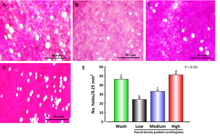
Figure 6: Penetrability of sperm as assessed by sperm-inner perivitelline layer (IPVL) interaction assay. Digital micrographs showing in vitro formation of holes on the surface of the IPVL by chicken sperm (4 x 10⁶ cells/mL) during incubation in motility buffer at 37 °C for 15 min. Micrographs represent IPVL penetrated by sperm having undergone traditional wash (A) and sperm from low- (B), medium- (C) and high- (D) quality groups from PDGC. Scale bar = 50 µm at 40X magnification. Data were subjected to one-way ANOVA, followed by the Tukey-Kramer test. Error bars indicate the standard error of the mean (SEM). Values are presented as the mean ± SEM (n = 5). a-d Values without a common superscript differed (P < 0.05). The number of penetration holes per 0.25 mm2 (E) differed between the sperm isolated from the different gradients of the density media. Sperm isolated into low-, medium- and high-quality groups by PDGC produced a low (24.40 ± 1.1), medium (33.20 ± 1.4) and high (51.20 ± 1.3) number of penetration holes, respectively. The sperm washed using the traditional method produced 46.40 ± 1.8 penetration holes, a value lower than the high-quality group but higher than the medium- and low-quality groups. Please click here to view a larger version of this figure.
