This protocol presents techniques and methodology necessary for the accurate delivery of magnetic nanoparticle hyperthermia using a sophisticated delivery and monitoring system.
Hyperthermia has long been used in the treatment of cancer. Techniques have varied from the intra-tumoral insertion of hot iron rods, to systemically delivered tumor antibody-targeted magnetic nanoparticles, at temperatures from 39 ˚C (fever-level) to 1,000 ˚C (electrocautery) and treatment times from seconds to hours. The temperature-time relationship (thermal dose) dictates the effect with high thermal doses resulting in the tissue ablation and lower thermal doses resulting in sublethal effects such as increased blood flow, accumulation of drugs and immune stimulation. One of the most promising current medical therapies is magnetic nanoparticle hyperthermia (mNPH). This technique involves activating magnetic nanoparticles, that can be delivered systemically or intratumorally, with a non-invasive, non-toxic alternating magnetic field. The size, construct and association of the magnetic nanoparticles and the frequency and field strength of the magnetic field are major heating determinants. We have developed sophisticated instrumentation and techniques for delivering reproducible magnetic nanoparticle hyperthermia in large and small animal models and cultured cells. This approach, using continuous, real time temperature monitoring in multiple locations, allows for the delivery of well-defined thermal doses to the target tissue (tumor) or cells while limiting non-target tissue heating. Precise control and monitoring of temperature, in multiple sites, and use of the industry standard algorithm (cumulative equivalent minutes at 43 ˚C /CEM43), allows for an accurate determination and quantification of thermal dose. Our system, which allows for a wide variety of temperatures, thermal doses, and biological effects, was developed through a combination of commercial acquisitions and inhouse engineering and biology developments. This system has been optimized in a manner that allows for the rapid conversion between ex vivo, in vitro, and in vivo techniques. The goal of this protocol is to demonstrate how to design, develop and implement an effective technique and system for delivering reproducible and accurate magnetic nanoparticle therapy (mNP) hyperthermia.
Hyperthermia has historically been used in cancer therapy, either alone or in combination with other treatments. Although it has a long history of use, the most advantageous method for delivering this treatment is still being debated and is dependent on the disease site and location. Methods for hyperthermia delivery include microwave, radiofrequency, focused ultrasound, laser, and metallic nanoparticles (such as gold or iron oxide)1,2,3,4. These methods of delivery can lead to a range of treatment temperatures from fever-level through to hundreds of degrees C. The biological effect of hyperthermia depends primarily on the temperatures used and the duration of the treatment5. For this manuscript and purpose, we are focusing on magnetic nanoparticle hyperthermia (mNPH). This method allows for focused, localized, well monitored, and controlled temperature changes, using non-toxic, FDA approved, iron oxide nanoparticles.
One pitfall of other hyperthermia modalities is a lack of precise cellular targeting; hyperthermia does not a have an inherently high therapeutic ratio, therefore, careful thermometry and targeting is necessary6. mNPH allows for systemic or intratumoral injection of mNPs, with heat only being generated where the mNPs are located, thus targeting the treatment to the tumor directly. mNPH can be effective when the magnetic nanoparticles are located inside or outside of the cell. For cancer therapy, the general overview of mNPH is that the magnetic nanoparticles are injected (intratumorally or intravenously), then an alternating magnetic field is applied, causing the nanoparticle magnetic poles to constantly realign, leading to a localized heating of the cells and tissue associated with the nanoparticles7,8. By adjusting the volume of nanoparticles and the frequency/strength of the alternating magnetic field (AMF), it is possible to carefully control the temperature generated within the tissue.
This treatment works well in tumors that are near the body surface, as deeper tumors require stronger AMF so the risk of eddy current heating increases9. There is evidence of hyperthermia being used clinically as a monotherapy, however, oftentimes hyperthermia is combined with radiation therapy or chemotherapy, leading to a more targeted anti-cancer effect10,11,12. Clinical evidence of hyperthermia working in combination with radiation therapy is reviewed in a previous publication13. Our lab has successfully treated a variety of animals, from mice to pigs and spontaneous canine cancers, using the mNPH method12,14,15. This protocol is designed for those interested in investigating the effects of localized hyperthermia treatment, either alone or in combination with other therapies.
One of the most important factors in hyperthermia is being able to measure and understand, in real time, the thermal dose being delivered to the target/tumor tissue. A standard way of calculating and comparing dose is through demonstration of the cumulative equivalent minutes of heating at 43 °C; this algorithm allows for the comparison of doses independent of the delivery system, maximum and minimum temperatures (within a specific range) and heat up/cool down parameters5,16. The CEM calculation works best for temperatures between 39-57 °C5. For example, in some of the studies we have performed, we have chosen a thermal dose of CEM43 30 (i.e., 30 min at 43 °C). Choosing this dose allowed us to look at a safe, effective, immunogenetic effects in vitro, both alone, and in combination with a single dose of radiation17.
With magnetic nanoparticle hyperthermia, there are several factors that need to be considered in building an appropriate delivery system. The instrumentation design includes important safety factors, such as the use of a chiller to ensure the magnetic field delivery equipment remains cool even when operated at high power, and fail-safe procedures that prevent the system from being turned on if all temperature, power assessment, and control systems have not been activated. Additionally, there are important biological factors that need to be considered for both in vivo and in vitro situations. When using cultured cells, it is necessary to treat in growth media and maintain at a consistent viable temperature to avoid physiological changes that could affect results. For individual nanoparticle types, it is important to know the specific absorption rate (SAR) when calculating AMF based heating parameters. Similarly, it is important to know the mNP/Fe concentration, in cells and tissues, that is necessary to achieve the desired heating. In vivo methods require even more attention to detail since the animal must be maintained under anesthesia during treatment and the animal’s core body temperature maintained at a normal level throughout the treatment. Allowing for the animal’s body temperature to drop, as happens under anesthesia, can affect the overall results, with respect to the thermal dose of the tissue being treated.
In this manuscript, we discuss the methods used to design and construct a versatile magnetic nanoparticle hyperthermia system, as well as important use factors that need to be considered. The system described allows for the robust, consistent, biologically appropriate, safe, and well-controlled delivery of magnetic nanoparticle hyperthermia. Finally, it should be noted that the mNPH studies we conduct often involve other therapies such as radiation, chemotherapy, and immunotherapy. For these results to be meaningful, it is important to determine how the delivered heat can affect the efficacy and/or safety-toxicity of other modalities (or vice versa) and the well-being of the animal. For this reason and the dosimetry and therapeutic situations previously mentioned, it is essential to pay strict attention to the magnetic nanoparticle hyperthermia dosing accuracy and the continuous core and target temperature measurements. The goal of this protocol is to provide a straightforward, consistent method and description for the delivery of safe and effective magnetic nanoparticle hyperthermia.
The Dartmouth College Animal Care and Use Program is accredited by the American Association for the Accreditation of Laboratory Animal Care (iAAALAC) and adheres to all UDSA and NIH (Office of Laboratory Animal Welfare) guidelines and regulations. All in vivo studies were approved by the Dartmouth College Institutional Animal Care and Use Committee (IACUC). Euthanasia procedure adhere to the 2020 AVMA Guidelines for the Euthanasia of Animals.
1. Instrumentation/design of the system
2. Hyperthermia in vitro
3. Hyperthermia in vivo
In vitro studies
Cells will only achieve and maintain the desired temperature and thermal dose if the amount and concentration of the magnetic nanoparticles/iron and the AMF are appropriately matched. When using magnetic nanoparticles to heat cells in vitro (and in vivo), it should be noted that to achieve hyperthermia in cells with internalized magnetic nanoparticles, a specific level of intracellular mNP/Fe will be necessary, and number and proximity of mNP loaded cells, to each other, will be necessary. If the level of mNP/Fe in the target cells/tissue is sufficient to achieve a heating effect, the magnetic field frequency and strength can be adjusted to achieve the desired temperature and effects. If plated properly, then further studies looking at genetic and molecular differences between different doses and timings can be pursued17. Figure 1 represents a schematic of the in vitro methods.
These in vitro methods can be used to investigate cellular mRNA and protein expression change. A recent example from our lab determined immunogenetic differences following CEM43 30 mNPH treatment, an 8 Gy radiation treatment, and the combination. We were able to identify similarities and differences in expression across immune and cytotoxic pathways to gain a better understanding into the mechanism behind the effects, and how they combine synergistically17. Every experiment utilizes a variety of environmentally and heated control samples. The controls will have different mRNA and protein expression levels as compared to those receiving hyperthermia treatment.
In vivo studies
In in vivo studies there are additional considerations. Regardless of the target thermal dose it is absolutely essential to maintain a physiologically acceptable core temperature in the animal being treated. This can be challenging with rodents under anesthesia as core temperature can be quickly lost (core temperature modulating techniques such as heating pads are often necessary). Lower than normal body temperatures can necessitate the need to push the AMF-mNPH too far, when trying to achieve a specific thermal dose in the tumor, resulting in unacceptable effects in the non-target tissue (non-target tissue eddy current heating is one such possibility). Even minor deviations in core body temperature can lead to undesirable physiological complications in the tumor or normal tissue. As mentioned previously, however worth repeating, for accurate, reproducible heating, it is essential to achieve a match between the mNP/Fe tissue concentration, AMF frequency, and field strength temperature monitoring parameters and target tissue size and depth. There must be a baseline concentration of mNPs within the tumor to allow for measurable heating. The level/ability of heat depends on not only mNP tissue concentration (mg Fe/g tissue) and their relative distribution within the tumor, but also the frequency of the AMF and subsequent field strength. Changes in any of the above can lead to different ranges of attainable temperatures within the tissue. Through many years of experience, we have optimized the concentration we use for preclinical tumor treatments and the frequency and field strength of the AMF system to allow for safe and effective activation. Because it is impossible to measure the temperature/thermal dose in all tissue sites, it is also essential to place as many fiber optical temperature probes as possible in strategic sites that allow for real-time efficacy and safety assessment, as seen in Figure 2. These probes allow for the recording of temperatures throughout the experiment, allowing for accurate dosimetry and thermal history of the experiment. Figure 3 demonstrates curves generated during an in vivo experiment, highlighting the capability to closely monitor temperature and adjust the system to maintain tumor temperatures within the desired range. Figure 4 summarizes the in vivo methods.
These in vivo methods, similar to the in vitro methods, can be used to investigate different cancer type, different hyperthermia doses, and with various combination treatments. For example, previous studies in our laboratory have investigated the combination of hyperthermia and chemotherapy12. We have also completed numerous hyperthermia and radiation experiments for the determination of efficacy and molecular mechanisms. The control mice for these experiments undergo all procedures except for the actual generation of hyperthermia. Figure 5 contains two volcano plots that demonstrate differentially expressed genes following in vitro and in vivo mNP hyperthermia treatment(mNPH). These figures are examples of how we use molecular techniques to monitor the hyperthermia effects.
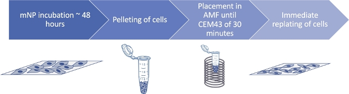
Figure 1: In vitro mNP hyperthermia schematic. This schematic demonstrates the method for in vitro magnetic nanoparticle hyperthermia. To ensure heating occurs, cells must be provided enough particles and time for adequate mNP uptake. Please click here to view a larger version of this figure.
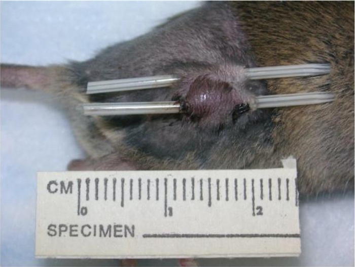
Figure 2: Placement of catheters for temperature monitoring. This figure demonstrates the placement of catheters that house the fiber optic temperature probes to record temperatures at different locations in the tumor and/or the tumor region. This figure is adapted from ref.19. Please click here to view a larger version of this figure.
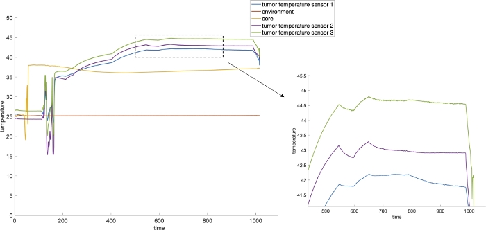
Figure 3: Real time temperature monitoring during treatment of a mouse tumor. This graph demonstrates the real time temperature readings that allow for monitoring the core body temperature, the environmental temperatures, and multiple temperatures within the tumor, during an in vivo experiment. The control of temperatures within the tumor are demonstrated through the minimal large-scale variations on the zoomed in portion of the figure. Please click here to view a larger version of this figure.
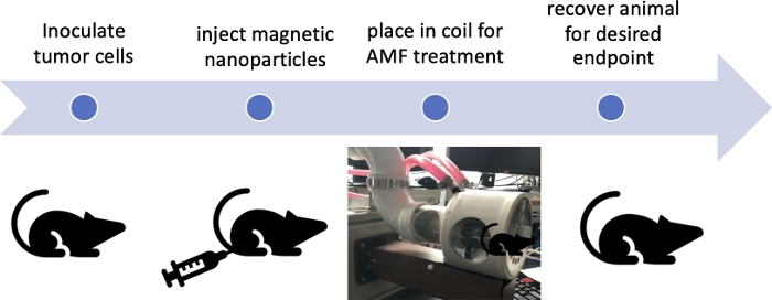
Figure 4: In vivo mNP hyperthermia schematic. This schematic demonstrates the method for in vivo magnetic nanoparticle hyperthermia. Injection of sufficient nanoparticles as well as enough time for distribution and absorption, ensures the ability to deliver the desired thermal dose. Please click here to view a larger version of this figure.
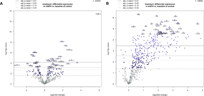
Figure 5: Differential gene expression. Differential gene expression following in vitro (A) and in vivo (B) mNP hyperthermia treatment. These volcano plots represent genetic changes on a log 2 x-axis, with significance on the y-axis, for both in vitro and in vivo mNPH methods. Each circle represents a different gene, with the 20 most significant differentially expressed genes labeled. The further the gene is from zero on the x-axis, the greater the fold change, and the higher the gene is on the y-axis, the lower the p-value. Although both had the same thermal dose, in vivo hyperthermia led to greater gene expression changes than in vitro. These plots are examples of the biological data that can be generated using the protocol described. The in vitro volcano plot has been adapted from ref.17. Please click here to view a larger version of this figure.
Supplementary File 1. Please click here to download this file.
The design and implementation of this system provides the ability to conduct accurate and reproducible in vitro and in vivo magnetic nanoparticle hyperthermia experiments. It is critical that the system is designed such that the AMF frequency and field strength are adequately matched to the magnetic nanoparticle type, concentration, and the tissue location and temperature desired. Additionally, the accurate monitoring of the temperature in real time is crucial for safety and the calculation of an accurate thermal dose (cumulative equivalent minutes at 43 °C/ CEM). The placement of probes as demonstrated in Figure 1, allows for the real time monitoring of thermal dose and core body temperature as seen in Figure 2.
The first step in accurate delivery of magnetic nanoparticle hyperthermia is building a safe system for animals and operators. All components of the system should also be well understood from an operational and delivery standpoint. In this situation, that means understanding the potential for AMF eddy currents and knowing where magnetic particles are located. The antennas, or coils, are a key factor in the shape and strength of the field, and the cooling system used is important to prevent coil overheating20. The field strength outside of the conductor is directly proportional to the current strength flowing through the conductor. The magnetic field strength at any point in the space surrounding the conductor is the vector sum of the fields produced by the conductors in the surrounding area. The magnetic field is produced at a right angle to the current flow and the strength decreases exponentially, as a function of the distance from the conductor, as per the Biot-Savart inverse square rule21. Thus, square tubing is used for in vivo hyperthermia for a more uniform field within the coil. Creating a magnetic field with the strength and volume needed for a potentially clinically relevant system, requires a high electrical current. Therefore, antenna designs must be able to accommodate significant electrical power levels. Also, AMF antennas must be designed so their inductance falls within the acceptable range of the power generator. At the frequencies typically used, most of the current flow is on the surface of the antenna conductor, meaning the surface affects the resistive heating which can be minimized by eliminating surface defects. This resistive heating also means that a coil cooling system is needed to ensure the coil and environment does not overheat.
A limitation of our system design is that it does not allow for a total range of frequencies and magnetic fields, but it does allow for fields to be generated that are appropriate for cells, rodents and large animals. Specifically, the maximum field strength available from any induction heating system is directly related to the current flow in the antenna (coil). AMF generators are rated in kilowatts, which are calculated by multiplying the available voltage by the available current (amperes). So, a 10kW system with a 500 V limit would have a maximum amperage of 20 A. The coils design will determine which limit is reached first, and thus the systems limit. The magnetic field strength created by any current decreases exponentially as a function of the distance from the conductor. Therefore, a larger diameter coil with the same geometry as a smaller diameter coil, run on the same system, would have a lower field strength at the center of the coil. Thus, the required magnetic field size and strength are limited by the capacity of the AMF generator. Building a larger coil and using more power leads to additional concerns, primarily eddy current heating.
There are several safety concerns that must be addressed when using this system to protect users, animals, and the system itself. First, adequate room ventilation must be maintained during the use of anesthesia. Second, all areas associated with the coil must be clear of metal and or conductors including high saline mixtures. Users must remove rings and other jewelry when working around the AMF, and samples should not contain any type of metal. Of most importance, people with pacemakers or other implanted devices or objects should consult with their doctor before working around the AMF. To protect the system, a fail-safe system should be used that ensures the generator and coil cooling needs are met before power is applied. Additionally, a thermal camera overview should be used to detect unintended heating.
For in vitro studies, the most important steps to follow are the concentration of iron in cells, the concentration of cells, AMF parameters, and thermal dose assessment. Cells can be treated/heated with magnetic nanoparticle hyperthermia by placing the magnetic nanoparticles in the supernatant, cells, or both. The amount of magnetic nanoparticle heating will depend on the level of magnetic nanoparticles/Fe. If the desire is to treat only cells with internalized iron, our experience is that individual cancer cells will only uptake a limited number of magnetic nanoparticles and that even when the uptake is optimal the cells must be aggregated/pelleted to create cell heating situation, even with optimized AMF. Maintaining the temperature of the media and cells at biologically relevant levels (when not being heated) is also important for accurate measurement of true heating. The 14-turn solenoid coil described here allows for biologically relevant temperatures to be maintained by submerging the samples in a thermally controlled water column.
For the in vivo studies, maintaining the animal core temperature and accurately measuring the temperature within the tumor are key factors. This animal containment system and design of the coil eliminates thermal drift in the animal’s environment due to coil/power settings and helps to maintain normal core body temperature. Maintaining the body core temperature is critical for meaningful experiment results. The rectal probe allows for real time monitoring of the animal’s core temperature. When under anesthesia, an animal’s core temperature inherently decreases. To address this situation, we developed an environmental heating system that delivers warm air around the animal containment vessel, allowing the core temperature to stay in the normal range. Maintaining normal core temperature is essential for ensuring accurate interpretation of hyperthermia treatment results, and the elimination of environmental factors. The placement of the temperature monitoring probes in multiple sites in the target tissue/tumor is important to get an accurate assessment of the temperature and thermal dose achieved. Because it is extremely difficult if not impossible to distribute magnetic nanoparticles homogeneously within a tumor, knowing the heating parameters in multiple sites is essential at achieving a consistent and accurate tissue/tumor thermal dose. It is important to note that the concentration for in vitro and in vivo studies is variable. This variation is because there are fewer boundaries in cell culture with cells having more access to the mNPs, so a lower concentration can be used. In vivo, a higher concentration is necessary due to the heterogenous nature of tumors and the complicated 3D morphology. Therefore, using the same concentration of particles in vivo and in vitro would lead to far fewer being taken up by cells.
This manuscript describes the parameters and instrumentation necessary to develop an effective and flexible alternating magnetic field generator and coil system for magnetic nanoparticle hyperthermia treatments. This system can be used for both in vitro and in vivo studies. The system is effective for localized/targeted hyperthermia and the sparing of normal tissue making it appealing, compared to other AMF-mNP hyperthermia systems. These hyperthermia treatments can be altered to investigate the effects of different doses, with a variety of nanoparticles or nanocarriers and adjunct treatments. Since tissue heating, especially magnetic nanoparticle heating, can be affected by so many variables, it is essential to understand the parameters in an investigation. If these criteria are met magnetic nanoparticle hyperthermia can address many molecular, cellular, and clinical situations, including independent and adjuvant tumor control. Although the methods described here require significant effort, if the guidelines are followed, the full potential of mNP hyperthermia can be realized.
The authors have nothing to disclose.
The study was funded by grant numbers: NCI P30 CA023108 and NCI U54 CA151662.
.25% Trypsin | Corning | 45000-664 | available from many companies |
1.5 mL tubes | Eppendorf | Eppendorf 22363204 | available from many companies |
B16F10 murine melanoma cells | American Type Culture Collection | CRL-6475 | |
C57/Bl6 mice | Charles river | 027C57BL/6 | 6-week-old female mice |
Chiller | Thermal Care | NQ 5 series | chiller that cools the coil |
Coolant fluid | Dow Chemical Company | Dowtherm SR-1 | antenna cooling fluid |
Fetal Bovine serum | Hyclone | SH30071 | available from many companies |
fiber optic probes, software and chassis | FISO | FISO evolution software used to read the temperatures | |
IR camera | Flir | infrared camera to monitor unintentional heating | |
iron oxide nanoparticles | micromod Partikeltechnologie GmbH | Bionized NanoFerrite | dextran coated iron oxide nanoparticles |
mouse coil, solenoid | Fluxtrol | custom built | |
penicillin/streptomycin | Corning | 45000-652 | available from many companies |
RF generator | Huttinger | TIG 10/300 | power source |
RPMI media | Corning | 45000-396 | available from many companies |