机械性敏感基于细胞的测定允许调查粘附细胞,具有广泛的,反映了力学可以在细胞生物学发挥核心作用的应用程序。这些应用程序通常专注于推动亚细胞过程或全细胞行为的基本机制。一方面,外部环境因素,如细 胞外基质组合物或基质硬度可显着地影响细胞的机械和生物反应。1同样可以使用许多类的药物活性化合物的后观察到,其中,效果是常其特征在于使用细胞培养模型2另一方面基因型性能,例如那些引起自发或实验诱导的基因突变,可诱导了与细胞骨架结构和功能的改变相关的细胞表型的显着的变化。3这些实施例短短数的许多可能的题目为哪些细胞机械表型是相关的,并且所有的这些都被有效地研究了micropost阵列。
在写这篇文章的时候,大约有200多篇已经发表了描述细胞micropost互动。这些作品讨论micropost偏转原理的理论问题,以及在其制造的实用说明。第一篇文章中描述的细胞和灵活micropost阵列的相互作用是出 版的谈及其同事在2003年4相反经典牵引力显微镜(TFM),其中连续的软衬底被用于估计nanonewton规模细胞收缩,Tan等人。描述使用由有机硅弹性体的多个紧密隔开的垂直梁的方法。该技术的主要优势出现两个主要的功能。首先,为了改变小区表观基板刚度1只需要改变micropost尺寸,同时保持基板组合物,否则恒定,从而避免了在表面拓扑结构和化学的差异。第二microposts像可以与力和空间分辨率个别粘着斑的数量级上离散地分析,并且可以减少所固有的由标准的TFM类似分析的分析挑战个人弹簧。
今天的micropost阵列的应用范围大大超过了力量的只是少数单个细胞的映射。例如,秋山报告使用的分离的背脉管组织从一个蛾毛虫作为致动器的一个micropost阵列,为了开发一种昆虫肌肉供电自主微型机器人。5
然而,microposts大部分发布的应用程序都集中在医疗条件,比如感染或癌症的研究。例如,micropost阵列已经用于研究力产生的奈瑟gonorrh捆绑IV型菌毛是与信号级联增强感染有关。6其他已经使用microposts研究并靶向细胞骨架的药物化合物治疗乳腺癌细胞OEA菌落。7
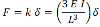
一个micropost的偏转采用经典梁理论常常被描述为与最终负载假设电池的悬臂只重视对micropost的最顶端。这里所施加的力 F引起的偏转δ取决于micropost的“抗弯刚性”k和由下式计算:
 (1)
(1)
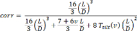
与E,I,和L是杨氏模量,转动惯量和梁的长度分别面积矩。然而,从这个公式的结果只能给力的,因为梁剪切加工和弯曲以及一般近似为底物变形者不被交流计数。考虑到microposts通常由从软材料如聚二甲基硅氧烷(PDMS)为基础的硅橡胶需要这些因素包括在内。 。舒恩等人证实,有这样的基础上micropost(L / D)和相应的聚合物的泊松比 V的纵横比的校正因子8它由下式给出。:
 (2)
(2)
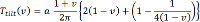
以 T 倾斜 (ⅴ)作为一个倾斜系数,其包括嵌合参数a = 1.3作为可以在同一篇文章中找到:
 (3)
(3)
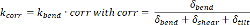
这意味着一个micropost的校正刚度ķ 压改正是纯弯曲刚度k = k 弯曲的产物和校正因子压改正计算公式如下:
 (4)
(4)
因此,细胞力的计算应采用公式(1)的更精致的变化,现在读来进行:
 (5)
(5)
修正的影响,只要是用于micropost尺寸典型值变得更为明显。例如,一个15微米长micropost具有圆形横截面,并且直径为5μm制成的基于PDMS的硅橡胶导致0.77的校正因子,因此,未校正的计算将23%高估施加的细胞的力。这变得更加严重为microposts具有较小的纵横比。
传统上,micropost图像分析也基于理想化的梁弯曲理论。在2005年率先使用MICR的组OPOST阵列出版适合micropost分析的图像分析软件9所述的软件需要软件许可证及用户必须采取三种图像的每个位置。分别来自micropost的顶部和底部的平面中的传输模式,另一种在荧光模式与染色的细胞。比较所述顶部和底部的位置为每个后micropost的软件确定的力矢量场,并计算相关的参数,如每交力。其它软件包存在,他们的分析原则在描述它们的相应条款简要地提到,但这些分析软件包通常是不公开的。10,11
设计用于映射细胞力的micropost阵列可分类为在正交micropost布局或六边形的,后者具有的所有邻居microposts之间等距的间隙的优点是。典型microposts公顷已经圆形截面和它们的尺寸范围从1.0微米至10微米的直径和2到50微米,长度4然而,microposts与椭圆形或正方形的横截面也有报道。12,13
使用基于PDMS的硅氧烷混合物作为micropost材料允许用于将纳米颗粒引入该混合物中。例如添加钴纳米棒使micropost的磁性活化,从而提供了潜在的实验设计的另一个自由度。14大部分组上产生平的刚性基板像盖玻璃或培养皿内其micropost阵列。然而,Mann和同事最近报道上形成可拉伸膜一个micropost阵列15这允许在研究活细胞的亚细胞动态响应在细胞收缩的术语细胞拉伸力到贴壁细胞的应用。
目前广泛使用的,最ESTAB用于制造micropost阵列lished过程中Sniadecki和同事。16-18的见地协议总之标准洁净室处理中描述被用于产生关于使用SU8光致抗蚀剂的硅晶片的顶部的微结构是基于软光刻。这之后是复制方法,其中在硅橡胶被浇铸在将它们转移到模具中的结构。在第二步骤中这些模具被用于复制使用的硅橡胶上的选择的衬底顶部的初始显微结构。然而,尽管大,越来越多的与他们申请公开,建立一个制造过程microposts的花费相当长的时间,即使对微工程专家;存在需要优化和适应特定实验室环境和micropost布局,得到可接受的质量水平的许多工艺步骤。
商业micropost阵列现已就绪-T邻的使用(“现成的架子”)格式具有一贯的高品质。因此,它们是替代所需的现场制作的复杂和冗长的制造工艺。本文的市售micropost阵列用于使用单明场显微术图像映射蜂窝力。更重要的是本文介绍和文件名为MechProfiler一个全功能的开源软件,可以下载作为补充材料,这份手稿。该软件的一个比较活跃的版本也可以在http://www.orthobiomech.ethz.ch找到。
一个“关闭的,现成的”检测和兼容的开源分析软件相结合,显着降低了进入障碍,以实现精确的TFM实验。研究人员无法获得任何无尘室设备或软件开发专业知识成功地分析细胞的力量。它使用户能够集中于mechanosensitivity分析输出,而不是技术本身,使之能够为更广泛的社区牵引力测量。此外,这是对铺平阵列micropost全自动筛选的方式迈出的重要一步。
该MechProfiler分析软件处理的文件格式TIFF,PNG,BMP和JPG图片。这些图像可以用荧光,相衬或明视场光学显微镜拍摄。该独立程序与游离Matlab的编译器运行时(见: 图12)一起运行,并底层算法允许简化图像处理,从而使用户能够处理具有单个或多个细胞的图像中约1分钟。此外,这些细胞既可以活的或“固定”。
该MechProfiler软件能够通过靠质量商业micropost阵列的再现性,大大提高了数据吞吐量分析,更具体地,默认“非偏转“的阵列中的每个交位置可以推定针对理想格(制造偏差为网格在用于该研究的阵列均小于100纳米)。
在短的一个打开选择的用于分析的图像文件,它们作物到感兴趣的区域中,定义覆盖细胞的职位或需要被丢弃,确定后的位置,计算该挠度/对理想格力,最后节省出口有可能所有的细胞特异性的数据,其中包括一个标准的办公电子表格。
