Here, we describe an easy-to-use methodology to generate 3D self-assembled cardiac microtissue arrays composed of pre-differentiated human-induced pluripotent stem cell-derived cardiomyocytes, cardiac fibroblasts, and endothelial cells. This user-friendly and low cell requiring technique to generate cardiac microtissues can be implemented for disease modeling and early stages of drug development.
Generation of human cardiomyocytes (CMs), cardiac fibroblasts (CFs), and endothelial cells (ECs) from induced pluripotent stem cells (iPSCs) has provided a unique opportunity to study the complex interplay among different cardiovascular cell types that drives tissue development and disease. In the area of cardiac tissue models, several sophisticated three-dimensional (3D) approaches use induced pluripotent stem cell-derived cardiomyocytes (iPSC-CMs) to mimic physiological relevance and native tissue environment with a combination of extracellular matrices and crosslinkers. However, these systems are complex to fabricate without microfabrication expertise and require several weeks to self-assemble. Most importantly, many of these systems lack vascular cells and cardiac fibroblasts that make up over 60% of the nonmyocytes in the human heart. Here we describe the derivation of all three cardiac cell types from iPSCs to fabricate cardiac microtissues. This facile replica molding technique allows cardiac microtissue culture in standard multi-well cell culture plates for several weeks. The platform allows user-defined control over microtissue sizes based on initial seeding density and requires less than 3 days for self-assembly to achieve observable cardiac microtissue contractions. Furthermore, the cardiac microtissues can be easily digested while maintaining high cell viability for single-cell interrogation with the use of flow cytometry and single-cell RNA sequencing (scRNA-seq). We envision that this in vitro model of cardiac microtissues will help accelerate validation studies in drug discovery and disease modeling.
Drug discovery and disease modeling in the field of cardiovascular research face several challenges due to a lack of clinically relevant samples and inadequate translational tools1. Highly complex pre-clinical models or oversimplified in vitro single-cell models do not exhibit pathophysiological conditions in a reproducible manner. Therefore, several miniaturized tissue-engineered platforms have evolved to help bridge the gap, with the goal of achieving a balance between ease of application in a high-throughput manner and faithful recapitulation of tissue function2,3. With the advent of induced pluripotent stem cell (iPSC) technology, tissue engineering tools can be applied to patient-specific cells with or without underlying cardiovascular disease state to answer research questions4,5,6. Such tissue engineered models with cellular composition similar to the heart tissue could be utilized in drug development efforts to test for cardiotoxicity and dysfunction induced by pathological changes in behavior of one or multiple cell types.
Self-assembled microtissues or organoids derived from human iPSCs are three-dimensional (3D) structures that are miniature tissue-like assemblies exhibiting functional similarities to their in vivo counterparts. There are several different approaches that allow formation of organoids in situ via directed differentiation of iPSCs or through the formation of embryoid bodies4. The resulting organoids are an indispensable tool to study morphogenetic processes that drive organogenesis. However, the presence of a variety of cell populations and differences in self-organization can lead to variability in outcomes between different organoids5. Alternatively, pre-differentiated cells that are self-assembled into microtissues with tissue-specific cell types to study local cell-cell interactions are excellent models, where it is feasible to isolate the self-assembled components. Particularly in human cardiac research, development of 3D cardiac microtissues with multicellular components has proven to be challenging when cells are derived from different patient lines or commercial sources.
To improve our mechanistic understanding of cell behaviors in a physiologically relevant, personalized, in vitro model, ideally all component cell types should be derived from the same patient line. In the context of a human heart, a truly representative cardiac in vitro model would capture the crosstalk among predominant cell types, namely, cardiomyocytes (CMs), endothelial cells (ECs), and cardiac fibroblasts (CFs)6,7. The faithful recapitulation of a myocardium not only requires biophysical stretch and electrophysiological stimulation, but also cell-cell signaling that arise from supporting cell types such as ECs and CFs8. CFs are involved in the synthesis of extracellular matrix and maintaining tissue structure; and in a pathological state, CFs can induce fibrosis and alter electrical conduction in the CMs9. Similarly, ECs can regulate contractile properties of CMs through paracrine signaling and supplying vital metabolic demands10. Hence, there is a need for human cardiac microtissues composed of all three major cell types to allow physiologically relevant high-throughput experiments to be conducted.
Here, we describe a bottom-up approach in fabrication of cardiac microtissues by derivation of human iPSC-derived cardiomyocytes (iPSC-CMs), iPSC-derived endothelial cells (iPSC-ECs), and iPSC-derived cardiac fibroblasts (iPSC-CFs) and their 3D culture in uniform cardiac microtissue arrays. This facile method of generating spontaneously beating cardiac microtissues can be utilized for disease modeling and rapid testing of drugs for functional and mechanistic understanding of heart physiology. Furthermore, such multicellular cardiac microtissue platforms could be exploited with genome editing techniques to emulate cardiac disease progression over time under chronic or acute culture conditions.
1. Medium, reagent, culture plate preparation
2. Cardiac differentiation and purification
NOTE: All iPSCs should be maintained at ~75% to 80% confluency prior to cardiomyocyte differentiation. iPSCs used for this protocol were derived from peripheral blood mononuclear cells (PBMCs) using Sendai virus reprogramming performed at the Stanford Cardiovascular Institute (SCVI) Biobank.
3. Endothelial cell differentiation and MACS
4. Cardiac fibroblast differentiation
5. Casting of cardiac microtissue molds and cell seeding
6. Fixation and permeabilization of cells and cardiac microtissues for immunostaining
7. Digestion of cardiac microtissues and preparation of cells for flow cytometry
8. Performing contraction analyses of spontaneously beating cardiac microtissues
Immunostaining and flow cytometry characterization of iPSC-derived CMs, ECs, and CFs
To generate cardiac microtissues composed of iPSC-CMs, iPSC-ECs, and iPSC-CFs, all three cell types are differentiated and characterized individually. In vitro differentiation of iPSCs to iPSC-CMs has improved over the past several years. However, the yield and purity of iPSC-CMs differ from line to line. The current protocol yields over 75% pure iPSC-CMs that spontaneously start beating around day 9 (Figure 1A). Further purification steps from day 9 to day 14 can improve iPSC-CM purity to over 80% as previously described12. Similarly, high-purity iPSC-ECs can be generated using previously published protocols13,14 that include the addition of several vascular growth factors that polarize endothelial progenitors arising from the mesoderm around days 4-5 (Figure 1B) to form phenotypically well-defined iPSC-ECs. iPSC-CFs are a highly heterogenous population based on their location and are characterized based on their morphology and expression of extracellular matrix proteins. Here, using published protocols15,16,17 with modifications, human iPSC-CFs are obtained from cardiac mesoderm progenitor cells (Figure 1C).
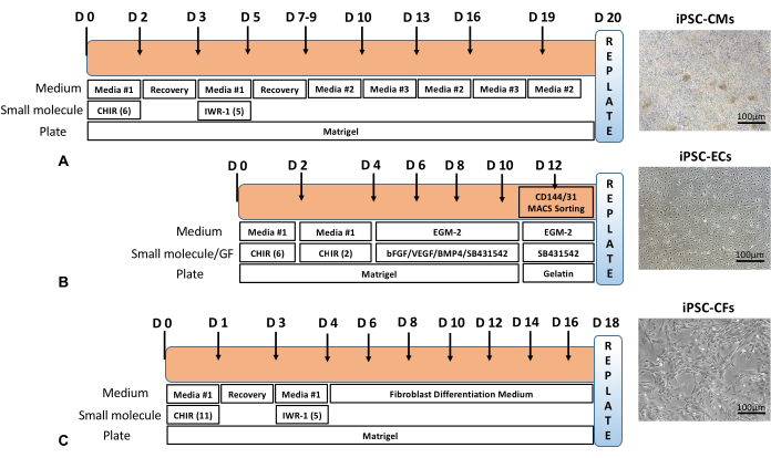
Figure 1: Differentiation timeline. Overall schematic of (A) iPSC-CM, (B) iPSC-EC, and (C) iPSC-CF differentiation timeline with representative phase contrast images of cells after differentiation and purification steps. Scale bar = 100 µm. Please click here to view a larger version of this figure.
The purity of iPSC-CMs, iPSC-ECs, and iPSC-CFs were determined by immunostaining and flow cytometry using cTnT2, platelet endothelial cell adhesion molecule (PECAM1/CD31) and vimentin (VIM), respectively (Figure 2A). Quantitative analysis showed a purified population containing over 90% cTnT2 cells at day 20. iPSC-ECs obtained at day 12 after MACS using CD31 beads were identified with immunostaining against PECAM1/CD31 endothelial cell surface marker. MACS yielded highly pure endothelial cells as evidenced by over 95% CD31+ cells at P0. However, it must be noted that the purity of endothelial cells decreased with higher passage numbers due to de-differentiation. Similarly, at day 20, flow cytometry analyses revealed that over 95% iPSC-CFs expressed the fibroblast marker VIM (Figure 2B).
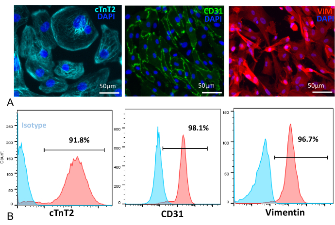
Figure 2: Immunofluorescence and flow cytometry characterization. (A) [Left to right panel] iPSC-CMs at day 25 stained with cTnT2 (cyan), iPSC-ECs stained with PECAM1/CD31 (green), iPSC-CFs stained with VIM (red), and nuclei stained with DAPI (blue). Scale bar = 50 µm. (B) [Left to right panel] Flow cytometry quantification showed a high percent purity of iPSC-CMs (91.8%), iPSC-ECs (98.1%), and iPSC-CFs (96.7%) following differentiation. Please click here to view a larger version of this figure.
Fabrication of cardiac microtissue cultures and size analyses
Single cell suspensions of iPSC-CMs, iPSC-ECs, and iPSC-CFs were mixed in 7:2:1 ratio and carefully dispensed into the cell seeding chamber of the sterilized agarose replica mold (Figure 3A,B). The cells uniformly settled inside the circular recesses in 2 h. Around day 3, the self-assembled cells organize into uniform sized spontaneously beating cardiac microtissues (Figure 3C). Arrays of different sized microtissues can be fabricated by tuning the final cell density (Figure 3D,E). Cardiac microtissues fabricated with a final cell density of 1 x 10⁶ cells/mL is ~300-350 µm in diameter, 2 x 10⁶ cells/mL is ~600 µm in diameter, and 4 x 10⁶ cells/mL is over 800 µm in diameter. Microtissue assembly was obtained with a cell density of 1 x 10⁶ cells/mL, which is typically used for experiments. These microtissue cultures can be maintained in culture for up to 6 weeks.
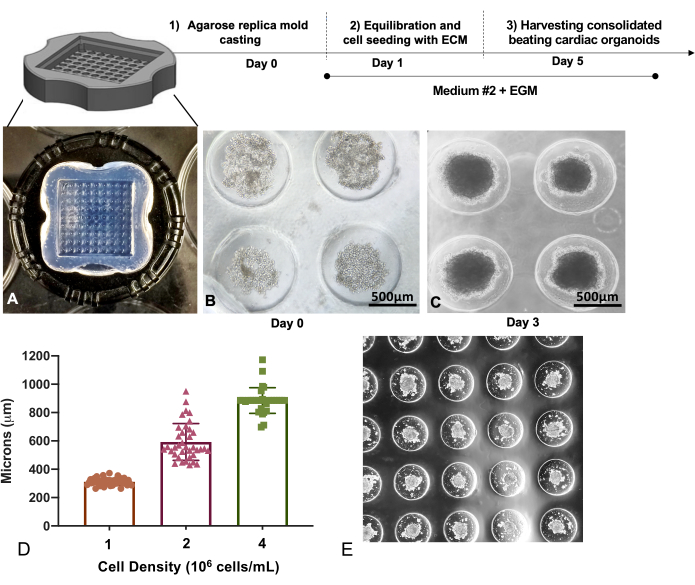
Figure 3: Replica molding technique to generate multicellular cardiac microtissues. (A) Replica molded agarose microwell trays of (B) iPSC-CM, iPSC-EC, and iPSC-CF mixtures captured inside the microwells for self-assembly. Scale bar 500 = µm. (C) Micrograph showing compaction of cardiac microtissues on day 3. Scale bar = 500 µm. (D) Cardiac microtissue sizes formed show a linear relationship with initial seeding densities, with higher cell densities resulting in larger microtissues. (E) A representative figure showing the self-assembled cardiac microtissues in the microwell array. Please click here to view a larger version of this figure.
Immunostaining and viability after enzymatic digestion
Immunofluorescence staining of day 12 post-fabrication using antibodies against cTnT2 for iPSC-CMs, CD31 for iPSC-ECs, and DDR2 for iPSC-CFs revealed a unique cell distribution in cardiac microtissues. iPSC-CMs, the heaviest of all three cell types, occupied the center, whereas iPSC-ECs were interspersed throughout the microtissues, and iPSC-CFs were observed to predominantly occupy the periphery (Figure 4A). Short and rapid digestion of the microtissues achieved using Dispase I and Liberase TL resulted in overall highly viable cell proportion (Figure 4B, top panel) with less than 5% apoptotic cells after 2 weeks in culture. This was followed by a brief 1 h exposure of cardiac microtissues to a high concentration (5 µM) of Doxorubicin, a chemotherapeutic drug that is known to induce dose-dependent cardiotoxicity (Figure 4B, bottom panel).
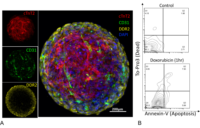
Figure 4: Immunostaining of cardiac microtissues and assessment of cell viability. (A) Confocal z-stack images of human cardiac microtissue stained for iPSC-CMs (cTnT2), iPSC-ECs (CD31), iPSC-CFs (DDR2), and nuclei stained with (DAPI). Scale bar = 200 µm. (B) Flow cytometry plots of cardiac microtissues enzymatically digested into single cell suspension and stained with Annexin V (apoptotic marker) and dead cell exclusion dye (To-Pro3). Digested single cell suspension showed a high cell viability (91%) with <5% apoptotic cell population (top panel), compared to single cell suspension treated with an apoptosis-inducing chemotherapeutic drug, Doxorubicin, for 1 h at 5 µM concentration (bottom panel). Please click here to view a larger version of this figure.
Computational contractility analysis
Contractility analyses of individual cardiac microtissues can be performed with the help of a MATLAB-based image analyses tool. Video recordings of spontaneously beating cardiac microtissues were obtained at 30 fps for analysis. As described previously11, the block-matching method employs motion tracking algorithm to capture movement of a block of pixels for the total frames acquired as a time series of motion vectors. The contractility of the microtissues and movement of the vectors generate a pseudo heatmap that illustrates mean or average contraction profile across the microtissue (Figure 5A). Contractile motion of the cardiac microtissues generates positive peaks that are measured as contraction velocity (blue circle), relaxation velocity (red triangle), and beat rate, the last of which is calculated as the time between two contraction cycles (Figure 5B). Furthermore, the contractility of the cardiac microtissues do not change significantly over 4 weeks in culture (Figure 5C).
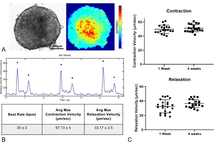
Figure 5: Contraction analysis of cardiac microtissues. (A) Phase contrast image and contraction map of cardiac microtissues 1 week after fabrication. Scale bar = 200 µm. (B) Cardiac microtissues show regular contraction and relaxation profiles and beat rates. Table shows representative values of beat rate, maximum contraction, and relaxation velocities. (C) Long-term culture of cardiac microtissues for up to 4 weeks does not significantly influence contractility parameters (n = 20/group). Please click here to view a larger version of this figure.
To generate cardiac microtissues from pre-differentiated iPSC-CMs, iPSC-ECs, and iPSC-CFs, it is essential to obtain a highly pure culture for better control of cell numbers after contact-inhibited cell compaction within the cardiac microtissues. Recently, Giacomelli et. al.18 have demonstrated the fabrication of cardiac microtissues using iPSC-CMs, iPSC-ECs, and iPSC-CFs. Cardiac microtissues generated using the described method consist of ~5,000 cells (70% iPSC-CMs, 15% iPSC-ECs, and 15% iPSC-CFs). In this method, both cardiomyocytes and endothelial cells were co-differentiated followed by separation of endothelial progenitors using CD34+ marker. The current protocol described here consists of 12,000 cells (70% iPSC-CMs, 20% iPSC-ECs, and 10% iPSC-CFs) per microtissue, which yields higher cell numbers per construct for downstream single cell analyses. Furthermore, since all three cell types are differentiated separately there is limited heterogeneity in cell populations that is unique to understanding cell-specific responses to treatments.
For cardiomyocyte differentiation, we and others have previously demonstrated the derivation of pure cardiomyocytes using chemically defined culture conditions12,19,20. Briefly, differentiation begins with mesendoderm induction, followed by modulation of Wnt/β-catenin signaling that promotes cardiac lineage specification. Further purification of the cardiomyocytes in a glucose-deprived medium allows the elimination of non-cardiomyocytes. Here, it is important to note that prolonged culture in purification medium can hamper the quality of cardiomyocytes. A recently published protocol shows the proliferative capacity of early-stage cardiomyocytes can be harnessed to obtain large number of cells. The expansion of human iPSC-CMs is induced with reintroduction of CHIR, a potent mitogen during the early proliferative phase21. Although the precise molecular mechanism is still unknown, this technique can significantly improve iPSC-CM yield by ten-fold or hundred-fold. Furthermore, to improve the fidelity of iPSC-CMs for disease modeling, they can be cultured in a maturation medium to enhance electrophysiological, mechanical, and structural maturation22. A thorough overview of iPSC-CM maturation techniques is reviewed elsewhere23.
Vascular endothelial cells have been generated from pluripotent stem cells with different purification variable efficiencies13,24,25. The current protocol provides high differentiation efficiency and selection of phenotypically stable iPSC-ECs with MACS26. The differentiation methodology to generate functional cardiac fibroblasts follows modulation of the Wnt pathway and fibroblast growth factor (FGF) signaling to generate iPSC-CFs17. In vivo CFs originate from epicardium, endocardium, and neural crest progenitors16,27,28,29. Here, the CF lineage is generated from committed mesodermal cardiac progenitors without generating epicardial cells as an intermediate. Overall, the protocols described here to generate iPSC-CMs, iPSC-ECs, and iPSC-CFs take into account the ease of reproducibility and high purity based on phenotypic characteristics. To obtain high quality microtissues, it is important to obtain >90% purity for each individual cell type. Higher purity in the cellular phenotype will also ensure detection of changes in cellular trajectory due to different treatments.
After successful derivation of all three cell types, the cells are carefully dispensed into the agarose seeding chamber to allow settling of the cells in the circular recesses. Critical parameters include achieving homogenous single cell suspension and preventing cell aggregates or clumps that may cause variability in size distribution of the cardiac microtissues. From an overall structure perspective, 3D constructs are often limited by diffusion of nutrients, and as the cell density increases, the size and thickness of the constructs also increase linearly. Tissue engineered constructs in the form of spherical structures have a uniform nutrient consumption rate due to isotropic diffusivity and fixed distance from the core to the surface30,31. The final size of the microtissue dictates the concentration gradient of nutrients and oxygen diffusion to the center. In a static culture system, constructs over 350-400 µm may lead to a necrotic cell core when cultured over longer periods of time. Hence, careful consideration should be given to seeding density. A limitation of cardiac microtissue model is that it does not allow cardiomyocyte alignment offered by methodologies that involve geometric confinement of single cells. Hence, quantification of parameters such as sarcomere alignment or length remains limited due to random multidimensional cellular assembly. Despite the limitation, the technique allows rapid dose-dependent assessment of drug or small molecule toxicities on cardiovascular cells32. In a recent study, we employed 3D microtissues composed of iPSC-CMs to model secondary iron overload-induced cardiomyopathy, and we observed a significant reduction in microtissue size due to a high concentration of iron treatment33.
With regard to immunostaining, unlike 2D cultures, longer permeabilization and primary antibody incubation duration is necessary to allow diffusion of antibodies throughout the microtissues. The incubation time for adequate antibody penetration may require further optimization for microtissues larger than ~400 µm in diameter. Another important aspect is the longer blocking step with serum proteins to reduce background fluorescence resulting from non-specific binding. Typically, a blocking serum containing high IgG levels is preferred, such as goat or donkey serum. In order to maintain structural integrity of the cardiac microtissues, we have not probed for proteins involved in specific cell-cell interactions and their maintenance over the culture period. For cellular phenotyping, the microtissues must be digested over a short period of time to ensure high cell viability and preservation of target extracellular antigens. A combination of enzymatic digestion and mechanical disruption with a wide-bore pipette tip allows for an efficient and mild treatment of the microtissues during the digestion process. High quality viable single cells obtained through this rapid digestion protocol can be used to elucidate complex biological cell-cell interactions with the help of scRNA-seq34,35,36.
In addition to morphological and structural characterization, it is important to assess functional properties of the cardiac microtissues to assess the efficacy, toxicity, and disease state of the tissue assembly in vitro. Quantitative analysis of tissue contraction is a relevant parameter to assess cardiac function. Several non-invasive video microscopy techniques combined with motion tracking algorithms have enabled real-time monitoring with a robust measure of cardiac tissue contraction linked to phenotypic characteristics. To quantify the changes in phenotype, the cardiac microtissues can be maintained in vitro for up to 4 weeks without significant changes in contractile parameters. Most software algorithms work on the principles of optical-flow and vector mapping, which are superimposed over captured series of video frames11,37,38. The optical flow analysis may lead to the detection of artifacts; hence, the user should avoid or minimize inadvertent surrounding disturbances that could result in sample movement or vibrations transmitted to the stage of the microscope. It must be noted that the contractility analyses may not detect passive tension or loading effects due to suspended form of the microtissues, in contrast to microtissues formed between flexible posts of pre-defined stiffness. However, in both cases, it is important to note that the contraction profiles of engineered cardiac muscle tissues do not achieve contraction forces generated at an organ level. The main advantages of image-based assays are that they are non-destructive and allow measurement of acute and chronic drug exposure without extensive calibration. These tools can be applied over a variety of 2D and 3D platforms to measure changes in cardiac contractility due to treatments or underlying genetic diseases, and to evaluate tissue maturation strategies. Future investigation of this microtissue model may involve combining electrophysiological measurements that will allow simultaneous recording of calcium- or voltage-sensitive dyes to obtain multiple independent recordings of each microtissue in an array in a high-throughput manner.
The authors have nothing to disclose.
We thank Dr. Amanda Chase for her helpful feedback on the manuscript. Funding support was provided by the Tobacco-Related Disease Research Program (TRDRP) of the University of California, T29FT0380 (D.T.) and 27IR-0012 (J.C.W.); American Heart Association 20POST35210896 (H.K.) and 17MERIT33610009 (J.C.W.); and National Institutes of Health (NIH) R01 HL126527, R01 HL123968, R01 HL150693, R01 HL141851, and NIH UH3 TR002588 (J.C.W).
12-well plates | Fisher Scientific | 08-772-29 | |
3D micro-molds | Microtissues | 12-81 format | |
6-well plates | Fisher Scientific | 08-772-1B | |
AutoMACS Rinsing Solution | Thermo Fisher Scientific | NC9104697 | |
B27 Supplement minus Insulin | Life Technologies | A1895601 | |
B27 Supplement plus Insulin | Life Technologies | 17504-044 | |
BD Cytofix | BD Biosciences | 554655 | |
BD Matrigel, hESC-qualified matrix | BD Biosciences | 354277 | |
Cardiac Troponin T Antibody | Miltenyi | 130-120-403 | |
CD144 (VE-Cadherin) MicroBeads | Miltenyi | 130-097-857 | |
CD31 Antibody | Miltenyi | 130-110-670 | |
CD31 Microbeads | Miltenyi | 130-091-935 | |
CHIR-99021 | Selleckchem | S2924 | |
DDR2 | Santa Cruz Biotechnology | sc-81707 | |
Dead Cell Apoptosis Kit with Annexin V FITC and PI | Thermo Fisher Scientific | V13242 | |
Dispase I | Millipore Sigma | 4942086001 | |
DMEM, high glucose (4.5g/L) no glutamine medium | 11960044 | ||
DMEM/F-12 basal medium | Gibco | 11320033 | |
Dulbecco's phosphate buffered saline (DPBS), no calcium, no magnesium | Life Technologies | 14190-136 | |
EGM2 BulletKit | Lonza | CC-3124 | |
Fetal bovine serum | Life Technologies | 10437 | |
FibroLife Serum-Free Fibroblast LifeFactors Kit | LifeLIne Cell Technology | LS-1010 | |
Glucose free RPMI medium | Life Technologies | 11879-020 | |
Goat serum | Life Technologies | 16210-064 | |
Human FGF-basic | Thermo Fisher Scientific | 13256029 | |
Human VEGF-165 | PeproTech | 100-20 | |
IWR-1-endo | Selleckchem | S7086 | |
Liberase TL | Millipore Sigma | 5401020001 | |
LS Sorting Columns | Miltenyi | 130-042-401 | |
MACS BSA Stock solution | Miltenyi | 130-091-376 | |
MACS Rinsing Buffer | Miltenyi | 130-091-222 | |
MidiMACS Separator | Miltenyi | 130-042-302 | |
RPMI medium | Life Technologies | 11835055 | |
SB431542 | Selleckchem | S1067 | |
TO-PRO 3 | Thermo Fisher Scientific | R37170 | |
Triton X-100 | Millipore Sigma | X100-100ML | |
TrypLE Select 10X | Thermo Fisher Scientific | red | |
Vimentin Alexa Fluor® 488-conjugated Antibody | R&D Systems | IC2105G |