In this protocol, AAV2 vector is produced by co-culturing Spodoptera frugiperda (Sf9) insect cells with baculovirus (BV)-AAV2-green fluorescent protein (GFP) or therapeutic gene and BV-AAV2-rep-cap infected Sf9 cells in suspension culture. AAV particles are released from the cells using detergent, clarified, purified by affinity column chromatography, and concentrated by tangential flow filtration.
Adeno-associated viruses (AAV) are promising vectors for gene therapy applications. Here, the AAV2 vector is produced by co-culture of Spodoptera frugiperda (Sf9) cells with Sf9 cells infected with baculovirus (BV)-AAV2-GFP (or therapeutic gene) and BV-AAV2-rep-cap in serum-free suspension culture. Cells are cultured in a flask in an orbital shaker or Wave bioreactor. To release the AAV particles, producer cells are lysed in buffer containing detergent, which is subsequently clarified by low-speed centrifugation and filtration. AAV particles are purified from the cell lysate using AVB Sepharose column chromatography, which binds AAV particles. Bound particles are washed with PBS to remove contaminants and eluted from the resin using sodium citrate buffer at pH 3.0. The acidic eluate is neutralized with alkaline Tris-HCl buffer (pH 8.0), diluted with phosphate-buffered saline (PBS), and further concentrated with tangential flow filtration (TFF). The protocol describes small-scale pre-clinical vector production compatible with scale-up to large-scale clinical-grade AAV manufacturing for human gene therapy applications.
Adeno-associated viruses (AAV) are non-enveloped human parvoviruses containing a single-stranded DNA of 4.6 kb. AAV vectors have several advantages over other viral vectors for gene therapy applications1,2,3,4. AAVs are naturally replication-incompetent, thereby, require a helper virus and host machinery for replication. AAVs do not cause any disease and have low immunogenicity in the infected host3,5. AAV can infect both quiescent and actively dividing cells and may persist as episome without integrating into the genome of the host cells (AAV rarely integrate into the host genome)1,3. These features have made AAV a desirable tool for gene therapy applications.
To generate an AAV gene transfer vector, the transgene cassette, including the therapeutic gene, is cloned between two internal terminal repeats (ITRs), which are typically derived from the AAV serotype 2. The maximum size from 5' ITR to 3' ITR, including the transgene sequence, is 4.6 kb6. Different capsids may have a different cell or tissue tropism. Therefore, capsids should be chosen based on the tissue or cell type intended to be targeted with the AAV vector7.
Recombinant AAV vectors are commonly produced in mammalian cell lines such as human embryonic kidney cells, HEK293 by transient transfection of the AAV gene transfer vector, AAV rep-cap, and helper virus plasmids2,3. However, there are several limitations for large-scale AAV production by transient transfection of adherent HEK293 cells. First, a large number of cell stacks or roller bottles are needed. Second, high-quality plasmid DNA and transfection reagents are needed, which increases the cost of manufacturing. Finally, when using adherent HEK293 cells, the serum is frequently needed for optimal production, complicating downstream processing1,2,3. An alternative method of AAV manufacturing involves using the insect cell line, Spodoptera frugiperda (Sf9) cells, and an insect virus called recombinant Autographa californica multicapsid nuclear polyhedrosis virus (AcMNPV or baculovirus)8,9,10. Sf9 cells are grown in serum-free suspension culture that is easy to scale up and is compatible with current good manufacturing practice (cGMP) production at a large scale, which does not require plasmid or transfection reagents. Moreover, the cost of the AAV production using the Sf9-baculovirus system is lower than the cost of using transient transfection of plasmids into HEK293 cells11.
The original rAAV production system using baculovirus-Sf9 cells used three baculoviruses: one baculovirus containing gene transfer cassette, the second baculovirus containing rep gene, and the third baculovirus containing serotype-specific capsid gene12,13. However, the baculovirus containing rep construct was genetically unstable upon multiple rounds of passages, which prevented amplification of the baculovirus for the large-scale AAV production. To resolve this issue, a novel rAAV vector system was developed, which contained two baculoviruses (TwoBac): one baculovirus containing the AAV gene transfer cassette and another baculovirus containing the AAV rep-cap genes together which are genetically more stable than the original system and more convenient to produce rAAV because of using TwoBac instead of three14,15. The OneBac system uses the AAV gene transfer cassette and the rep-cap genes in a single baculovirus which is more convenient to produce the rAAV because of using one baculovirus instead of using TwoBac or ThreeBac2,16,17. In our study, the TwoBac system was used for optimization.
The baculovirus system for AAV production also has limitations: baculovirus particles are unstable for long-term storage in serum-free medium11, and if the baculovirus titer is low, a large volume of baculovirus supernatant is needed, which may become toxic to the growth of Sf9 cells during AAV production (personal observation). The use of titer-less infected-cell preservation and scale-up (TIPS) cells, or baculovirus-infected insect cells (BIIC), provides a good option for AAV production in which baculovirus-infected Sf9 cells are prepared, cryopreserved, and subsequently used for infection of fresh Sf9 cells. Another advantage is the increased stability of baculovirus (BV) in Sf9 cells after cryopreservation10,11.
Two types of TIPS cells are generated to enable AAV production: the first one by infection of Sf9 cells with the BV-AAV2-GFP or therapeutic gene, and the second one by infection of Sf9 cell with BV-AAV2-rep-cap. TIPS cells are cryopreserved in small and ready-to-use aliquots. AAV vectors are produced in serum-free suspension culture in a flask placed in an orbital shaker or Wave bioreactor by co-culturing TIPS cells that produce baculoviruses and fresh Sf9 cells. Sf9 cells are infected by baculoviruses that carry the AAV2-GFP vector and the rep-cap sequences to generate AAV. Four to five days later, when AAV yields are the highest, the producer cells are lysed with detergent to release the AAV particles. The cell lysate is subsequently clarified by low-speed centrifugation and filtration. AAV particles are purified from the lysate by AVB Sepharose column chromatography. Finally, AAV vectors are concentrated using TFF. The protocol describes the production of AAV at a small scale, useful for research and pre-clinical studies. However, the methods are scalable and compatible with manufacturing clinical-grade AAV vectors for gene therapy applications.
See Figure 1 for an illustration summarizing the protocol.
1. Generation of baculovirus-infected TIPS cells
2. Production of AAV vector
3. Lysis of cells and release of AAV
4. Purification of AAV vector using affinity column chromatography system
5. Concentration and diafiltration of AAV vector using tangential flow filtration (TFF)
6. Infection of AAV samples into the target cells to evaluate the presence of AAV in the purification steps
Here, the representative results of process development for the production and purification of AAV vectors using the Sf9 insect cell system are shown. The method includes co-culture of Sf9 cells with baculovirus-infected TIPS cells, feeding the cells with growth medium, harvesting and lysis of the producer cells to release the AAV particles, clarification of the cell lysate with nuclease treatment, centrifugation and filtration, purification of AAV using AVB Sepharose affinity chromatography, and concentration with TFF (Figure 1).
TIPS cells are generated by infection of BV-AAV2-GFP or therapeutic gene and BV-AAV2-rep-cap into Sf9 cells separately. Most of the Sf9 cells become infected with the baculovirus in 3-4 days due to the multiple rounds of infection, evidenced by baculovirus glycoprotein gp64 expression in (Figure 2) and the cells show an increase in diameter (Figure 3). TIPS cells are harvested 3-4 days post-infection and cryopreserved. Sf9 cells are co-cultured with the TIPS cells that secrete baculovirus particles in the culture medium that infect naive Sf9 cells. Baculovirus is replication-competent; therefore, the number of infected cells rapidly increases by multiple round infections with newly produced baculovirus particles that are secreted into the culture medium8,9. The cells show an increase in diameter, cytopathic effect, and around half of the cells die in 5 days post-infection, which are the signs of completion of AAV production (Figure 4).
The AAV producer cells are harvested by low-speed centrifugation, lysed with buffer containing detergent to release the AAV into the cell lysate. This is then treated with nuclease for digestion of DNA and RNA to reduce viscosity, filtered through a 0.8 µm and 0.2 µm membrane, and subsequently purified and concentrated. The cell lysate is loaded onto a AVB Sepharose column using a chromatography system. The AVB Sepharose resin binds AAV2 particles due to its affinity to the capsid proteins. Wash buffer is run through the AVB Sepharose column to remove unbound and loosely bound materials until the ultraviolet (UV) absorbance curve (280 nm) becomes stable at the baseline. Since AAV particles strongly bind AVB Sepharose, no significant number of AAV2 particles is detected during the washing. The AAV particles are eluted with acidic buffer (pH 3.0), which dissociates the interaction between AAV particles and AVB Sepharose resin. To prevent the pH-mediated degradation of the AAV by the acidic solution, the eluant is neutralized with an alkaline buffer (pH 8.0). A peak of protein is seen while elution with acidic buffer corresponding to the AAV fraction (Figure 5 and Figure 6). Purified AAV is diluted 10-fold with PBS, concentrated and buffer exchanged with a TFF system. In this example, total AAV particles in cell lysate (560 mL) are 1.1 x 10¹⁴ vector genome (vg), after AVB Sepharose chromatography purification (25 mL) are 4.1 x 10¹³ vg, and after concentration with TFF (25 mL) are 2.4 x 10¹³ vg. The purified AAV samples show three distinct capsid proteins, VP1, VP2, and VP3, after SDS-PAGE and silver staining (Figure 7)
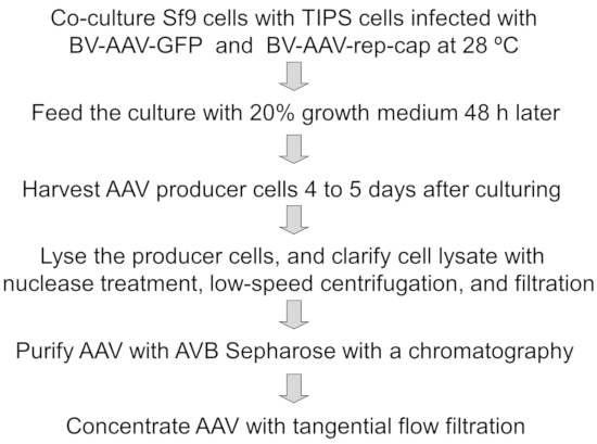
Figure 1: A schematic diagram for the production and purification of AAV Vector. Please click here to view a larger version of this figure.
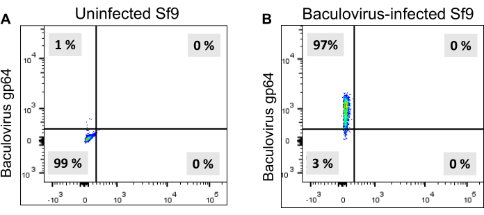
Figure 2: Flow cytometry analysis of baculovirus gp64 expression in Sf9 cells. Baculovirus infected Sf9 cells are stained with a mouse anti-baculovirus gp64 antibody containing a fluorescent dye, which is detected by flow cytometry. (A) Uninfected Sf9 cells. (B) Baculovirus infected Sf9 cells are showing gp64 expression in most of the cells. Please click here to view a larger version of this figure.
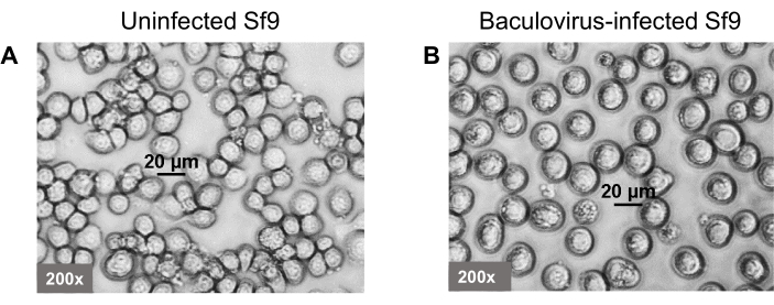
Figure 3: Morphology of the baculovirus-infected Sf9 (TIPS) cells. Baculovirus is infected into the Sf9 cells to produce the TIPS cells. (A) Uninfected Sf9 cells. (B) Baculovirus infected Sf9 (TIPS) cells. Cells are shown at 200x magnification. Please click here to view a larger version of this figure.
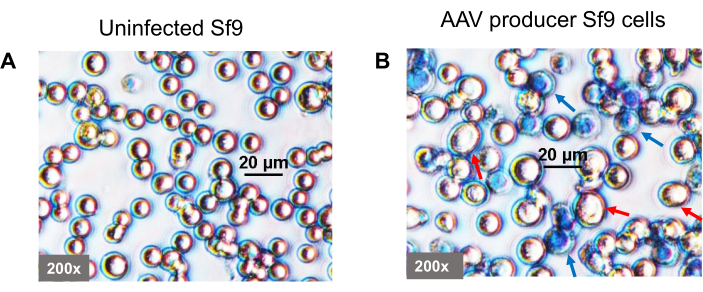
Figure 4: Morphology of the baculovirus-infected Sf9 cells during AAV production. TIPS cells secrete baculoviruses that infect co-cultured naïve Sf9 cells during AAV production. (A) Uninfected Sf9 cells. (B) Baculovirus infected Sf9 cells show an increase in diameter. Five days post-infection, almost half of the cells die (visualized under an inverted phase microscope after trypan blue staining). Red arrows indicate live cells, and blue arrows indicate dead cells. Cells are shown at 200x magnification. Please click here to view a larger version of this figure.
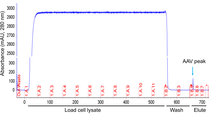
Figure 5: AAV purification using AVB Sepharose column chromatography. A chromatogram shows the absorbance of protein samples at 280 nm during sample loading on column, washing, and elution. The chromatogram has been modified to fit in the figure. Please click here to view a larger version of this figure.
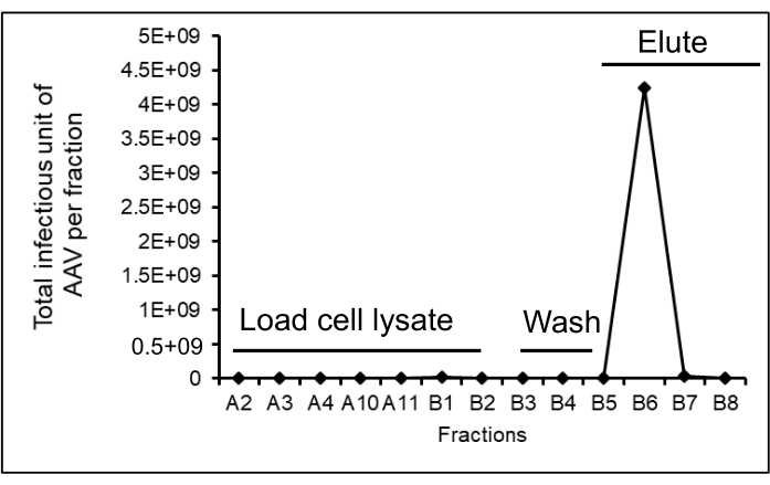
Figure 6: The number of infectious AAV particles in the flow-through during loading onto the column, washing, and elution. A total of 560 mL of AAV samples are loaded on a 10 mL AVB Sepharose column. The fraction volume for the column load is 50 mL, column wash is 50 mL, and elution is 25 mL. AAV titers are measured after infection of HT1080 cells with the column run-through samples while loading on column, washing, and elution to investigate the presence of AAV at each step of the purification. The total purified AAV yield is 4.3 x 10⁹ infectious units. Each diamond-shaped symbol represents the infectious units (IU) of AAV in each fraction. Please click here to view a larger version of this figure.
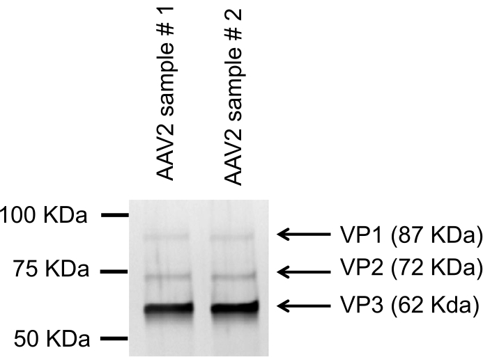
Figure 7. SDS-PAGE and silver staining of pure AAV vector showing capsid proteins. The reduced AAV samples are run on SDS-PAGE, and silver staining is performed. Three distinct bands of AAV capsid proteins, VP1, VP2, and VP3, are visible. Please click here to view a larger version of this figure.
The parameters used in this protocol for the process development of production, purification, and concentration of AAV vectors can be applied to both small and large-scale manufacturing of AAV vectors for gene therapy applications. The entire upstream and downstream process can be performed in a closed system compatible with the current Good Manufacturing Practices (cGMP). The major advantages of the Sf9-baculovirus system are scalability for large-scale GMP-grade AAV production at an affordable cost. The system does not need expensive plasmids and transfection reagents, which are needed for the production of AAV using HEK293 cells. It has been reported that both HEK293 cells and Sf9 cells yield similar quality AAV vectors (Eric D. Horowitz, ASGCT 2018, Abstract #100). The major challenge is that this system requires the generation of baculoviruses and TIPS cells that need a significant time and effort.
In this protocol, two types of TIPS cells (TwoBac system) were used: one TIPS cell containing baculovirus with AAV-gene transfer cassette and another TIPS cell containing AAV-rep-cap. The OneBac system2,16,17 can generate TIPS cells containing both AAV gene transfer cassette and rep-cap genes in a single baculovirus. The OneBac system provides an alternative approach to produce the rAAV using only one set of TIPS cells rather than two as described in the TwoBac system, which would further simplify the protocol.
This protocol describes AAV production in Sf9 cells. It is important to optimize the ratio of the baculovirus-infected TIPS cells to producer naïve Sf9 cells to obtain a good AAV yield. If this ratio is sub-optimal, the yield of AAV will be reduced11. For example, if more AAV ITR containing gene transfer vectors are generated than the capsids in the producer cells due to the sub-optimal ratio of TIPS and producer cells, all of the available gene transfer vectors will not get enough capsids to produce full AAV particles. On the other hand, if more capsids are generated than the AAV gene transfer vectors in the producer cells, all capsids will not get AAV ITR containing gene transfer vectors that result in empty AAV particles.
As the cells multiply, the nutrients of the culture medium become exhausted, and metabolic waste products accumulate. Therefore, supplementation of 20% fresh growth medium into the cell culture 2 days after co-culture of TIPS cells and Sf9 cells can increase AAV titers significantly (Amine A. Kamen, National Research Council Canada, personal communication).
The purity of AAV particles is a critical factor for achieving effective transduction of target cells without any cytotoxicity for both in vitro and in vivo studies3,5. Therefore, it is important to include a chromatography step that can selectively purify AAV particles and eliminate impurities such as host cell proteins and debris, genomic and baculoviral DNA, and aggregated and fragmented vectors. While loading AAV supernatant onto a AVB Sepharose column, it is critical to check the flow-through samples for the presence of AAV particles that may pass through the column without binding to the resin. If that is the case, (1) a lower run speed that results in a longer residence time will be necessary for binding the AAV particles, (2) the amount of loaded sample onto the column should be reduced, and/or (3) the volume of the AVB Sepharose should be increased, so that the binding capacity of the column never exceeds the number of AAV particles. The ability of AVB Sepharose resin to bind AAV particles at a high flow rate and with high affinity and capacity is important to reduce purification time. The major limitation of AVB Sepharose is that it binds the capsid of AAV serotypes 1, 2, and 5. Therefore, different chromatography resins should be tested and used for the purification of other AAV serotypes18. The downstream purification protocol described herein can also be used to purify AAV produced in HEK 293 cells.
This protocol can purify AAV from cell lysate but cannot remove empty particles. A few articles have described methods that can distinguish empty vs. full AAV particles in purified AAV stocks19,20. However, we believe that the AAV production should be optimized at the upstream process level to minimize the generation of empty particles. If the ratio of AAV gene transfer vector and capsid production in producer cells are not optimal, more empty particles may generate.
In addition to binding full AAV particles, AVB Sepharose medium binds fragmented AAV particles or capsid proteins which can be removed using TFF. Most of the low molecular weight particles are eliminated from AAV samples by including the TFF step downstream of the column chromatography. In addition, TFF is used to perform a buffer exchange/diafiltration and to concentrate the AAV10,21.
Although ultracentrifugation of the AAV lysate with cesium chloride or iodixanol gradient is the preferred method for small-scale and pre-clinical grade AAV purification, this method is not scalable and less suitable for large-scale purification of AAV21,22.
In conclusion, this protocol for the process development of production and purification of AAV will be useful for small-scale pre-clinical and large-scale manufacturing of recombinant AAV for the gene therapy of inherited genetic diseases.
The authors have nothing to disclose.
We would like to thank Dr. Robert M. Kotin (National Heart, Blood and Lung Institute, NIH) for generously providing us the AAV plasmids and Danielle Steele and Rebecca Ernst (Cincinnati Children's Hospital) for their technical assistance. This work is supported by the Start-Up fund from Cincinnati Children's Research Foundation to M.N.
1 N Sodium Hydroxide | Sigma-Aldrich | 1.09137 | For Akta Avant cleaning |
2 L flasks | ThermoFisher Scientific | 431281 | Flask for suspension culture |
50 ml Conical tube | ThermoFisher Scientific | 14-959-49A | For collection of supernatants |
24-well plate | ThermoFisher Scientific | 07-200-80 | Adherent cell culture plate |
250 mL flasks | ThermoFisher Scientific | 238071 | Flask for suspension culture |
Akta Avant 150 with Unicorn Software | Cytiva | 28976337 | Chromatography system |
AVB Sepharose High Performance | Cytiva | 28411210 | Chromatography medium |
Baculovirus-AAV-2 GFP | In-house | non-catalog | AAV transfer vector |
Baculovirus-AAV-2 rep-cap | In-house | non-catalog | AAV packaging vector |
Nuclease | Sigma-Aldrich | E1014 | Enzyme to degrade DNA and RNA |
Blocking buffer | Santa Cruz Biotechnologies | 516214 | Blocking to prevent non-specific antibody binding to cells |
Cell lysis buffer | In-house | Non-catalog item | 20 mM Tris-HCl (pH 8.0), 150 mM NaCl, 0.5 % Titron X-100 |
Cellbag, 2 L and 10 L | Cytiva | 28937662 | Bioreactor bag |
Cleaning buffer | In-house | Non-catalog item | 100 mM citric acid (pH 2.1) |
Cryovial | Thomas Scientific | 1222C24 | For cryopreservation |
DMEM | Sigma-Aldrich | D6429 | Growth media for cell lines |
Elution buffer | In-house | Non-catalog item | 50 mM sodium citrate buffer (pH 3.0) |
Ethanol | Sigma-Aldrich | E7073 | For disinfection and storage of the chromatography |
Filtration unit | Pall Corporation | 12941 | Membrane filter |
HT1080 cell line | ATCC | CCL-121 | Fibroblast cell line |
HyClone™ SFX-Insect culture media | Cytiva | SH30278.02 | Serum-free insect cell growth medium |
Peristaltic Pump | Pall Corporation | Non-catalog item | TFF pump |
MaxQ 8000 orbital shaker incubator | ThermoFisher Scientific | Non-catalog item | Shaker for suspension culture |
Microscope | Nikon | Non-catalog item | Cell monitoring and counting |
Mouse anti-baculovirus gp64 PE antibody | Santa Cruz Biotechnologies | 65498 PE | Monitoring baculovirus infection in Sf9 cells |
Oxygen tank | Praxair | Non-catalog item | 40 % Oxygen supply is needed for Sf9 cell growth |
PBS | ThermoFisher Scientific | 20012027 | Wash buffer |
Silver Staining kit | ThermoFisher Scientific | LC6100 | Staining AAV capsid proteins |
Sf9 cells | ThermoFisher Scientific | 11496-015 | Insect cells |
Steile water | In-house | Non-catalog item | For Akta Avant cleaning |
Table top centrifuge | ThermoFisher Scientific | 75253839/433607 | For clarification of Baculovirus supernatant |
Tangential Flow Filtration (TFF) cartridge | Pall Corporation | OA100C12 | TFF cartridge to concentrate the AAV |
Tris-HCl, pH 8.0 | ThermoFisher | 15568025 | Alkaline buffer to neutralize the eluted AAV |
WAVE Bioreactor System 20/50 | Cytiva | 28-9378-00 | Bioreactor |