Summary

Implantation of Engineered Tissue in the Rat Heart

Published: June 24, 2009
doi:

Summary

Here, we describe a cardiac surgical procedure to implant engineered tissue in the atrioventricular (AV)-groove of an adult Lewis rat.

Abstract

Rodent surgery is often an important component in assessing the utility of engineered tissues. A wide variety of surgical procedures can be performed in common laboratory rats or mice and these quite frequently serve as an intermediate step between bench-top experiments and large animal testing or human trials. Given that rodents provide an established, cost-effective, and physiologically-relevant model system in which to test novel combinations of scaffolding materials and cells, they are particularly well-suited for cardiovascular tissue engineering studies. Presently, we describe an open-heart surgical procedure to implant engineered tissue containing myogenic progenitor cells in the atrioventricular (AV) groove of a rat heart. These implants are intended to create an electrical conduit between the right atrium and right ventricle with the ultimate goal of providing an alternative treatment to conventional pacemaker implantation in pediatric patients with complete heart block[1]. The engineered tissue is implanted in the AV-groove by means of a thoracotomy. For our purposes, Lewis rats are anesthetized and invasively ventilated to maintain positive airway pressure during the sterile surgical procedure. The approach to the heart is performed by a right thoracotomy through an antero-lateral incision at the 5th intercostal space. The tissue construct is fixed in the AV groove using a single 7-0 Prolene suture and positioned between the right ventricle and atrium at the ventral portion of the heart. The epicardium is partially removed to allow direct contact between the recipient myocardial cells and those contained in the engineered tissue. Following implantation, the chest wall is closed in layers, any pneumothorax is evacuated, and the animal is extubated and treated with analgesic.

Protocol

Part 1: Induction of general anesthesia

  1. A 200-250 g Lewis rat is anesthetized with 50 mg/kg Ketamine and 4 mg/kg Xylazine injected with a 30 G needle into the intraperitoneal cavity. The upper abdomen and right chest are clipped to remove hair and the incision site thoroughly cleaned with 70% ethanol and then a povidone-iodine topical antiseptic solution.
  2. The animal is intubated endotracheally using a 16 G intravenous catheter connected to a small animal respirator (Harvard Apparatus). The respirator is connected to a vaporizer to supply 0.5 to 1.0% Isoflurane (Baxter) mixed with 100% oxygen to maintain general anesthesia. Respiration rate and tidal volume is adjusted according to the body weight of the rat. The animal is placed on a heating pad (Hood Thermo-Pad) to maintain body temperature during the surgical procedure.
  3. To monitor heart rate and oxygen saturation throughout the surgical procedure a pulsoximeter device (Nellcor) is clipped onto one of the back limbs.

Part 2: Thoracotomy and engineered tissue implantation

  1. The surgical site is prepared with sterile surgical draping that is held in place with towel clamps.
  2. An incision is made from the antero-lateral to the lateral wall of the right chest, just above the 5th intercostal space. The interior of the chest is accessed through the 5th intercostal space and a small retractor (Roboz) is positioned to maximize the view.
  3. The pericardium is cut just above the appendix of the right atrium and right ventricle from the base toward the apex of the heart. Afterward, the AV groove is identified and the epicardial layer adjacent to the aorto-atrio-ventricular triangle is gently teased apart.
  4. A 7-0 Prolene suture (Ethicon) is placed through the basal shoulder of the right ventricle. A 2 x 2 x 2 mm piece is dissected from the engineered tissue construct and positioned within the AV groove and gently tied down with the Prolene suture.
  5. The surgical field is checked for bleeding and two 4-0 Vicryl sutures (Ethicon) are placed between the upper and lower intercostal space for closure of the rib-cage.
  6. Any apparent pneumothorax is excavated by expanding the lungs by way of the ventilator.
  7. The chest wall is then closed in layers.

Part 3: Recovery

  1. While the animal recovers from the surgery, the incision site is infiltrated with 5 mL 0.25% Bupivacaine[2].
  2. The animal is extubated following weaning from the respirator and is returned to a cage with fresh bedding once it is fully awake and sternal. The rat is subsequently administered 0.01 mg/kg Buprenorphine analgesia subcutaneously every 12 hours for 3 days.

Part 4: Representative results

If performed properly, the described surgical implantation of engineered tissue in the AV-groove of the heart (Figure 1) results in greater than 90% recovery of the animals.

Figure 1
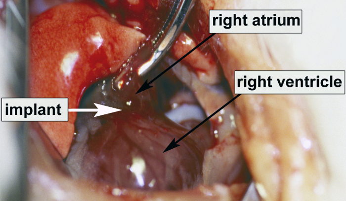
Figure 1. A photograph of the exposed heart showing the site of implantation of engineered tissue.

Discussion

For a favorable outcome to the surgical procedure, it is critical to properly intubate and invasively ventilate the animal. If the rat is simply sedated and allowed to spontaneously breathe, both lungs will inevitably collapse while the thorax is opened due to common pleura in rodents. Before closing the chest, any pneumothorax needs to be completely eliminated and the lungs fully expanded to reduce atelectasis. Otherwise, weaning from the respirator and recovery will be unsuccessful and the animal will become hypercapnic and/or hypoxic[3]. Removal of epicardial tissue within the AV groove is potentially hazardous and could result in massive venous bleeding from the right atrium due to the delicate nature of the rat atrial wall. While placing the Prolene suture in step 2.4, it is essential to be aware of and avoid the epicardial coronary branches. The animal should be kept warm on a heading pad during the recovery period or its body temperature will fall abruptly.

Disclosures

The authors have nothing to disclose.

Acknowledgements

This work is supported by research grants from the National Institutes of Health (HL068915; HL088206) and contributions to the Cardiac Conduction Fund at Children’s Hospital Boston.

Materials

Material Name Type Company Catalogue Number Comment
7×4 Thermo Pad 4/box Equipment Hood Thermo-Pad, Summerland, BC Canada MODEL 704R  
Inspira ASV Ventilator Equipment Harvard Apparatus, Holliston, MA 01746 USA    
Handheld Pulse Oximeter Equipment Nellcor, Boulder, CO 80301 USA N20PA  
Retractor Equipment Roboz, Gaithersburg, MD 20898 USA RS-6514  
Isoflurane (Forane) Reagent Baxter 1001936040  
Ketamine HCl Reagent HOSPIRA Inc., Lake Forest, IL 60045 RL-0065  
Xylazine (AnaSed) Reagent Lloyd LB15705A  
Buprenorphine HCL Inject Reagent Bedford Labs, Bedford, OH 44146 USA Div-BUP-P00  
Bupivacaine 0.25% Reagent HOSPIRA Inc., Lake Forest, IL 60045    
check_url/kr/1139?article_type=t

Play Video

Cite This Article
Sill, B., Alpatov, I. V., Pacak, C. A., Cowan, D. B. Implantation of Engineered Tissue in the Rat Heart. J. Vis. Exp. (28), e1139, doi:10.3791/1139 (2009).

View Video