Summary

تقييم عصبي عضلي وظيفة عن طريق العصب عن طريق الجلد الكهربائية تحفيز

Published: September 13, 2015
doi:

Summary

We present a protocol to assess changes in neuromuscular function. Percutaneous electrical nerve stimulation is a non-invasive method that evokes muscular responses. Electrophysiological and mechanical properties of these responses permit the evaluation of neuromuscular function from brain to muscle (supra-spinal, spinal and peripheral levels).

Abstract

Percutaneous electrical nerve stimulation is a non-invasive method commonly used to evaluate neuromuscular function from brain to muscle (supra-spinal, spinal and peripheral levels). The present protocol describes how this method can be used to stimulate the posterior tibial nerve that activates plantar flexor muscles. Percutaneous electrical nerve stimulation consists of inducing an electrical stimulus to a motor nerve to evoke a muscular response. Direct (M-wave) and/or indirect (H-reflex) electrophysiological responses can be recorded at rest using surface electromyography. Mechanical (twitch torque) responses can be quantified with a force/torque ergometer. M-wave and twitch torque reflect neuromuscular transmission and excitation-contraction coupling, whereas H-reflex provides an index of spinal excitability. EMG activity and mechanical (superimposed twitch) responses can also be recorded during maximal voluntary contractions to evaluate voluntary activation level. Percutaneous nerve stimulation provides an assessment of neuromuscular function in humans, and is highly beneficial especially for studies evaluating neuromuscular plasticity following acute (fatigue) or chronic (training/detraining) exercise.

Introduction

يستخدم بشكل واسع عن طريق الجلد تحفيز العصب الكهربائية لتقييم العصبية والعضلية وظيفة 1. يتكون المبدأ الأساسي لإحداث حافز الكهربائية إلى العصب المحرك الطرفية لتستثير تقلص العضلات. الميكانيكي (قياس عزم الدوران) والكهربية (النشاط electromyographic) ردود تسجل في وقت واحد. عزم الدوران، وسجلت في المفصل النظر فيها، ويتم تقييم باستخدام مقياس العمل. وقد أظهرت electromyographic (EMG) الإشارات المسجلة باستخدام أقطاب سطح لتمثيل نشاط العضلات 2. هذه الطريقة غير الغازية ليست مؤلمة وتنفيذها بسهولة أكبر من التسجيلات في العضل. كل من الأقطاب الكهربائية أحادي وثنائي القطب يمكن استخدامها. وقد تبين تكوين القطب القطب إلى أن تكون أكثر حساسية للتغيرات في نشاط العضلات والتي يمكن أن تكون مفيدة للعضلات الصغيرة. ومع ذلك، فقد ثبت أقطاب القطبين لتكون أكثر فعالية في تحسين ص إشارة إلى الضوضاءATIO 4 و هي الأكثر شيوعا كطريقة لتسجيل وقياس نشاط الوحدة الحركية. فإن المنهجية المبينة أدناه يركز على التسجيلات القطبين. النشاط EMG هو مؤشر على فعالية وسلامة الجهاز العصبي العضلي. استخدام التحفيز العصبي عن طريق الجلد يقدم مزيدا من الإيضاحات وظيفة العصبية والعضلية، والتغيرات في أي مستوى العضلات، والعمود الفقري، أو فوق الشوكي (الشكل 1).

الشكل 1
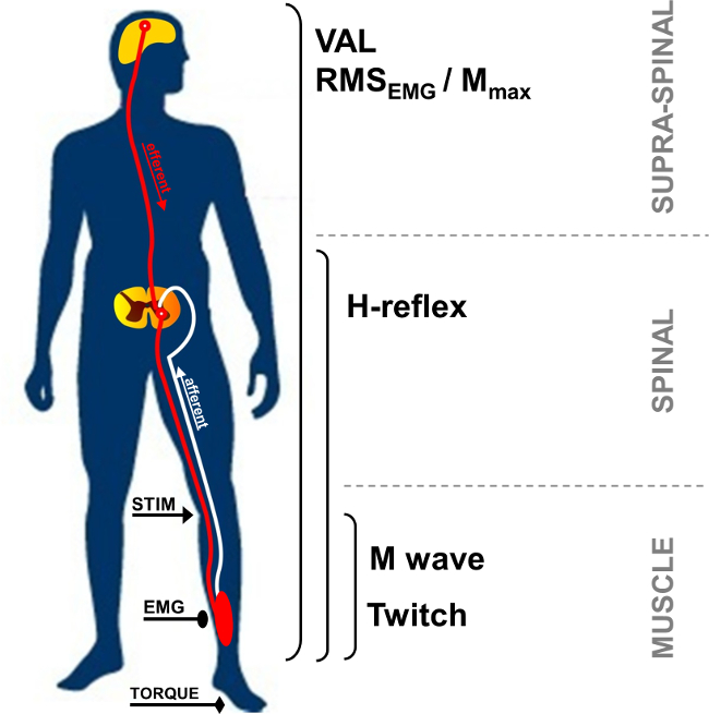
الشكل 1: لمحة عامة عن القياسات العصبية والعضلية STIM: تحفيز العصب. EMG: الكهربائي. فال: الطوعي مستوى التنشيط. RMS: جذر متوسط ​​مربع. M الأقصى: القصوى M-سعة الموجة.

في بقية، وإمكانية عمل العضلات مجمع، كما دعا M الموجة، هي استجابة قصيرة الكمون لوحظ بعد الحرفية التحفيز، وتمثل كتلة العضلات منفعل من قبل أكتيف المباشر أوجه من المحاور الحركية مما يؤدي إلى العضلات (الشكل 2، رقم 3). M-موجة السعة تزيد مع كثافة حتى تصل إلى القاع من قيمته القصوى. هذا الرد، ودعا M ماكس، تمثل خلاصة متزامن من جميع الوحدات الحركية و / أو إمكانات العمل الألياف العضلية المسجلة في إطار الأقطاب EMG السطحية 5. يستخدم تطور السعة أو موجة منطقة ذروة إلى ذروة لتحديد التعديلات من انتقال العصبية والعضلية 6. تغييرات في ردود الميكانيكية المرتبطة M الموجة، أي ذروة نشل عزم الدوران / القوة، قد يعزى إلى تغيرات في استثارة العضلات و / أو داخل الألياف العضلية (7). وتقدم الجمعية من M ماكس السعة ونشل ذروة عزم الدوران السعة (حزب العمال نسبة / M) مؤشر الكفاءة الكهربائية لعضلة أي الاستجابة الميكانيكية لأمر محرك كهربائي معين.

52974 / 52974fig2.jpg "/>
الشكل 2: السيارات والممرات انعكاسية تفعيلها من خلال تحفيز العصب التحفيز الكهربائي لالمختلط (السيارات / الحسي) العصبية (STIM) يؤدي الى الاستقطاب في كل من محور عصبي السيارات والأولى أ ارد اطلاق النار. الاستقطاب من الأولى أ afferents نحو الحبل الشوكي ينشط في العصبون الحركي ألفا، وهذا بدوره يثير رد فعل منعكس-H (المسار 1 + 2 + 3). تبعا لشدة التحفيز، محور عصبي محرك الاستقطاب يثير استجابة العضلات مباشرة: M-موجة (مسار 3). في أقصى كثافة M الموجة، يتم إنشاء تيار أيضا معاكس للمسيرة (3 ') ويصطدم مع كرة ارادي (2). هذا التصادم جزئيا أو كليا يلغي ردا-H لا ارادي.

وH-رد الفعل هو استجابة الكهربية المستخدمة لتقييم التغيرات في IA-α العصبون الحركي المشبك 9. يمكن تقييم هذه المعلمة في الراحة أو أثناء الانقباضات الطوعية. يمثل H-المنعكس البديل من منعكس تمتد (الشكل 2، نوmber 1-3). وH-المنعكس ينشط الوحدات الحركية تجنيد monosynaptically التي كتبها الأولى أ مسارات ارد 10،11، ويمكن أن يخضع للتأثيرات الطرفية والمركزية 12. ومن المعروف أن طريقة تستحضر H-العاكسة لديها مصداقية داخل تخضع عالية لتقييم استثارة العمود الفقري في بقية 13،14 وخلال تقلصات متساوي القياس 15.

خلال الانكماش الطوعي، وحجم محرك الأقراص العصبي الطوعي يمكن تقييمها باستخدام اتساع إشارة EMG، كميا باستخدام عموما ساحة متوسط ​​الجذر (RMS). يستخدم RMS EMG عادة وسيلة لقياس مستوى الإثارة للنظام المحرك خلال الانكماش الطوعي (الشكل 1). بسبب التباين داخل وبين تخضع 16، RMS EMG لابد من تطبيع باستخدام EMG المسجلة خلال العضلات محددة القصوى الانكماش الطوعي (RMS EMGmax). وبالإضافة إلى ذلك، لأن التغييرات في إشارة EMG قد بمطلوب ه يرجع إلى تغييرات على مستوى الطرفية، وتطبيع باستخدام المعلمة الطرفية مثل M-موجة لتقييم فقط عنصرا مركزيا في إشارة EMG. ويمكن القيام بذلك عن طريق قسمة RMS EMG من السعة القصوى أو RMS Mmax من M-الموجة. التطبيع باستخدام RMS Mmax (أي RMS EMG / RMS Mmax) هو الأسلوب المفضل لأنه يأخذ في الاعتبار احتمال تغيير مدة M-موجة 17.

ويمكن أيضا أن يتم تقييم الأوامر الحركية عن طريق حساب مستوى تفعيل الطوعي (VAL). يستخدم هذا الأسلوب أسلوب نشل الاستيفاء 18 عن طريق إضافة والتحفيز الكهربائي في M كثافة كحد أقصى خلال الانكماش الطوعي القصوى. تتم مقارنة عزم الدوران الإضافي الناجم عن تحفيز العصب لنشل التحكم التي تنتجها تحفيز العصب متطابقة في عضلة استرخاء potentiated 19. لتقييم الأقصى فال، وinterpo نشل الأصليتقنية lation صفها ميرتون 18 ينطوي على التحفيز واحد محرف على الانكماش الطوعي. في الآونة الأخيرة، أصبح استخدام التحفيز إقران أكثر شعبية لأن الزيادات عزم الدوران أثار أكبر، اكتشفت بسهولة أكثر، وأقل تغيرا مقارنة مع ردود التحفيز واحدة 20. يوفر VAL مؤشر قدرة الجهاز العصبي المركزي لتفعيل الحد الأقصى للعضلات العاملة 21. حاليا، تقييم VAL باستخدام تقنية نشل الاستيفاء هو الأسلوب الأكثر قيمة لتقييم مستوى تنشيط العضلات 22. وعلاوة على ذلك، وعزم دوران الذروة المقررة باستخدام مقياس العمل هو الأكثر درس بشكل صحيح قوة اختبار المعلمة الاستخدام المعمول بها في مجال البحوث والمرافق الصحية 23.

تحفيز العصب الكهربائية يمكن استخدامها في مجموعة متنوعة من مجموعات العضلات (مثل العضلات القابضة الكوع، العضلات القابضة في المعصم، الباسطة في الركبة، والعضلات القابضة أخمصي). ومع ذلك، يجعل الأعصاب الوصول للتقنية صعبة في بعض المجموعات العضلات. العضلات الباسطة أخمصي، وخاصة الرؤوس ربلة (النعلية وgastrocnemii) العضلات، ويتم التحقيق بشكل متكرر في الأدب 24. في الواقع، وتشارك هذه العضلات في الحركة وتبرير المصالح الخاصة. المسافة بين الموقع التحفيز وأقطاب تسجيل تتيح التعرف على موجات أثار مختلفة من الرؤوس الربلية العضلات. الجزء السطحي من العصب الخلفي قصبي في المبأبضية وعدد كبير من مغزل يجعل من الاسهل لتسجيل ردود الفعل المنعكس مقارنة العضلات الأخرى 24. لهذه الأسباب، تركز منهجية لا ارادي تعرض حاليا على مجموعة ثلاثية الرؤوس الربلية العضلات (النعلية والساق). وبالتالي فإن الهدف من هذا البروتوكول هو وصف عن طريق الجلد تقنية تحفيز العصب للتحقيق وظيفة العصبية والعضلية في ربلة ثلاثية الرؤوس.

Protocol

الإجراءات التجريبية أوجز حصلت على موافقة الأخلاقية المؤسسي وفقا للإعلان هلسنكي. تم جمع البيانات من أحد المشاركين التمثيلي الذي كان على بينة من الإجراءات وأعطى الموافقة المسبقة عن المكتوب. 1. إعداد الصك <l…

Representative Results

زيادة كثافة التحفيز يؤدي إلى تطور مختلف من سعة الاستجابة بين H- وM-الأمواج. في بقية، وH-المنعكس يصل الحد الأقصى لقيمة قبل أن تغيب تماما من إشارة EMG، في حين M موجة يزيد تدريجيا حتى تصل إلى القاع في شدة القصوى (انظر الشكل 4 لتصوير بياني لM-موجة والشكل 6</…

Discussion

تحفيز العصب عن طريق الجلد يمكن القياس الكمي لخصائص عديدة من الجهاز العصبي العضلي ليس فقط لفهم الأساسية للمراقبة وظيفة حركي في البشر صحية، ولكن أيضا لتكون قادرة على تحليل التعديلات حادة أو مزمنة من خلال التعب أو تدريب 17. وهذا مفيد جدا وخصوصا لبروتوكولات مجهد، ح?…

Disclosures

The authors have nothing to disclose.

Acknowledgements

The authors have no acknowledgements.

Materials

Biodex dynamometer Biodex Medical System Inc., New York, USA www.biodex.com
MP150 Data Acquisition System Biopac Systems Inc., Goleta, USA
Acknowledge 4.1.0 software Biopac Systems Inc., Goleta, USA www.biopac.com
DS7A constant current high voltage stimulator Digitimer, Hertfordshire, UK www.digitimer.com
Silver chloride surface electrodes Control Graphique Medical, Brie-Comte-Robert, France
Computer
1 Cable for connecting the Biodex to the MP150
1 Cable for connecting the Digitimer to the MP150
1 Cable for connecting the MP150 to the computer

References

  1. Desmedt, J. E., Hainaut, K. Kinetics of myofilament activation in potentiated contraction staircase phenomenon in human skeletal muscle. Nature. 217 (5128), 529-532 (1968).
  2. Bouisset, S., Maton, B. Quantitative relationship between surface EMG and intramuscular electromyographic activity in voluntary movement. American Journal of Physical Medicine. 51 (6), 285-295 (1972).
  3. Gabriel, D. A. Effects of monopolar and bipolar electrode configurations on surface EMG spike analysis. Medical Engineering and Physics. 33 (9), 1079-1085 (2011).
  4. Merletti, R., Rainoldi, A., Farina, D. Surface electromyography for noninvasive characterization of muscle. Exercise and Sport Sciences Reviews. 29 (1), 20-25 (2001).
  5. Lepers, R. Aetiology and time course of neuromuscular fatigue during prolonged cycling exercises. Science, & Motricité. 52, 83-107 (2004).
  6. Baudry, S., Klass, M., Pasquet, B., Duchateau, J. Age related fatigability of the ankle dorsiflexor muscles during concentric and eccentric contractions. European Journal of Applied Physiology. 100 (5), 515-525 (2007).
  7. Place, N., Yamada, T., Bruton, J. D., Westerblad, H. Muscle fatigue From observations in humans to underlying mechanisms studied in intact single muscle fibres. European Journal of Applied Physiology. 110 (1), 1-15 (2010).
  8. Scaglioni, G., Narici, M. V., Maffiuletti, N. A., Pensini, M., Martin, A. Effect of ageing on the electrical and mechanical properties of human soleus motor units activated by the H reflex and M wave. The Journal of Physiology. 548 (Pt. 2), 649-661 (2003).
  9. Schieppati, M. The Hoffmann reflex a means of assessing spinal reflex excitability and its descending control in man. Progress in Neurobiology. 28 (4), 345-376 (1987).
  10. Pierrot Deseilligny, E., Burke, D. . The circuitry of the human spinal cord: its role in motor control and movement disorders. , (2005).
  11. Duclay, J., Pasquet, B., Martin, A., Duchateau, J. Specific modulation of corticospinal and spinal excitabilities during maximal voluntary isometric shortening and lengthening contractions in synergist muscles. The Journal of Physiology. 589 (Pt. 11), 2901-2916 (2011).
  12. Grosprêtre, S., Papaxanthis, C., Martin, A. Modulation of spinal excitability by a sub threshold stimulation of M1 area during muscle lengthening. 신경과학. 263, 60-71 (2014).
  13. Mynark, R. G. Reliability of the soleus H reflex from supine to standing in young and elderly. Clinical Neurophysiology. 116 (6), 1400-1404 (2005).
  14. Palmieri, R. M., Hoffman, M. A., Ingersoll, C. D. Intersession reliability for H reflex measurements arising from the soleus peroneal and tibialis anterior musculature. The International Journal of Neuroscience. 112 (7), 841-850 (2002).
  15. Chen, Y. S., Zhou, S., Cartwright, C., Crowley, Z., Baglin, R., Wang, F. Test retest reliability of the soleus H reflex is affected by joint positions and muscle force levels. Journal of Electromyography and Kinesiology. 20 (5), 987-987 (2010).
  16. Lehman, G. J., McGill, S. M. The importance of normalization in the interpretation of surface electromyography A proof of principle. Journal of Manipulative and Physiological Therapeutics. 22 (7), 444-446 (1999).
  17. Lepers, R. Interest and limits of percutaneous nerve electrical stimulation in the evaluation of muscle fatigue. Science, & Motricité. 70 (70), 31-37 (2010).
  18. Merton, P. A. Voluntary strength and fatigue. The Journal of Physiology. 123, 553-564 (1954).
  19. Gandevia, S. C. Spinal and supraspinal factors in human muscle fatigue. Physiological Reviews. 81 (4), 1725-1789 (2001).
  20. Shield, A., Zhou, S. Assessing voluntary muscle activation with the twitch interpolation technique. Sports Medicine. 34 (4), 253-267 (2004).
  21. Rozand, V., Pageaux, B., Marcora, S. M., Papaxanthis, C., Lepers, R. Does mental exertion alter maximal muscle activation. Frontiers in Human Neuroscience. 8, 755 (2014).
  22. Place, N., Maffiuletti, N. A., Martin, A., Lepers, R. Assessment of the reliability of central and peripheral fatigue after sustained maximal voluntary contraction of the quadriceps muscle. Muscle and Nerve. 35 (4), 486-495 (2007).
  23. Kannus, P. Isokinetic evaluation of muscular performance: implications for muscle testing and rehabilitation. International Journal of Sports Medicine. 15, S11-S18 (1994).
  24. Tucker, K. J., Tuncer, M., Türker, K. S. A review of the H reflex and M wave in the human triceps surae. Human Movement Science. 24 (5-6), 667-688 (2005).
  25. Taylor, N. A., Sanders, R. H., Howick, E. I., Stanley, S. N. Static and dynamic assessment of the Biodex dynamometer. European Journal of Applied Physiology and Occupational Physiology. 62 (3), 180-188 (1991).
  26. Sale, D., Quinlan, J., Marsh, E., McComas, A. J., Belanger, A. Y. Influence of joint position on ankle plantarflexion in humans. Journal of Applied Physiology. 52 (6), 1636-1642 (1982).
  27. Cattagni, T., Martin, A., Scaglioni, G. Is spinal excitability of the triceps surae mainly affected by muscle activity or body position. Journal of Neurophysiology. 111 (12), 2525-2532 (2014).
  28. Gerilovsky, L., Tsvetinov, P., Trenkova, G. Peripheral effects on the amplitude of monopolar and bipolar H-reflex potentials from the soleus muscle. Experimental Brain Research. 76 (1), 173-181 (1989).
  29. Schieppati, M. The Hoffmann reflex a means of assessing spinal reflex excitability and its descending control in man. Progress in Neurobiology. 28 (4), 345-376 (1987).
  30. Hermens, H. J., Freriks, B., Disselhorst Klug, C., Rau, G. Development of recommendations for SEMG sensors and sensor placement procedures. Journal of Electromyography and Kinesiology. 10 (5), 361-374 (2000).
  31. Kamen, G., Sison, S. V., Du, C. C., Patten, C. Motor unit discharge behavior in older adults during maximal effort contractions. Journal of Applied Physiology. 79 (6), 1908-1913 (1995).
  32. Neyroud, D., Rüttimann, J., et al. Comparison of neuromuscular adjustments associated with sustained isometric contractions of four different muscle groups. Journal of Applied Physiology. 114, 1426-1434 (2013).
  33. Rupp, T., Girard, O., Perrey, S. Redetermination of the optimal stimulation intensity modifies resting H-reflex recovery after a sustained moderate-intensity muscle contraction. Muscle and Nerve. 41 (May), 642-650 (2010).
  34. Zehr, E. P. Considerations for use of the Hoffmann reflex in exercise studies. European Journal of Applied Physiology. 86 (6), 455-468 (2002).
  35. Gondin, J., Duclay, J., Martin, A. Soleus and gastrocnemii evoked V wave responses increase after neuromuscular electrical stimulation training. Journal of Neurophysiology. 95 (6), 3328-3335 (2006).
  36. Rochette, L., Hunter, S. K., Place, N., Lepers, R. Activation varies among the knee extensor muscles during a submaximal fatiguing contraction in the seated and supine postures. Journal of Applied Physiology. 95 (4), 1515-1522 (2003).
  37. Fuglevand, A. J., Zackowski, K. M., Huey, K. A., Enoka, R. M. Impairment of neuromuscular propagation during human fatiguing contractions at submaximal forces. The Journal of Physiology. 460, 549-572 (1993).
  38. Vandervoort, A. A., McComas, A. J. Contractile changes in opposing muscles of the human ankle joint with aging. Journal of Applied Physiology. 61 (1), 361-367 (1986).
  39. Grosprêtre, S., Martin, A. Conditioning effect of transcranial magnetic stimulation evoking motor evoked potential on V wave response. Physiological Reports. 2 (11), e12191 (2014).
  40. Allen, G. M., Gandevia, S. C., McKenzie, D. K. Reliability of measurements of muscle strength and voluntary activation using twitch interpolation. Muscle and Nerve. 18 (6), 593-600 (1995).
  41. Cooper, M. A., Herda, T. J., Walter Herda, A. A., Costa, P. B., Ryan, E. D., Cramer, J. T. The reliability of the interpolated twitch technique during submaximal and maximal isometric muscle actions. Journal of Strength and Conditioning Research. 27 (10), 2909-2913 (2013).
  42. Froyd, C., Millet, G. Y., Noakes, T. D. The development of peripheral fatigue and short term recovery during self paced high intensity exercise. The Journal of Physiology. 591 (Pt 5), 1339-1346 (2013).
  43. Pierrot Deseilligny, E., Morin, C., Bergego, C., Tankov, N. Pattern of group I fibre projections from ankle flexor and extensor muscles in man. Experimental Brain Research. 42 (3-4), 337-350 (1981).
  44. Brooke, J. D., McIlroy, W. E., et al. Modulation of H reflexes in human tibialis anterior muscle with passive movement. Brain Research. 766 (1-2), 236-239 (1997).
  45. Hultborn, H., Meunier, S., Morin, C., Pierrot Deseilligny, E. Assessing changes in presynaptic inhibition of I a fibres a study in man and the cat. The Journal of Physiology. 389, 729-756 (1987).
  46. Meunier, S., Pierrot Deseilligny, E. Cortical control of presynaptic inhibition of Ia afferents in humans. Experimental Brain Research. 119 (4), 415-426 (1998).
  47. Aymard, C., Baret, M., Katz, R., Lafitte, C., Pénicaud, A., Raoul, S. Modulation of presynaptic inhibition of la afferents during voluntary wrist flexion and extension in man. Experimental Brain Research. 137 (1), 127-131 (2001).
  48. Abbruzzese, G., Trompetto, C., Schieppati, M. The excitability of the human motor cortex increases during execution and mental imagination of sequential but not repetitive finger movements. Experimental Brain Research. 111 (3), 465-472 (1996).
  49. Garland, S. J., Klass, M., Duchateau, J. Cortical and spinal modulation of antagonist coactivation during a submaximal fatiguing contraction in humans. Journal of Neurophysiology. 99, 554-563 (2008).
  50. Rodriguez Falces, J., Place, N. Recruitment order of quadriceps motor units Femoral nerve vs direct quadriceps stimulation. European Journal of Applied Physiology. 113, 3069-3077 (2013).
  51. Rodriguez Falces, J., Maffiuletti, N. A., Place, N. Spatial distribution of motor units recruited during electrical stimulation of the quadriceps muscle versus the femoral nerve. Muscle and Nerve. 48 (November), 752-761 (2013).
  52. Bathien, N., Morin, C. Comparing variations of spinal reflexes during intensive and selective attention (author’s transl). Physiology, & Behavior. 9 (4), 533-538 (1972).
  53. Earles, D. R., Koceja, D. M., Shively, C. W. Environmental changes in soleus H reflex excitability in young and elderly subjects. The International Journal of Neuroscience. 105 (1-4), 1-13 (2000).
  54. Paquet, N., Hui Chan, C. W. Human soleus H reflex excitability is decreased by dynamic head and body tilts. Journal of Vestibular Research Equilibrium, & Orientation. 9 (5), 379-383 (1999).
  55. Miyahara, T., Hagiya, N., Ohyama, T., Nakamura, Y. Modulation of human soleus H reflex in association with voluntary clenching of the teeth. Journal of Neurophysiology. 76 (3), 2033-2041 (1996).
  56. Pinniger, G. J., Nordlund, M. M., Steele, J. R., Cresswell, a. GH reflex modulation during passive lengthening and shortening of the human triceps surae. Journal of Physiology. 534 (Pt 3), 913-923 (2001).
  57. Tallent, J., Goodall, S., Hortobágyi, T., St Clair Gibson, A., French, D. N., Howatson, G. Repeatability of corticospinal and spinal measures during lengthening and shortening contractions in the human tibialis anterior muscle). PLoS ONE. 7 (4), e35930 (2012).
  58. Grospretre, S., Martin, A. H. reflex and spinal excitability methodological considerations. Journal of Neurophysiology. 107 (6), 1649-1654 (2012).
  59. Hugon, M. Methodology of the Hoffmann reflex in man. New Developments in Electromyography and Chemical Neurophysiology. 3m, 277-293 (1973).
  60. Bigland Ritchie, B., Zijdewind, I., Thomas, C. K. Muscle fatigue induced by stimulation with and without doublets. Muscle and Nerve. 23 (9), 1348-1355 (2000).
  61. Kent Braun, J. A., Le Blanc, R. Quantitation of central activation failure during maximal voluntary contractions in humans. Muscle and Nerve. 19 (7), 861-869 (1996).
  62. Herbert, R. D., Gandevia, S. C. Twitch interpolation in human muscles mechanisms and implications for measurement of voluntary activation. Journal of Neurophysiology. 82, 2271-2283 (1999).
  63. Miller, M., Downham, D., Lexell, J. Superimposed single impulse and pulse train electrical stimulation A quantitative assessment during submaximal isometric knee extension in young healthy men. Muscle and Nerve. 22 (8), 1038-1046 (1999).
  64. Button, D. C., Behm, D. G. The effect of stimulus anticipation on the interpolated twitch technique. Journal of Sports Science and Medicine. 7 (4), 520-524 (2008).
  65. Goss, D. a., Hoffman, R. L., Clark, B. C. Utilizing Transcranial Magnetic Stimulation to Study the Human Neuromuscular System. Journal of Visualized Experiments. (59), e3387 (2012).
  66. Sartori, L., Betti, S., Castiello, U. Corticospinal excitability modulation during action observation. Journal Of Visualized Experiments: Jove. (82), 51001 (2013).
  67. Rozand, V., Lebon, F., Papaxanthis, C., Lepers, R. Does a mental training session induce neuromuscular fatigue. Medicine and Science in Sports and Exercise. 46 (10), 1981-1989 (2014).
  68. Rozand, V., Cattagni, T., Theurel, J., Martin, A., Lepers, R. Neuromuscular fatigue following isometric contractions with similar torque time integral. International Journal of Sports Medicine. 36, 35-40 (2015).
  69. Belanger, A. Y., McComas, A. J. Extent of motor unit activation during effort. Journal of Applied Physiology. 51 (5), 1131-1135 (1981).
  70. Morse, C. I., Thom, J. M., Davis, M. G., Fox, K. R., Birch, K. M., Narici, M. V. Reduced plantarflexor specific torque in the elderly is associated with a lower activation capacity. European Journal of Applied Physiology. 92 (1-2), 219-226 (2004).
  71. Dalton, B. H., McNeil, C. J., Doherty, T. J., Rice, C. L. Age related reductions in the estimated numbers of motor units are minimal in the human soleus. Muscle and Nerve. 38 (3), 1108-1115 (2008).
  72. Hunter, S. K., Todd, G., Butler, J. E., Gandevia, S. C., Taylor, J. L. Recovery from supraspinal fatigue is slowed in old adults after fatiguing maximal isometric contractions. Journal of Applied Physiology. 105 (4), 1199-1209 (2008).
  73. Jakobi, J. M., Rice, C. L. Voluntary muscle activation varies with age and muscle group. Journal of Applied Physiology. 93 (2), 457-462 (2002).
  74. Lepers, R., Millet, G. Y., Maffiuletti, N. a Effect of cycling cadence on contractile and neural properties of knee extensors. Medicine and Science in Sports and Exercise. 33 (11), 1882-1888 (2001).
  75. Duchateau, J., Hainaut, K. Isometric or dynamic training differential effects on mechanical properties of a human muscle. Journal of Applied Physiology. 56 (2), 296-301 (1984).
  76. Millet, G. Y., Martin, V., Martin, A., Vergès, S. Electrical stimulation for testing neuromuscular function From sport to pathology. European Journal of Applied Physiology. 111, 2489-2500 (2011).
  77. Cattagni, T., Scaglioni, G., Laroche, D., Van Hoecke, J., Gremeaux, V., Martin, A. Ankle muscle strength discriminates fallers from non fallers. Frontiers in Aging Neuroscience. 6, 336 (2014).
  78. Horstman, A. M., Beltman, M. J., et al. Intrinsic muscle strength and voluntary activation of both lower limbs and functional performance after stroke. Clinical Physiology and Functional Imaging. 28 (4), 251-261 (2008).
  79. Sica, R. E., Herskovits, E., Aguilera, N., Poch, G. An electrophysiological investigation of skeletal muscle in Parkinson’s disease. Journal of the Neurological Sciences. 18 (4), 411-420 (1973).
  80. Knikou, M., Mummidisetty, C. K. Locomotor Training Improves Premotoneuronal Control after Chronic Spinal Cord Injury. Journal of Neurophysiology. 111 (11), 2264-2275 (2014).

Play Video

Cite This Article
Rozand, V., Grosprêtre, S., Stapley, P. J., Lepers, R. Assessment of Neuromuscular Function Using Percutaneous Electrical Nerve Stimulation. J. Vis. Exp. (103), e52974, doi:10.3791/52974 (2015).

View Video