Summary

경피적 전기 신경 자극을 사용하여 신경 근육 기능의 평가

Published: September 13, 2015
doi:

Summary

We present a protocol to assess changes in neuromuscular function. Percutaneous electrical nerve stimulation is a non-invasive method that evokes muscular responses. Electrophysiological and mechanical properties of these responses permit the evaluation of neuromuscular function from brain to muscle (supra-spinal, spinal and peripheral levels).

Abstract

Percutaneous electrical nerve stimulation is a non-invasive method commonly used to evaluate neuromuscular function from brain to muscle (supra-spinal, spinal and peripheral levels). The present protocol describes how this method can be used to stimulate the posterior tibial nerve that activates plantar flexor muscles. Percutaneous electrical nerve stimulation consists of inducing an electrical stimulus to a motor nerve to evoke a muscular response. Direct (M-wave) and/or indirect (H-reflex) electrophysiological responses can be recorded at rest using surface electromyography. Mechanical (twitch torque) responses can be quantified with a force/torque ergometer. M-wave and twitch torque reflect neuromuscular transmission and excitation-contraction coupling, whereas H-reflex provides an index of spinal excitability. EMG activity and mechanical (superimposed twitch) responses can also be recorded during maximal voluntary contractions to evaluate voluntary activation level. Percutaneous nerve stimulation provides an assessment of neuromuscular function in humans, and is highly beneficial especially for studies evaluating neuromuscular plasticity following acute (fatigue) or chronic (training/detraining) exercise.

Introduction

경피적 전기 신경 자극이 널리 신경근 1 기능을 평가하기 위해 사용된다. 기본 원리는 근육의 수축을 연상 말초 운동 신경에 전기 자극을 유도으로 구성되어 있습니다. 기계 (토크 측정) 및 전기 생리학 (근전도 활동) 응답을 동시에 기록됩니다. 고려 관절에 기록 토크는, 에르고 미터를 이용하여 평가된다. 표면 전극을 사용하여 기록 근전도 (EMG) 신호 (2)의 근육 활동을 나타내는 것으로 입증되었다. 이 비 침습적 방법은 고통보다 쉽게​​ 근육 녹음보다 구현되지 않습니다. 모두 모노 폴라 및 바이폴라 전극을 사용할 수있다. 모노 폴라 전극 구성은 작은 근육에 유용 할 수 근육 활동 (3)의 변화에보다 민감한 것으로 밝혀졌다. 그러나, 바이폴라 전극 신호대 R 개선에 더욱 효과적인 것으로 밝혀졌다페이지에 계속 4, 가장 일반적으로 기록 및 모터 유닛의 활성을 정량하는 방법으로 사용된다. 후술하는 방법은 바이폴라 녹화에 초점을 맞출 것이다. EMG 활성은 신경 근육 시스템의 효능 및 무결성의 지표이다. 경피 신경 자극의 사용은 신경 근육 기능에 더 통찰력, 근육 척추, 또는 문헌 – 척추 레벨 (그림 1)에서, 변화를 제공합니다.

그림 1
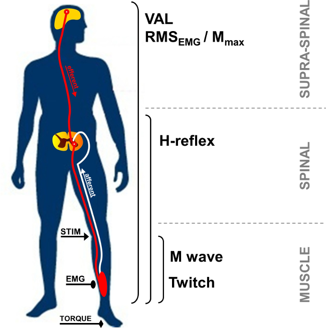
그림 1 :. 신경 근육 측정의 개요 STIM : 신경 자극. EMG : 근전도. 발 : 자원 봉사 활성화 수준. RMS : 루트가 광장을 의미한다. M 최대 : 최대한 M 파 진폭.

휴지, 또한, M 파라고 복합 근 활동 전위는 자극 인공물 후에 관찰 짧은 지연 응답이며, 직접 액티브 의한 흥분성 근육량을 나타낸다 근육 (그림 2, 3 번)에 이르는 모터 축삭의 ATION. M 파 진폭은 최대 값의 고원에 도달 할 때까지 강도가 증가한다. M 최대 호출이 응답은, 표면 EMG 전극 (5) 아래에 기록 된 모든 모터 유닛 및 / 또는 근육 섬유 활동 전위의 동기 합산을 나타낸다. 피크 – 투 – 피크 진폭 또는 파 영역의 진화 신경근 변속기 (6)의 변경을 식별하는 데 사용된다. M 파, 피크 트 토크 / 힘과 관련된 기계적인 반응의 변화로 인해 근육의 흥분성 및 / 또는 근육 섬유 (7) 내에서의 변화 일 수 있습니다. M의 최대 피크 진폭과 트 토크 진폭 (백금 / M 비)의 관계, 즉 전기 모터에 주어진 명령에 대한 응답 기계적 근육 (8)의 전기 효율의 지표를 제공한다.

52974 / 52974fig2.jpg "/>
그림 2 :. 모터 및 신경 자극에 의해 활성화 재귀 경로 혼합 (모터 / 감각) 신경 (STIM)의 전기 자극은 모터 축삭 및 IA의 구 심성 발사 모두의 탈분극을 유도한다. 척수가 다시 H-반사 반응 (경로 1 + 2 + 3)을 불러 일으키는 알파 motoneuron를 활성화쪽으로 IA의 탈분극은 구 심성. M 파 (경로 3) : 자극의 강도에 따라, 모터 축삭 탈분극은 직접 근육 반응을 불러 일으킨다. 최대 M 파의 강도에, antidromic 현재도 (3 ') 생성되고 반사 발리 (2)와 충돌. 이 충돌은 부분적으로 또는 완전히 H 반사 반응을 취소합니다.

H 반사는 IA-α의 motoneuron 9 시냅스의 변화를 평가하기 위해 사용되는 전기 생리학적인 반응이다. 이 매개 변수는 정지 또는 자발적 수축하는 동안 평가 될 수있다. H 반사는 스트레치 반사의 변형을 나타냅니다 (그림 2, 뉴mber 1-3). H 반사는 monosynaptically IA의 구 심성 경로 (10, 11)에 의해 보충 모터 장치를 활성화하고 주변과 중앙의 영향 (12)를 실시 할 수있다. H 반사를 불러 일으키는 방법은 나머지 13, 14에서와 아이소 메트릭 수축 15시 척추 흥분을 평가하는 높은 내 주제 신뢰성을하는 것으로 알려져있다.

자발적인 수축 동안 자발적 신경 드라이브의 크기는 일반적으로 평균 제곱근을 이용하여 정량화, EMG 신호의 진폭을 이용하여 평가 될 수있다 (RMS). RMS EMG 자발적인 수축은 일반적으로 (도 1) 중 모터 시스템의 여기 레벨을 정량하는 방법을 사용한다. 때문에 인트라 간 주제 변동 (16), RMS EMG는 근육 별 최대 자발적 수축 (RMS EMGmax) 동안 기록 된 EMG를 사용하여 정규화해야합니다. 또한 때문에에게 EMG 신호의 변화가있다 ㄴ이러한 M 파 같은 파라미터를 이용하여 주변 둘레 레벨에 정규화 변경으로 인해 전자가 EMG 신호의 핵심 구성 요소를 평가할 필요가있다. 이는 최대 진폭 또는 M 파의 RMS Mmax 순으로 RMS EMG 분할함으로써 수행 될 수있다. RMS Mmax 순을 사용하여 정규화는 고려 M 파의 지속 시간 (17)의 가능한 변화를 가져 오기 때문에 선호하는 방법입니다 (즉 EMG / RMS Mmax 순는 RMS).

모터 커맨드는 자발적 활성 수준 (VAL)을 산출함으로써 평가할 수있다. 이 방법은 최대 자율 수축 중에 M의 최대 강도로 전기 자극을 중첩 트 보간 기법 (18)을 사용한다. 신경 자극에 의해 유도되는 여분 토크가 이완 된 근육 19 potentiated 동일 신경 자극에 의해 생성 된 제어 트와 비교된다. 최대 발, 원래 트 interpo을 평가하려면머튼 (18)에 의해 설명 LATION 기술은 자발적 수축을 통해 보간 하나의 자극을 포함한다. 유발 토크 증가가 단일 자극 반응 (20)에 비해 더 큰 용이하게 검출하고, 이하 가변 때문에 최근에는 한 쌍의 자극의 사용은 더욱 인기를 끌고있다. VAL은 작동 근육 (21)을 최대로 활성화하는 중추 신경계의 능력의 지표를 제공한다. 현재 VAL은 트 보간 기법을 사용하여 근육의 활성화 (22)의 레벨을 평가하는 가장 중요한 평가 방법이다. 또한, 작업 계를 사용하여 평가 피크 토크는 연구 및 임상에서의 사용 (23)의 적용을 가장 적절히 조사 강도 테스트 파라미터이다.

전기 신경 자극은 근육 그룹 (예를 들면 팔꿈치 굴근, 손목 굴근, 무릎 신전근, 족저 굴근)의 다양성에 사용될 수있다. 그러나, 신경 접근성하게일부 근육 그룹의 어려운 기술. 발바닥 굴곡 근육, 특히 삼두근 surae (가자미근과 gastrocnemii) 근육은 자주 문학 (24)에 조사된다. 사실,이 근육들은 특별한 관심을 정당화, 운동에 참여하고 있습니다. 자극 부위와, 기록 전극 간의 거리는 삼두근 surae 근육의 다른 유발 파도의 식별을 허용한다. 오금의 후방 경골 신경의 피상적 인 부분과 스핀들의 많은 수는 쉽게 다른 근육 (24)에 비해 반사 응답을 기록 할 수 있습니다. 이러한 이유로, 현재 제시된 반사 방법은 근육의 삼두근 surae 그룹 (가자미근과 비복근)에 초점을 맞추고있다. 이 프로토콜의 목적은 삼두근 surae 신경근의 기능을 조사하기 위해 경피 신경 자극 기법을 설명하는 것이다.

Protocol

실험 절차는 기관 윤리 승인을받은 헬싱키 선언에 따라 있습니다 설명했다. 데이터는 절차를 알고 있었다 그의 서면 동의서를 준 대표 참가자로부터 수집 하였다. 1. 장비 준비 면도에 의한 전극의 위치에 피부를 청소하고, 낮은 임피던스 (<5 kΩ의)를 얻기 위해 알코올로 먼지를 제거합니다. 가자미 근육의 내측 복사뼈에 대퇴골의 내측 condylis 사이의 라인의 …

Representative Results

증가 자극 강도와 M-H- 파도 사이 응답 진폭 다른 진화 리드. M 파가 점진적으로 최대 강도의 고원에 도달 할 때까지 증가하면서 휴식, H 반사가, 근전도 신호로부터 완전히 결석되기 전에 최대 값에 도달 (진화를위한 그래픽 M-파의 묘사와 그림 6은 그림 4 참조 M-파도와 강도 H 반사)의. 가자미 근육의 경우, 자극 발병과 M 파 사이의 대기 시간은 약 10 밀리 초 (그림 4A)과 H 파?…

Discussion

경피적 신경 자극은 신경 근육 시스템 건강한 인간 neuromotor 기능의 기본 컨트롤을 이해뿐만 아니라, 피로 (17) 훈련을 통해 급성 또는 만성 적응을 분석 할 수있을뿐만 아니라 수많은 특성의 정량화 할 수 있습니다. 특히 측정이 빠른 복구 (42)의 영향을 피하기 위해 연습 종료 후 가능한 빨리 수행되어야 피로 프로토콜에 매우 유익하다.

수많은 연구가 삼두근 s…

Disclosures

The authors have nothing to disclose.

Acknowledgements

The authors have no acknowledgements.

Materials

Biodex dynamometer Biodex Medical System Inc., New York, USA www.biodex.com
MP150 Data Acquisition System Biopac Systems Inc., Goleta, USA
Acknowledge 4.1.0 software Biopac Systems Inc., Goleta, USA www.biopac.com
DS7A constant current high voltage stimulator Digitimer, Hertfordshire, UK www.digitimer.com
Silver chloride surface electrodes Control Graphique Medical, Brie-Comte-Robert, France
Computer
1 Cable for connecting the Biodex to the MP150
1 Cable for connecting the Digitimer to the MP150
1 Cable for connecting the MP150 to the computer

References

  1. Desmedt, J. E., Hainaut, K. Kinetics of myofilament activation in potentiated contraction staircase phenomenon in human skeletal muscle. Nature. 217 (5128), 529-532 (1968).
  2. Bouisset, S., Maton, B. Quantitative relationship between surface EMG and intramuscular electromyographic activity in voluntary movement. American Journal of Physical Medicine. 51 (6), 285-295 (1972).
  3. Gabriel, D. A. Effects of monopolar and bipolar electrode configurations on surface EMG spike analysis. Medical Engineering and Physics. 33 (9), 1079-1085 (2011).
  4. Merletti, R., Rainoldi, A., Farina, D. Surface electromyography for noninvasive characterization of muscle. Exercise and Sport Sciences Reviews. 29 (1), 20-25 (2001).
  5. Lepers, R. Aetiology and time course of neuromuscular fatigue during prolonged cycling exercises. Science, & Motricité. 52, 83-107 (2004).
  6. Baudry, S., Klass, M., Pasquet, B., Duchateau, J. Age related fatigability of the ankle dorsiflexor muscles during concentric and eccentric contractions. European Journal of Applied Physiology. 100 (5), 515-525 (2007).
  7. Place, N., Yamada, T., Bruton, J. D., Westerblad, H. Muscle fatigue From observations in humans to underlying mechanisms studied in intact single muscle fibres. European Journal of Applied Physiology. 110 (1), 1-15 (2010).
  8. Scaglioni, G., Narici, M. V., Maffiuletti, N. A., Pensini, M., Martin, A. Effect of ageing on the electrical and mechanical properties of human soleus motor units activated by the H reflex and M wave. The Journal of Physiology. 548 (Pt. 2), 649-661 (2003).
  9. Schieppati, M. The Hoffmann reflex a means of assessing spinal reflex excitability and its descending control in man. Progress in Neurobiology. 28 (4), 345-376 (1987).
  10. Pierrot Deseilligny, E., Burke, D. . The circuitry of the human spinal cord: its role in motor control and movement disorders. , (2005).
  11. Duclay, J., Pasquet, B., Martin, A., Duchateau, J. Specific modulation of corticospinal and spinal excitabilities during maximal voluntary isometric shortening and lengthening contractions in synergist muscles. The Journal of Physiology. 589 (Pt. 11), 2901-2916 (2011).
  12. Grosprêtre, S., Papaxanthis, C., Martin, A. Modulation of spinal excitability by a sub threshold stimulation of M1 area during muscle lengthening. 신경과학. 263, 60-71 (2014).
  13. Mynark, R. G. Reliability of the soleus H reflex from supine to standing in young and elderly. Clinical Neurophysiology. 116 (6), 1400-1404 (2005).
  14. Palmieri, R. M., Hoffman, M. A., Ingersoll, C. D. Intersession reliability for H reflex measurements arising from the soleus peroneal and tibialis anterior musculature. The International Journal of Neuroscience. 112 (7), 841-850 (2002).
  15. Chen, Y. S., Zhou, S., Cartwright, C., Crowley, Z., Baglin, R., Wang, F. Test retest reliability of the soleus H reflex is affected by joint positions and muscle force levels. Journal of Electromyography and Kinesiology. 20 (5), 987-987 (2010).
  16. Lehman, G. J., McGill, S. M. The importance of normalization in the interpretation of surface electromyography A proof of principle. Journal of Manipulative and Physiological Therapeutics. 22 (7), 444-446 (1999).
  17. Lepers, R. Interest and limits of percutaneous nerve electrical stimulation in the evaluation of muscle fatigue. Science, & Motricité. 70 (70), 31-37 (2010).
  18. Merton, P. A. Voluntary strength and fatigue. The Journal of Physiology. 123, 553-564 (1954).
  19. Gandevia, S. C. Spinal and supraspinal factors in human muscle fatigue. Physiological Reviews. 81 (4), 1725-1789 (2001).
  20. Shield, A., Zhou, S. Assessing voluntary muscle activation with the twitch interpolation technique. Sports Medicine. 34 (4), 253-267 (2004).
  21. Rozand, V., Pageaux, B., Marcora, S. M., Papaxanthis, C., Lepers, R. Does mental exertion alter maximal muscle activation. Frontiers in Human Neuroscience. 8, 755 (2014).
  22. Place, N., Maffiuletti, N. A., Martin, A., Lepers, R. Assessment of the reliability of central and peripheral fatigue after sustained maximal voluntary contraction of the quadriceps muscle. Muscle and Nerve. 35 (4), 486-495 (2007).
  23. Kannus, P. Isokinetic evaluation of muscular performance: implications for muscle testing and rehabilitation. International Journal of Sports Medicine. 15, S11-S18 (1994).
  24. Tucker, K. J., Tuncer, M., Türker, K. S. A review of the H reflex and M wave in the human triceps surae. Human Movement Science. 24 (5-6), 667-688 (2005).
  25. Taylor, N. A., Sanders, R. H., Howick, E. I., Stanley, S. N. Static and dynamic assessment of the Biodex dynamometer. European Journal of Applied Physiology and Occupational Physiology. 62 (3), 180-188 (1991).
  26. Sale, D., Quinlan, J., Marsh, E., McComas, A. J., Belanger, A. Y. Influence of joint position on ankle plantarflexion in humans. Journal of Applied Physiology. 52 (6), 1636-1642 (1982).
  27. Cattagni, T., Martin, A., Scaglioni, G. Is spinal excitability of the triceps surae mainly affected by muscle activity or body position. Journal of Neurophysiology. 111 (12), 2525-2532 (2014).
  28. Gerilovsky, L., Tsvetinov, P., Trenkova, G. Peripheral effects on the amplitude of monopolar and bipolar H-reflex potentials from the soleus muscle. Experimental Brain Research. 76 (1), 173-181 (1989).
  29. Schieppati, M. The Hoffmann reflex a means of assessing spinal reflex excitability and its descending control in man. Progress in Neurobiology. 28 (4), 345-376 (1987).
  30. Hermens, H. J., Freriks, B., Disselhorst Klug, C., Rau, G. Development of recommendations for SEMG sensors and sensor placement procedures. Journal of Electromyography and Kinesiology. 10 (5), 361-374 (2000).
  31. Kamen, G., Sison, S. V., Du, C. C., Patten, C. Motor unit discharge behavior in older adults during maximal effort contractions. Journal of Applied Physiology. 79 (6), 1908-1913 (1995).
  32. Neyroud, D., Rüttimann, J., et al. Comparison of neuromuscular adjustments associated with sustained isometric contractions of four different muscle groups. Journal of Applied Physiology. 114, 1426-1434 (2013).
  33. Rupp, T., Girard, O., Perrey, S. Redetermination of the optimal stimulation intensity modifies resting H-reflex recovery after a sustained moderate-intensity muscle contraction. Muscle and Nerve. 41 (May), 642-650 (2010).
  34. Zehr, E. P. Considerations for use of the Hoffmann reflex in exercise studies. European Journal of Applied Physiology. 86 (6), 455-468 (2002).
  35. Gondin, J., Duclay, J., Martin, A. Soleus and gastrocnemii evoked V wave responses increase after neuromuscular electrical stimulation training. Journal of Neurophysiology. 95 (6), 3328-3335 (2006).
  36. Rochette, L., Hunter, S. K., Place, N., Lepers, R. Activation varies among the knee extensor muscles during a submaximal fatiguing contraction in the seated and supine postures. Journal of Applied Physiology. 95 (4), 1515-1522 (2003).
  37. Fuglevand, A. J., Zackowski, K. M., Huey, K. A., Enoka, R. M. Impairment of neuromuscular propagation during human fatiguing contractions at submaximal forces. The Journal of Physiology. 460, 549-572 (1993).
  38. Vandervoort, A. A., McComas, A. J. Contractile changes in opposing muscles of the human ankle joint with aging. Journal of Applied Physiology. 61 (1), 361-367 (1986).
  39. Grosprêtre, S., Martin, A. Conditioning effect of transcranial magnetic stimulation evoking motor evoked potential on V wave response. Physiological Reports. 2 (11), e12191 (2014).
  40. Allen, G. M., Gandevia, S. C., McKenzie, D. K. Reliability of measurements of muscle strength and voluntary activation using twitch interpolation. Muscle and Nerve. 18 (6), 593-600 (1995).
  41. Cooper, M. A., Herda, T. J., Walter Herda, A. A., Costa, P. B., Ryan, E. D., Cramer, J. T. The reliability of the interpolated twitch technique during submaximal and maximal isometric muscle actions. Journal of Strength and Conditioning Research. 27 (10), 2909-2913 (2013).
  42. Froyd, C., Millet, G. Y., Noakes, T. D. The development of peripheral fatigue and short term recovery during self paced high intensity exercise. The Journal of Physiology. 591 (Pt 5), 1339-1346 (2013).
  43. Pierrot Deseilligny, E., Morin, C., Bergego, C., Tankov, N. Pattern of group I fibre projections from ankle flexor and extensor muscles in man. Experimental Brain Research. 42 (3-4), 337-350 (1981).
  44. Brooke, J. D., McIlroy, W. E., et al. Modulation of H reflexes in human tibialis anterior muscle with passive movement. Brain Research. 766 (1-2), 236-239 (1997).
  45. Hultborn, H., Meunier, S., Morin, C., Pierrot Deseilligny, E. Assessing changes in presynaptic inhibition of I a fibres a study in man and the cat. The Journal of Physiology. 389, 729-756 (1987).
  46. Meunier, S., Pierrot Deseilligny, E. Cortical control of presynaptic inhibition of Ia afferents in humans. Experimental Brain Research. 119 (4), 415-426 (1998).
  47. Aymard, C., Baret, M., Katz, R., Lafitte, C., Pénicaud, A., Raoul, S. Modulation of presynaptic inhibition of la afferents during voluntary wrist flexion and extension in man. Experimental Brain Research. 137 (1), 127-131 (2001).
  48. Abbruzzese, G., Trompetto, C., Schieppati, M. The excitability of the human motor cortex increases during execution and mental imagination of sequential but not repetitive finger movements. Experimental Brain Research. 111 (3), 465-472 (1996).
  49. Garland, S. J., Klass, M., Duchateau, J. Cortical and spinal modulation of antagonist coactivation during a submaximal fatiguing contraction in humans. Journal of Neurophysiology. 99, 554-563 (2008).
  50. Rodriguez Falces, J., Place, N. Recruitment order of quadriceps motor units Femoral nerve vs direct quadriceps stimulation. European Journal of Applied Physiology. 113, 3069-3077 (2013).
  51. Rodriguez Falces, J., Maffiuletti, N. A., Place, N. Spatial distribution of motor units recruited during electrical stimulation of the quadriceps muscle versus the femoral nerve. Muscle and Nerve. 48 (November), 752-761 (2013).
  52. Bathien, N., Morin, C. Comparing variations of spinal reflexes during intensive and selective attention (author’s transl). Physiology, & Behavior. 9 (4), 533-538 (1972).
  53. Earles, D. R., Koceja, D. M., Shively, C. W. Environmental changes in soleus H reflex excitability in young and elderly subjects. The International Journal of Neuroscience. 105 (1-4), 1-13 (2000).
  54. Paquet, N., Hui Chan, C. W. Human soleus H reflex excitability is decreased by dynamic head and body tilts. Journal of Vestibular Research Equilibrium, & Orientation. 9 (5), 379-383 (1999).
  55. Miyahara, T., Hagiya, N., Ohyama, T., Nakamura, Y. Modulation of human soleus H reflex in association with voluntary clenching of the teeth. Journal of Neurophysiology. 76 (3), 2033-2041 (1996).
  56. Pinniger, G. J., Nordlund, M. M., Steele, J. R., Cresswell, a. GH reflex modulation during passive lengthening and shortening of the human triceps surae. Journal of Physiology. 534 (Pt 3), 913-923 (2001).
  57. Tallent, J., Goodall, S., Hortobágyi, T., St Clair Gibson, A., French, D. N., Howatson, G. Repeatability of corticospinal and spinal measures during lengthening and shortening contractions in the human tibialis anterior muscle). PLoS ONE. 7 (4), e35930 (2012).
  58. Grospretre, S., Martin, A. H. reflex and spinal excitability methodological considerations. Journal of Neurophysiology. 107 (6), 1649-1654 (2012).
  59. Hugon, M. Methodology of the Hoffmann reflex in man. New Developments in Electromyography and Chemical Neurophysiology. 3m, 277-293 (1973).
  60. Bigland Ritchie, B., Zijdewind, I., Thomas, C. K. Muscle fatigue induced by stimulation with and without doublets. Muscle and Nerve. 23 (9), 1348-1355 (2000).
  61. Kent Braun, J. A., Le Blanc, R. Quantitation of central activation failure during maximal voluntary contractions in humans. Muscle and Nerve. 19 (7), 861-869 (1996).
  62. Herbert, R. D., Gandevia, S. C. Twitch interpolation in human muscles mechanisms and implications for measurement of voluntary activation. Journal of Neurophysiology. 82, 2271-2283 (1999).
  63. Miller, M., Downham, D., Lexell, J. Superimposed single impulse and pulse train electrical stimulation A quantitative assessment during submaximal isometric knee extension in young healthy men. Muscle and Nerve. 22 (8), 1038-1046 (1999).
  64. Button, D. C., Behm, D. G. The effect of stimulus anticipation on the interpolated twitch technique. Journal of Sports Science and Medicine. 7 (4), 520-524 (2008).
  65. Goss, D. a., Hoffman, R. L., Clark, B. C. Utilizing Transcranial Magnetic Stimulation to Study the Human Neuromuscular System. Journal of Visualized Experiments. (59), e3387 (2012).
  66. Sartori, L., Betti, S., Castiello, U. Corticospinal excitability modulation during action observation. Journal Of Visualized Experiments: Jove. (82), 51001 (2013).
  67. Rozand, V., Lebon, F., Papaxanthis, C., Lepers, R. Does a mental training session induce neuromuscular fatigue. Medicine and Science in Sports and Exercise. 46 (10), 1981-1989 (2014).
  68. Rozand, V., Cattagni, T., Theurel, J., Martin, A., Lepers, R. Neuromuscular fatigue following isometric contractions with similar torque time integral. International Journal of Sports Medicine. 36, 35-40 (2015).
  69. Belanger, A. Y., McComas, A. J. Extent of motor unit activation during effort. Journal of Applied Physiology. 51 (5), 1131-1135 (1981).
  70. Morse, C. I., Thom, J. M., Davis, M. G., Fox, K. R., Birch, K. M., Narici, M. V. Reduced plantarflexor specific torque in the elderly is associated with a lower activation capacity. European Journal of Applied Physiology. 92 (1-2), 219-226 (2004).
  71. Dalton, B. H., McNeil, C. J., Doherty, T. J., Rice, C. L. Age related reductions in the estimated numbers of motor units are minimal in the human soleus. Muscle and Nerve. 38 (3), 1108-1115 (2008).
  72. Hunter, S. K., Todd, G., Butler, J. E., Gandevia, S. C., Taylor, J. L. Recovery from supraspinal fatigue is slowed in old adults after fatiguing maximal isometric contractions. Journal of Applied Physiology. 105 (4), 1199-1209 (2008).
  73. Jakobi, J. M., Rice, C. L. Voluntary muscle activation varies with age and muscle group. Journal of Applied Physiology. 93 (2), 457-462 (2002).
  74. Lepers, R., Millet, G. Y., Maffiuletti, N. a Effect of cycling cadence on contractile and neural properties of knee extensors. Medicine and Science in Sports and Exercise. 33 (11), 1882-1888 (2001).
  75. Duchateau, J., Hainaut, K. Isometric or dynamic training differential effects on mechanical properties of a human muscle. Journal of Applied Physiology. 56 (2), 296-301 (1984).
  76. Millet, G. Y., Martin, V., Martin, A., Vergès, S. Electrical stimulation for testing neuromuscular function From sport to pathology. European Journal of Applied Physiology. 111, 2489-2500 (2011).
  77. Cattagni, T., Scaglioni, G., Laroche, D., Van Hoecke, J., Gremeaux, V., Martin, A. Ankle muscle strength discriminates fallers from non fallers. Frontiers in Aging Neuroscience. 6, 336 (2014).
  78. Horstman, A. M., Beltman, M. J., et al. Intrinsic muscle strength and voluntary activation of both lower limbs and functional performance after stroke. Clinical Physiology and Functional Imaging. 28 (4), 251-261 (2008).
  79. Sica, R. E., Herskovits, E., Aguilera, N., Poch, G. An electrophysiological investigation of skeletal muscle in Parkinson’s disease. Journal of the Neurological Sciences. 18 (4), 411-420 (1973).
  80. Knikou, M., Mummidisetty, C. K. Locomotor Training Improves Premotoneuronal Control after Chronic Spinal Cord Injury. Journal of Neurophysiology. 111 (11), 2264-2275 (2014).

Play Video

Cite This Article
Rozand, V., Grosprêtre, S., Stapley, P. J., Lepers, R. Assessment of Neuromuscular Function Using Percutaneous Electrical Nerve Stimulation. J. Vis. Exp. (103), e52974, doi:10.3791/52974 (2015).

View Video