Summary

Sıkıştırılmış Kolajen kıyılmış Doku: Tek aşamalı Rekonstrüktif Onarım için Cep içeren Biotransplant

Published: February 24, 2016
doi:

Summary

Doku mühendisliği genellikle doku rejenerasyonu için Otogreft yaratmak için in vitro genişlemesinde içerir. Bu çalışmada, genişleme, doku, yenilenme ve in vivo olarak yeniden yapılanma için bir yöntem olup vücut dışında hücreleri ve biyolojik malzemelerin işlenmesi en aza indirmek için geliştirilmiştir.

Abstract

Conventional techniques for cell expansion and transplantation of autologous cells for tissue engineering purposes can take place in specially equipped human cell culture facilities. These methods include isolation of cells in single cell suspension and several laborious and time-consuming events before transplantation back to the patient. Previous studies suggest that the body itself could be used as a bioreactor for cell expansion and regeneration of tissue in order to minimize ex vivo manipulations of tissues and cells before transplanting to the patient. The aim of this study was to demonstrate a method for tissue harvesting, isolation of continuous epithelium, mincing of the epithelium into small pieces and incorporating them into a three-layered biomaterial. The three-layered biomaterial then served as a delivery vehicle, to allow surgical handling, exchange of nutrition across the transplant, and a controlled degradation. The biomaterial consisted of two outer layers of collagen and a core of a mechanically stable and slowly degradable polymer. The minced epithelium was incorporated into one of the collagen layers before transplantation. By mincing the epithelial tissue into small pieces, the pieces could be spread and thereby the propagation of cells was stimulated. After the initial take of the transplants, cell expansion and reorganization would take place and extracellular matrix mature to allow ingrowth of capillaries and nerves and further maturation of the extracellular matrix. The technique minimizes ex vivo manipulations and allow cell harvesting, preparation of autograft, and transplantation to the patient as a simple one-stage intervention. In the future, tissue expansion could be initiated around a 3D mold inside the body itself, according to the specific needs of the patient. Additionally, the technique could be performed in an ordinary surgical setting without the need for sophisticated cell culturing facilities.

Introduction

Deri ve ürogenital sistem için transplantasyon en doku mühendisliği çalışmaları özel donanımlı hücre kültürleme tesislerde 1,2 sağlıklı doku ve hücre genişlemesi otolog hücre hasat içerir.

Hasta otogreft almaya hazır olduğunda hücre genişlemesi sonra hücreler, genellikle daha sonra kullanılmak üzere saklanır. Azot dondurucular -150 ° C veya daha düşük düşük sıcaklıklarda uzun süreli depolama izin. donma süreci dikkatli ve hücreleri kaybetmemek için kontrol olmalıdır. hücre ölümü biri risk hücre zarlarının yırtılmasına yol açabilir çözdürme işlemi sırasında hücre içi su kristalleşme olduğunu. Hücre dondurma, genellikle hücre, fetal sığır serumu ve dimetil sülfoksit yüksek konsantrasyonu kullanılarak, yavaş ve kontrollü bir soğutma (-1 ° C başına dakika) ile gerçekleştirilir. Çözündükten sonra, hücreler dondurma orta çıkarılması ve hücre kültürü plastik ya da ilgili kültürlenmesiyle yeniden işlenmesi gerekenhastaya geri transplantasyon öncesi biyomalzeme.

Yukarıda belirtilen adımları zaman alıcı ve zahmetli olan, 3 masraflı. Buna ek olarak, hasta transplantasyon için öngörülen hücrelerin in vitro işleme son derece düzenlenir ve iyi eğitimli ve akredite personel ve laboratuarlar 4 gerektirir. Sonuçta, teknik sadece teknik olarak gelişmiş merkezlerin çok az sayıda kurulabilir ve sık cerrahi hastalıklarda daha geniş bir kullanım şüpheli, güvenli ve güvenilir bir üretim süreci temin etmek.

Laboratuar ortamında hücre kültürü içinde sınırlamalarını aşmak için, in vivo olarak, hücre genişlemesi için kıyılmış doku nakli kavramı biyoreaktör olarak vücut kendisini kullanarak sokulur. Bu amaçlar için, otogreft tercihen i organı nihai yeniden düzenleme için gerekli olan şekle göre olan bir 3 boyutlu kalıp üzerinde nakledilen olacaktır5-7 nterest.

Başlangıçta, kıyılmış epitel nakli fikri o epiteli bir yara kenarlarından nasıl büyür açıklanan 1958 yılında Meek tarafından sunuldu. O cildin küçük bir parça (Şekil 1) 8 dik yönde iki parça kesilerek 100% oranında ve böylece hücre genişlemesi için potansiyel marjlarını artırmak ve olduğunu göstermektedir. Teori cilt transplantasyon 9 fileli kısmi kalınlıkta deri grefti kullanımı ile desteklenen ve deride modelleri 10 yara iyileşmesi olmuştur.

Şekil 1
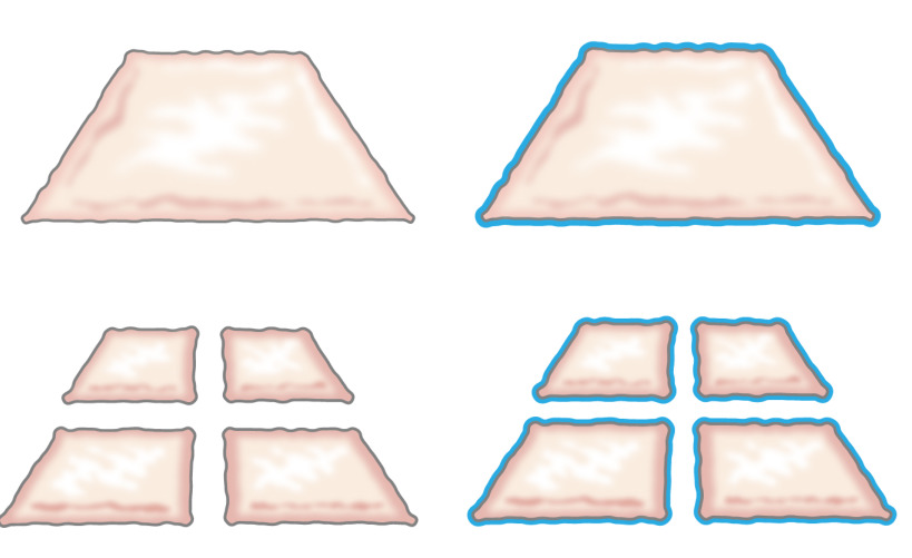
Şekil 1:. Meek teori Meek teorisine göre, epitel bir yara kenarlarından büyür. kıyma teknolojisi ile maruz alanını arttırarak, kıyılmış doku birçok noktalardan yaralar epithelializes.

Bu çalışma hipo dayanmaktadırAynı ilke, bir kalıp etrafında kıyılmış epitel yerleştirerek deri altı dokuya uygulanabilir tezi. (Iç vücuttan yabancı cisim (kalıp) yara alanı kaplar ve ayıran sürekli neoepithelium oluşturmak için epitel hücreleri yara alanları (göç) kapak, kıyılmış nakli (yeniden) den seferber olur ve bölme (genişletmek) Şekil 2).

şekil 2
Şekil 2:. Meek teorisine göre, in vivo vücut içi doku genişlemesi için kıyılmış epiteli ile 3D kalıp Karikatür deri altı dokuya bir kalıp üzerine yerleştirilir ve daha sonra nakledilen kıyılmış doku kullanarak, hipotez epitel hücreleri göç olduğunu kıyılmış doku kenarları, yeniden, ve yara alanı kaplar ve iç gövde arasında yabancı cisim (kalıp) ayıran bir kesintisiz neoepithelium oluşturacak şekilde genişler.

Önceki in vivo çalışmalar umut verici sonuçlar gösterse de rejenere epitel iyi 7 mekanik travma dayanacak, böylece daha fazla iyileştirmeler otogrefti takviye ile elde edilebilir. kolay besin ve atık ürünlerin yayılması, olasılık kalıbına 3D bir şekilde ve cerrahi işleme kolaylığı: Bu amaçla, başarılı bir biyomalzeme önemli bir önkoşul gibi, tespit edilmiştir. Sonuçlar bu ihtiyaçların kıyılmış dokuya kompozit biyo materyal ekleyerek karşılanabileceğini yapılmıştır.

kıyılmış dokudan oluşan bir iskele geliştirmeyi amaçlayan mevcut çalışma biyolojik olarak parçalanabilir bir kumaş takviye çekirdek içeren kollajen plastik sıkıştırılmış. Bu sayede, canlı hücreler kıyılmış doku parçacıklarının göç olabilir ve orijinal epiteli (deri veya ürotelyum) morfolojik özellikleri karakteristiği ile çoğalırlar. Plastik sıkıştırma kullanarak, iskele azaltmak olduyaklaşık 420 um kıyılmış parçacıkları olarak 1 cm arasında D üst katman kolajen kaplı edildi. Çekirdek kumaşın herhangi bir polimer olabilir, ancak kapsayan kollajen tabakaların 11, birbirleriyle amacıyla hidrofilik yüzey ile değiştirilmesi gereken olabilir.

yöntem kıyılmış mesane mukozasını veya domuz kıyılmış cilt kültürü için bir iskele olarak kullanarak iki plastik sıkıştırılmış kollajen jel içinde poli (ε-kaprolakton) oluşan bir örgü örgü (PCL) dahil ederek gelişmiş bir iskele bütünlüğü sağlanmış. Yapı, iyi konsolide hibrid yapısı üstünde tabakalı, çok katmanlı ürotelyum ve skuamöz deri epitelyumunun başarılı oluşumu gösteren in vitro en fazla 6 hafta içinde, hücre kültür koşullarında muhafaza edilmiştir. yapı işlemek için kolay oldu ve mesane büyütme amaçlı veya cilt kusurlarının örtülmesini yerine dikilir olabilir. Doku iskele tüm parçaları FDA onaylı ve teknik vardırDoku kıyma hasat, plastik sıkıştırma ve tek aşamalı müdahale olarak hastaya geri nakli tek aşamalı işlemleri için kullanılabilir. Prosedür herhangi bir genel cerrahi ünitesinde steril koşullar altında doku genişletme ve yeniden inşası için yapılabildi.

Protocol

Tüm hayvan protokolleri Hayvanlar Stockholm İl Komitesi tarafından önceden onaylanmış ve tüm işlemler hayvan kullanımı yanı sıra, ilgili federal yasalar için düzenlemeler uymuştur. 1. Hayvan Prosedürleri Cerrahi Hayvan Hazırlama steril koşullar altında çalışması için gerekli tüm malzeme ve enstrümanlar ile cerrahi tablo hazırlayın. enfeksiyon riskini azaltmak ve hayatta kalma cerrahi koşulları optimize etmek için steril koşullar altında sa…

Representative Results

Bu çalışma, kollajen ve kıyılmış doku plastik sıkıştırma kullanarak nakli için bir biyo materyal üretmek için nasıl gösteren bir yöntem sunar. Mesane mukozasının ve cilt hasat edilmiş ve daha sonra mekanik olarak küçük parçacıklar (Şekil 3) içine kıyılmış edilebilir. Plastik sıkıştırma ile, kıyılmış partiküller bir kollajen jel (Şekil 4) dış tabakaları içinde mekanik olarak güçlü olan bir merkezi olarak yerleş…

Discussion

Bu çalışma, cerrahi masada transplantasyon için otolog doku ile mesane duvar yamaları üretmek için kolay kullanımlı bir yaklaşım sunuyor. Yamalar ve plastik sıkıştırma ile kombinasyon halinde, dış yüzeylerde kıyılmış doku olmayan orta ve kollajen biyolojik olarak parçalanabilir bir polimer örgü kombinasyonu ile oluşturulur. Plastik sıkıştırma yöntemi daha önce başka yazarlar tarafından tanımlanan ve kolajen jellerin 12,13 sıvının hızla dışarı itilmesini olarak tanıml…

Disclosures

The authors have nothing to disclose.

Acknowledgements

The authors thank the Swedish Society for Medical Research, the Promobilia Foundation, the Crown Princess Lovisa Foundation, the Freemason Foundation for Children’s Welfare, the Swedish Society of Medicine, the Solstickan Foundation, Karolinska Institutet, and the Stockholm City Council for financial support.

Materials

Silicone catheter 10-French Preparing the animal for surgery , Section 1
DMEM 10X Gibco 31885-023 Plastic compression section 4
24 well plates Falcon 08-772-1 Plastic compression section 4
3'3,'5-Triiodothyronine Sigma-Aldrich IRMM469   In vitro culture; Section 5
4% PFA Labmed Solutions 200-001-8 Immunocytochemistry; Section 6
70% ethanol Histolab Immunocytochemistry; Section 6
ABC Elite kit: Biotin -Streptavidin detection kit Vector PK6102 Immunocytochemistry; Section 6
Absolute ethanol Histolab 1399.01 Immunocytochemistry; Section 6
Adenine Sigma-Aldrich A8626   In vitro culture; Section 5
Atropine 25 μg/kg  Temgesic, RB Pharmaceuticals, Great Britain Preparing the animal for surgery , Section 1
Azaperone 2 mg/kg  Stresnil, Janssen-Cilag, Pharma, Austria Preparing the animal for surgery , Section 1
Biosafety Level 2 hood  Plastic compression; Section 4
Blocking solution:  Normal serum from the same species as the secondary secondary antibody was generated in. Vector The blocking solution depends of the  origin of  first antibody Immunocytochemistry; Section 6
Buprenorphine 45 μg/kg Atropin, Mylan Inc, Canonsburg, PA Preparing the animal for surgery , Section 1
Carprofen 3 mg/kg    Rimadyl, Orion Pharma, Sweden Preparing the animal for surgery , Section 1
Chlorhexidine gluconate  Hibiscrub 40 mg/mL, Regent Medical, England Preparing the animal for surgery , Section 1
Cholera toxin  Sigma-Aldrich C8052   In vitro culture; Section 5
Coplin jar: staining jar for boiling Histolab 6150 Immunocytochemistry; Section 6
Stainless mold  (33x22x10 mm) custom made Plastic compression; Section 4
DMEM Gibco 3188-5023 Plastic compression section 4. Keep on ice  when using it in plastic compression
Epidermal growth factor Sigma-Aldrich E9644   In vitro culture; Section 5
Ethilon (non-absorbable monofilament for skin sutures) Ethicon Surgery, Section 1
Fetal bovine serum (FBS) Gibco 10437-036 Plastic compression section 4
Forceps (Adison with tooth) Preparing the animal for surgery , Section 1
Gauze (Gazin Mullkompresse)  Preparing the animal for surgery , Section 1
Ham´s F12 Gibco 31765-027 Plastic compression section 4
Hematoxylin Histolab 1820 Immunocytochemistry; Section 6
Humidity chamber DALAB Immunocytochemistry; Section 6
Hydrocortisone Sigma-Aldrich H0888   In vitro culture; Section 5
Hydrogen peroxide Solution 30% Sigma-Aldrich H1009 Immunocytochemistry; Section 6
Insulin Sigma-Aldrich I3536   In vitro culture; Section 5
Isoflurane  Isoflurane, Baxter, Deerfield, IL Preparing the animal for surgery , Section 1
Lidocaine 5 mg/ml Xylocaine, AstraZeneca, Sweden Preparing the animal for surgery , Section 1
Lucose 25 mg/mL  Baxter, Deerfield, IL Preparing the animal for surgery , Section 1
Marker pen pap pen Sigma-Aldrich Z377821-1EA Immunocytochemistry; Section 6
Medetomidine 25 μg/kg  Domitor, Orion Pharma, Sweden Preparing the animal for surgery , Section 1
Mincing device Applied Tissue Technologies LLC  Minced tissue preparation, section 2
Monocryl (absorbable monofilament) Ethicon Surgery, Section 1
NaCl Sigma-Aldrich S7653 Immunocytochemistry; Section 6
NaOH 1N Merck Millipore 106462 Plastic compression section 4 and cell culture
Nylon mesh, 110 uM thick pore size 0.04 sqmm Plastic compression; Section 4
Oculentum simplex APL: ointment for eye protection APL Vnr 336164 Surgery, Section 1
PBS Gibco 14190-094 Plastic compression section 4
Penicillin-Streptomycin Gibco 15140-122 Plastic compression section 4
Phenobarbiturate 15 mg/kg  Pentobarbital, APL, Sweden Preparing the animal for surgery , Section 1
PLGA Knitted fabric Plastic compression; Section 4
Rat-tail collagen First LINK, Ltd, UK 60-30-810 Plastic compression section 4, keep on ice
Scalpel blade – 15 Preparing the animal for surgery , Section 1
Shaving shears Preparing the animal for surgery , Section 1
Stainless stell mesh, 400 uM thick pore size  Plastic compression; Section 4
Steril gloves Preparing the animal for surgery , Section 1
Sterile gowns Preparing the animal for surgery , Section 1
Sterile drapes
Sterilium Bode Chemie HAMBURG Preparing the animal for surgery , Section 1
Suture Thread Ethilon Preparing the animal for surgery , Section 1
TE-solution (antigen unmasking solution) consist of 10 mM Tris and 1 mM EDTA, pH 9.0 10 mM Tris/1 mM EDTA,  adjust pH to  9.0
Tiletamine hypochloride 2,5 mg/kg Preparing the animal for surgery , Section 1
Transferrin  Sigma-Aldrich T8158   In vitro culture; Section 5
Trizma Base, H2NC  Sigma-Aldrich T6066 Immunocytochemistry; Section 6
Vector VIP kit: Enzyme  peroxidase substrate  kit Vector SK4600 Immunocytochemistry; Section 6
Vicryl (absorbable braded) Ethicon Surgery, Section 1
Tris buffer pH 7.6 (washing buffer) TE solution: Make 10X  (0,5M Tris, 1,5M NaCl) by mixing: 60,6 g Tris (Trizma Base, H2NC(CH2OH)3, M=121.14 g/mol), add 800 ml  distilled water adjust the pH till 7.6, add 87,7 g NaCl and fill to 1000 ml with  distilled water. Dilute to 1X with distilled water.
X-tra solv (solvent) DALAB 41-5213-810 Immunocytochemistry; Section 6. Use under fume hood
Zolazepam hypochloride Zoletil, Virbac, France Preparing the animal for surgery , Section 1
Depilatory wax strips Veet Preparing the animal for surgery , Section 1
Pentobarbital sodium Lundbeck Termination, Section 3

References

  1. Rheinwald, J. G., Green, H. Serial cultivation of strains of human epidermal keratinocytes: the formation of keratinizing colonies from single cells. Cell. 6, 331-343 (1975).
  2. Fossum, M., Nordenskjold, A., Kratz, G. Engineering of multilayered urinary tissue in vitro. Tissue Engineering. 10, 175-180 (2004).
  3. Salmikangas, P., et al. Manufacturing, characterization and control of cell-based medicinal products: challenging paradigms toward commercial use. Regen Med. 10, 65-78 (2015).
  4. Fossum, M., et al. Minced skin for tissue engineering of epithelialized subcutaneous tunnels. Tissue Engineering. Part A. 15, 2085-2092 (2009).
  5. Fossum, M., et al. Minced urothelium to create epithelialized subcutaneous conduits. The Journal of Urology. 184, 757-761 (2010).
  6. Reinfeldt Engberg, G., Lundberg, J., Chamorro, C. L., Nordenskjold, A., Fossum, M. Transplantation of autologous minced bladder mucosa for a one-step reconstruction of a tissue engineered bladder conduit. BioMed Research International. 2013, 212734 (2013).
  7. Meek, C. P. Successful microdermagrafting using the Meek-Wall microdermatome. Am J Surg. 96, 557-558 (1958).
  8. Tanner, J. C., Vandeput, J., Olley, J. F. The Mesh skin graft. Plastic and Reconstructive Surgery. 34, 287-292 (1964).
  9. Svensjo, T., et al. Autologous skin transplantation: comparison of minced skin to other techniques. The Journal of Surgical Research. 103, 19-29 (2002).
  10. Ajalloueian, F., Zeiai, S., Rojas, R., Fossum, M., Hilborn, J. One-stage tissue engineering of bladder wall patches for an easy-to-use approach at the surgical table. Tissue Engineering. Part C, Methods. 19, 688-696 (2013).
  11. Engelhardt, E. M., et al. A collagen-poly(lactic acid-co-varepsilon-caprolactone) hybrid scaffold for bladder tissue regeneration. Biomaterials. 32, 3969-3976 (2011).
  12. Brown, R. A., Wiseman, M., Chuo, C. B., Cheema, U., Nazhat, S. N. Ultrarapid engineering of biomimetic materials and tissues: fabrication of nano- and microstructures by plastic compression. Adv Funct Mater. 15, 1762-1770 (2005).
  13. Fumagalli Romario, U., Puccetti, F., Elmore, U., Massaron, S., Rosati, R. Self-gripping mesh versus staple fixation in laparoscopic inguinal hernia repair: a prospective comparison. Surg Endosc. 27, 1798-1802 (2013).
  14. Muangman, P., et al. Complex Wound Management Utilizing an Artificial Dermal Matrix. Annals of Plastic Surgery. 57, 199-202 (2006).
  15. Ajalloueian, F., Zeiai, S., Fossum, M., Hilborn, J. G. Constructs of electrospun PLGA, compressed collagen and minced urothelium for minimally manipulated autologous bladder tissue expansion. Biomaterials. 35, 5741-5748 (2014).
  16. Orabi, H., AbouShwareb, T., Zhang, Y., Yoo, J. J., Atala, A. Cell-seeded tubularized scaffolds for reconstruction of long urethral defects: a preclinical study. Eur Urol. 63, 531-538 (2013).
  17. Blais, M., Parenteau-Bareil, R., Cadau, S., Berthod, F. Concise review: tissue-engineered skin and nerve regeneration in burn treatment. Stem Cells Transl Med. 2, 545-551 (2013).
  18. Serpooshan, V., Muja, N., Marelli, B., Nazhat, S. N. Fibroblast contractility and growth in plastic compressed collagen gel scaffolds with microstructures correlated with hydraulic permeability. J Biomed Mater Res A. 96, 609-620 (2011).
check_url/kr/53061?article_type=t

Play Video

Cite This Article
Chamorro, C. I., Zeiai, S., Reinfeldt Engberg, G., Fossum, M. Minced Tissue in Compressed Collagen: A Cell-containing Biotransplant for Single-staged Reconstructive Repair. J. Vis. Exp. (108), e53061, doi:10.3791/53061 (2016).

View Video