This study describes a simplified surgical procedure and technique for performing cavernous nerve stimulation with the isolation of the nerve-electrode complex using silicone glue and intracavernous pressure measurement.
The stimulation of the cavernous nerve (CN) and measurement of intracavernous pressure (ICP) have been used extensively to test and evaluate therapies for erectile dysfunction. However, the methods used vary between laboratories, and pitfalls still exist. The goal of this study was to describe a surgical technique that would provide a reliable and reproducible model. By exposing the ischiocavernosus muscle at its point of insertion on the ischial tuberosity, the penile crus could be cannulated with minimal dissection and injury to the structures involved in erectile function. Repeated stimulation of the CN, without the need for lifting and drying, was achieved by using a 125 µm bipolar silver electrode and biocompatible silicon glue to isolate the electrode-nerve complex. This method prevents neuropraxia by reducing stretching and drying the nerve and provides complete isolation of the nerve, negating electrical leakage and preventing stimulation of alternative pathways.
In vivo study of erectile function in experimental animals started in 1863 with the pioneering experimental work of Eckhard1. Electrostimulation of the pelvic nerves was used to induce the increased ICP in dogs. Throughout the 20th century, similar experimental protocols were used in larger animals such as dogs, monkeys, cats and rabbits. Evaluating erectile function in a rat was first developed by Quinlan et al. in 19892. The method has since been modified and updated by several other groups34. Today, the rat is the most widely used animal model for studying the pathology of erectile dysfunction and evaluating emerging treatment options. The main steps of the procedure include, recording systemic blood pressure using a line in the carotid artery, cannulation of the penile crus to measure ICP, and stimulation of the CN to induce an increase in the ICP. Although several researchers have refined the model, its reproducibility remains a problem, and variable results have been reported by different laboratories. Several pitfalls still persist.
Previous articles5,6,7,8,9,10 describe the use of full penile exposure with degloving of the penis for cavernous body cannulation. This is not an optimal approach as manipulation and disruptive dissection causes injury to structures, which are essential to erectile function. The dissection of the CN has been well described10,11, but stimulation of the nerve is not optimal due to multiple factors that could affect experimental results. The technique of CN stimulation includes lifting the nerve from the surrounding tissue by pulling on the bipolar hook electrode, which is positioned around the nerve, and drying the nerve before each stimulation. This can lead to various degrees of nerve damage and electrical current leakage, resulting in a diminished response or false increase in the ICP through stimulation of alternative pathways e.g., pelvic floor muscles, bladder and gastrointestinal tract12. All of these factors limit reproducibility.
During our study, we observed that both the depth and type of anesthesia have a profound effect on the ICP. The anesthetics used are sodium pentobarbital, ketamine/xylazine or ketamine/midazolam injection, or isoflurane/oxygen inhalation.
Here we describe a simplified surgical method and provide data in support of standardization of the experimental protocol.
Animals were housed in the University of Southern Denmark Animal Care Facility as per institutional guidelines. All animal experiments were carried out in accordance with the National Institutes of Health guide for the care and use of laboratory animals. This is an acute, non-survival surgery procedure.
1. Preparation of Tubing, Electrode, and Instruments for the Surgical Procedure
2. Preparation of the Animal
3. Presurgical Preparation
4. Ischiocavernosus Muscle Dissection for the ICP Measurement
5. CN Dissection for Stimulation
6. Cavernous Body Cannulation for the ICP Measurement
7. Stimulation of the CN
The use of this protocol with the recommended stimulation settings, under inhalation anesthesia with isoflurane 2.0% oxygen 0.8 L/min, should produce results as shown in Figure 11 and Figure12, where there is multiple back to back stimulations between 75-80 mm Hg. Figure 13 shows the same stable response over a 20-min stimulation with the response stable at 73-77 mm Hg. Test the line for ICP measurement by flushing the tube and tapping on the crus (Figure 9). The rapid response back to baseline is the hallmark of a well-placed line. If the integrity of the tunica albuginea is damaged, the test would result in lower peak pressure and slow response back to baseline after flushing and tapping, and a diminished response when stimulating (Figure 14). There would also be leakage of the heparinized NaCl when flushing and bleeding during stimulations.
The types and levels of anesthesia as well as the use of oxygen had a major impact on ICP. Figure 15 demonstrates the effect of the different levels of isoflurane on ICP, with both a decreased response and a less stable plateau. With isoflurane at 2%, there was a stable response in the ICP measurement with multiple stimulations on 78 mm Hg. Increasing the concentration of isoflurane to 3.5%, however, resulted in a rapid 50% decrease to 34 mm Hg in multiple, subsequent stimulations. The same effect was observed when switching the isoflurane from 2.0% to 3.0%, where a 19% decrease in response was observed, and from 2.5% to 5%, where an even more rapid decrease in response of 70% was seen. Blood pressure remained stable throughout all stimulations. In rats anesthetized using the isoflurane/oxygen anesthesia during surgery and initial stimulations, who then received 25% of the recommended dose of fentanyl/midazolam (while the isoflurane was discontinued), there was a similarly stable response but it increased by 25% during the fentanyl/midazolam anesthesia compared to isoflurane (Figure 16).
Administration of oxygen through a nose cone increased the oxygen saturation in the blood from 61-75% to 99-100% in about 20 s. When the oxygenation was stopped, the same decrease was seen over approximately 1 min. Blood pressure was stable throughout the stimulations, but oxygen administration through a nose cone (0.8 L/min) had a large effect on the maximum ICP measurement, reducing it by 35-45% in back-to-back stimulations (Figure 17).
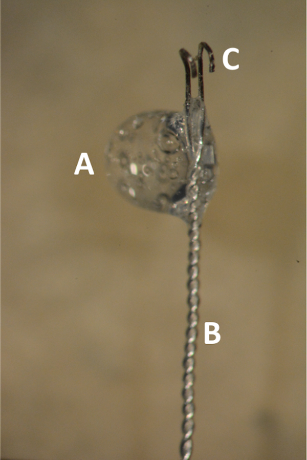
Figure 1. The bipolar Teflon coated silver electrode. (A) Glue bubble in the transition zone between the coated and uncoated electrode. (B) Distal 2 cm of the electrode tightly braded. (C) Parallel uncoated hooks 1 mm apart. Please click here to view a larger version of this figure.
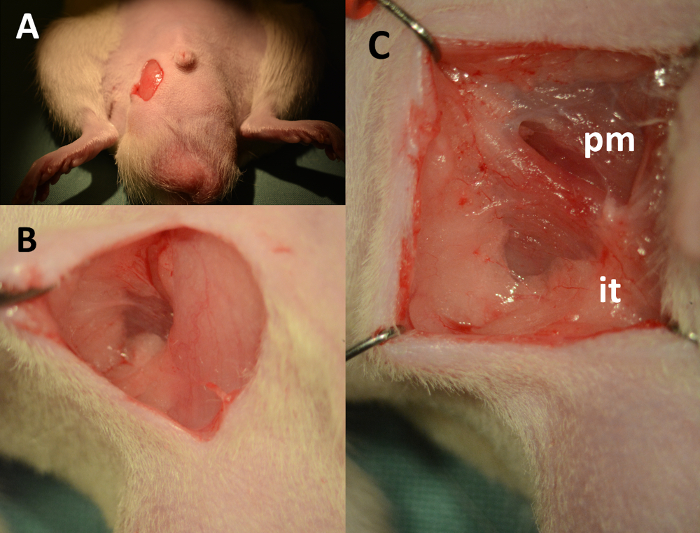
Figure 2. Dissection of the cavernous body – landmarks. (A) 1 cm vertical skin incision, downward starting 2 mm lateral from the base of the penis. (B) Fascia lateral to the scrotum separated using cotton-tipped swabs. (C) View of the operating field after placement of retractors (pm: pyramidalis muscle, it: insertion point of crus to the ischial tuberosity). Please click here to view a larger version of this figure.
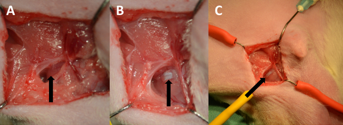
Figure 3. Exposing the tunica albuginea. (A) The ischiocavernosus muscle (arrow). (B) Tunica albuginea (arrow). (C) Low power magnification showing the course of the cavernous body. Please click here to view a larger version of this figure.
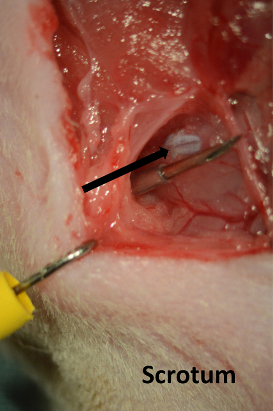
Figure 4. Line for cavernous pressure recording. Needle introduced through the skin on the perineum running parallel with cavernous body (arrow). Please click here to view a larger version of this figure.
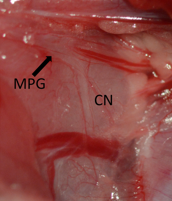
Figure 5. Exposure of the MPG marked by arrow and CN running vertical on the dorsolateral aspect of the prostate. Major pelvic ganglion (MPG) marked by arrow, cavernous nerve (CN). Please click here to view a larger version of this figure.
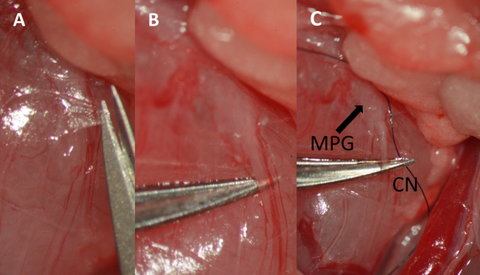
Figure 6. Dissection of the cavernous nerve. (A) Cutting the fascia overlying the cavernous nerve with micro scissors. (B) Separating the nerve from the underlying tissue using micro forceps. (C) Placing a 9-0 ligature underneath the nerve. Major pelvic ganglion (MPG) marked by arrow, cavernous nerve (CN). Please click here to view a larger version of this figure.
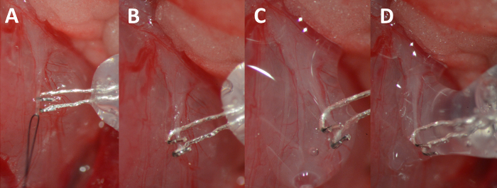
Figure 7. Hooking of the nerve. (A) Elevating the nerve by gently pulling on the suture. (B) Nerve resting in the hooks of the electrode. (C) Nerve and electrode complex isolated with the biocompatible silicon glue. (D) Additional glue bubble added to completely isolate the nerve-electrode complex. Please click here to view a larger version of this figure.
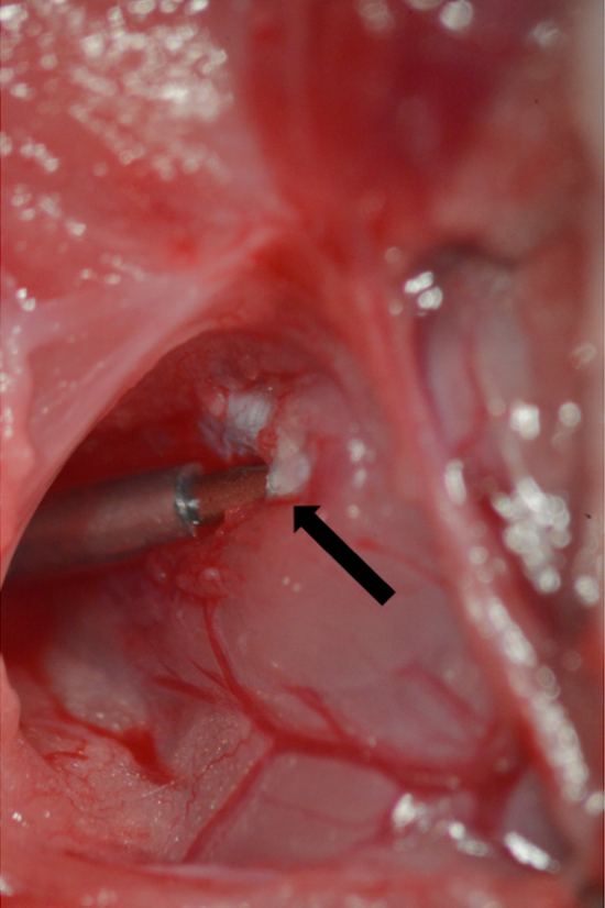
Figure 8. Cannulation of the tunica albuginea. A 23 G needle connected to PE-50 tubing inserted into the tunica albuginea. Point of insertion marked by arrow. Please click here to view a larger version of this figure.
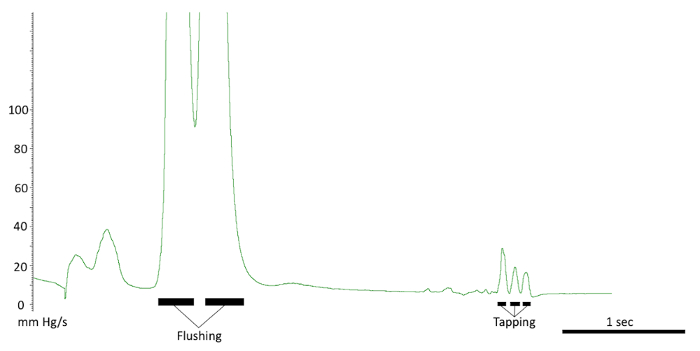
Figure 9. Testing the intracavernous line. The responses seen with a correct line placement. Note the flushing of the line and response to tapping on the crus. Also, note the quick pressure drop back to baseline. Please click here to view a larger version of this figure.
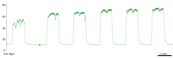
Figure 10. Initial responses to cavernous nerve stimulation. Diminished first response. First stimulation of 50 mm Hg and a fluctuating plateau. Second and third stimulation of 66 mm Hg. The following measurements were recorded at the normal level at 73 mm Hg. Please click here to view a larger version of this figure.
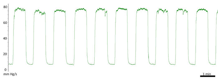
Figure 11. Repeated cavernous nerve stimulation and intracavernous pressure recording. Showing the stability of the results using this protocol. Ten back-to-back stimulations between 75-78 mm Hg. Please click here to view a larger version of this figure.
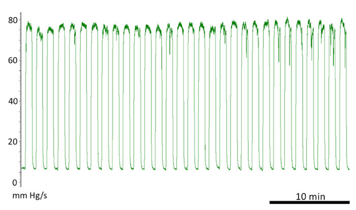
Figure 12. Cavernous nerve stimulation and pressure recording. Approximately 30 back-to-back stimulations with < 6 mm Hg variability in the pressure. Please click here to view a larger version of this figure.
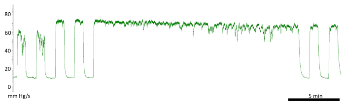
Figure 13. Continuous stimulation lasting 20 min. Increased fluctuation at the end, but the subsequent stimulations, after a 1 min rest, produced a stable response. Please click here to view a larger version of this figure.
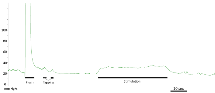
Figure 14. Leaking tunica albuginea. Prolonged response back to baseline after flushing and tapping. Decreased response after stimulation. Please click here to view a larger version of this figure.
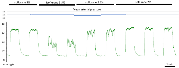
Figure 15. The effect of anesthesia dose on intracavernous pressure. Decreased and more fluctuating response on increasing isoflurane compared to the stable response on 2% in the first two, and the last three stimulations. The blue trace on top showing constant mean arterial pressure. Please click here to view a larger version of this figure.
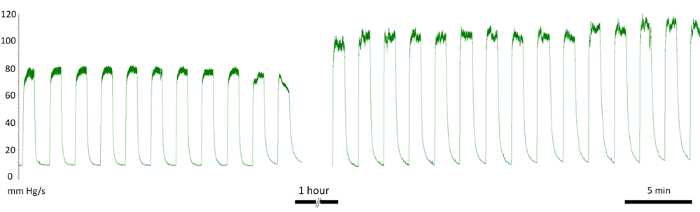
Figure 16. The effect of anesthesia type on intracavernous pressure. The initial stimulations performed under isoflurane anesthesia show a pressure increase of 80 mm Hg, once fentanyl/midazolam was administered there was an increase in response to 110 mm Hg. Please click here to view a larger version of this figure.
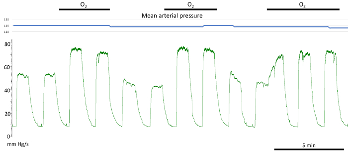
Figure 17. The effect of oxygen administration through the nose cone. Discontinuing oxygen administration through the nose cone resulted in a significant reduction in cavernous pressure increase with cavernous nerve stimulation. No effect on the mean arterial pressure was noted (blue trace above). Please click here to view a larger version of this figure.
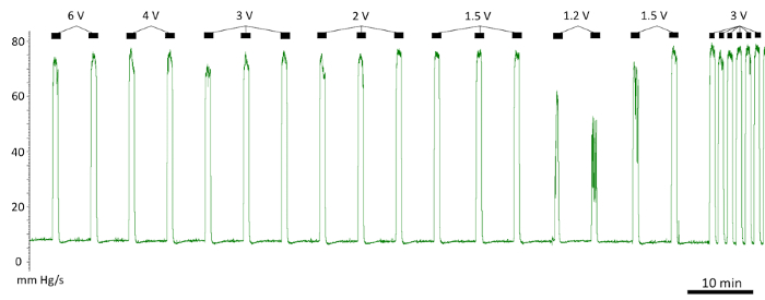
Figure 18. The effect of voltage on pressure response to stimulation. The voltage between 1.5-6 V produced an identical pressure response. The response diminished below 1.5 V. Please click here to view a larger version of this figure.
The principal goal of this study was to describe a simplified surgical technique of penile crus cannulation for ICP recording and isolation of the CN for electrostimulation. We introduced modifications to the dissection of the cavernous body to simplify the surgery and provide reproducible recordings of the increase of the ICP with CN stimulation. With a 1 cm vertical skin incision, lateral to the base of the penis, using the palpable ischial tuberosity as guidance, we achieved good exposure of the ischiocavernosus muscle and tunica albuginea. This procedure is quicker (under 15 min) than those described in the literature and causes minimal tissue disruption2,3,4.
The currently used technique of CN stimulation includes hooking, lifting, and drying of the CN before each electrostimulation is applied10. This technique does not guarantee that the conditions remain the same for each stimulation. Also, lifting of the nerve requires the use of a micromanipulator and repeated stretching and releasing of the nerve, which could lead to neuropraxia. Compared to the current CN stimulation technique, the use of a biocompatible silicon glue to isolate the nerve-electrode complex reduces the frequency of the nerve stretching to two instances; once to place the bipolar electrode around the nerve, and once to isolate the nerve and the electrode using glue. Subsequently, multiple neurostimulations could be performed without the need for nerve manipulations.
Insertion of the needle into the cavernous body for pressure recording constitutes a critical step. New users of this technique should practice this part before starting experiments. When deploying the needle into the crus, it is critical that the crus is stretched and the needle is deployed parallel to its course. The other critical step is the nerve handling during its dissection from the surrounding tissue and placement of the electrode underneath. While dissection itself is not a difficult task, any stretch or crush of the nerve could result in damage. Our modification of this step makes the need for a micromanipulator unnecessary and simplifies nerve handling. The use of the biocompatible silicon glue to isolate the nerve and provide stable and reliable contact between the electrode and the nerve reduces the necessary manipulation. Silicone glue could also be used in other animal models where in vivo neurostimulation is applied.
The stimulation parameters listed in numerous studies vary from 14-20 Hz and 1.5-12 V11,13,14. This technique showed that stimulation using a 1.5 mA, 16 Hz, 3 V, and 5 ms pulse produced a full response. With increasing stimulation parameters, the ICP does not increase (Figure 18). This suggests that stimulation using any parameters, which are above the threshold for triggering arterial (cavernous artery), arteriolar, and sinusoidal smooth muscle relaxation, are sufficient to trigger the reflex and would result in a full physiological response. The subsequent increase in the frequency, amplitude, or pulse width does not lead to a stronger physiological response and it could deplete neurotransmitters or even cause nerve injury. With the above-described parameters, we were able to achieve a reproducible response with a rest period as short as 30 s more than 40 times in a row.
The depth of anesthesia clearly affects the physiological response. With inhalation anesthesia, it could be well controlled, however the peak response is approximately 25% lower, when compared to fentanyl/midazolam anesthesia administered by injection. We observed that the dose of isoflurane could be maintained at the level determined by the minimum isoflurane concentration necessary to eliminate toe pinch response. Most investigators, use injection anesthesia with sodium pentobarbital, fentanyl/midazolam, ketamine/xylazine, or ketamine/midazolam15.
None of the published articles mentioned the use of controlled oxygenation. Results from this study show that this had a major impact on ICP. Therefore, it is important that, regardless of type of anesthesia used, the animal receives oxygen through the nose cone.
The use of rats is advantageous due to their size and resilience. Both in previously published studies and confirmed in this research, Sprague Dawley rats have been shown to have an intact response to CN stimulation in the range of 6-12 weeks old and weighing 200-550 g. Using mice is more challenging, but beneficial due to the availability of transgenic technology12.
The authors have nothing to disclose.
The authors have no acknowledgements.
Adson forceps | Fine Science Tool | 11006-12 | |
Dumont #7 forceps | Fine Science Tool | 11271-30 | |
Dumont #5 forceps | Fine Science Tool | 11273-20 | |
ToughCut Mayo scissor | Fine Science Tool | 14110-15 | |
Miniature Vannas Spring scissor | Fine Science Tool | 15006-09 | |
Ultra Fine Hemostat | Fine Science Tool | 13020-12 | |
Crile Hemostat | Fine Science Tool | 13004-14 | |
Kwik-Sil Adhesive | World Precision Instruments | KWIK-SIL | |
Teflon coated silver wire 0.125 mm | World Precision Instruments | AGT0510 | |
Elastic wire retractors | Custom made | ||
Scalpel blade | Fine Science Tool | 10023-00 | |
PE-50 tubing | Warner Instruments | 64-0753 | |
23 G Needle | Kruuse | 121272 | |
SD-9 Square Pulse Stimulator | Somatco | 1077/183 | |
Blood pressure transducer and cable | World Precision Instruments | BLPR2 | |
Raucotupf Cotton-tipped Applicators | Lohmann-Raucher | 11966 | |
Pro-ophta Ocular Sticks | Lohmann-Raucher | 16515 | |
NaCl 0,9 % 100 mL | Local pharmacy | ||
Heparin | Local pharmacy | ||
25 mL Syringe | Odense University Hospital | ||
Vet eye ointment – Viscotears | Local pharmacy | ||
silver wires | Science Products GmbH, Heidelberg, Germany | ||
Silicon Glue | Kwik-Sil, World Precision Instruments, Sarasota, FL, USA |