This protocol describes a new intraoperative imaging technique that uses a ruthenium complex as a source of chemiluminescent light emission, thereby producing high signal-to-noise ratios during in vivo imaging. Intraoperative imaging is an expanding field that could revolutionize the way that surgical procedures are performed.
Intraoperative imaging techniques have the potential to make surgical interventions safer and more effective; for these reasons, such techniques are quickly moving into the operating room. Here, we present a new approach that utilizes a technique not yet explored for intraoperative imaging: chemiluminescent imaging. This method employs a ruthenium-based chemiluminescent reporter along with a custom-built nebulizing system to produce ex vivo or in vivo images with high signal-to-noise ratios. The ruthenium-based reporter produces light following exposure to an aqueous oxidizing solution and re-reduction within the surrounding tissue. This method has allowed us to detect reporter concentrations as low as 6.9 pmol/cm2. In this work, we present a visual guide to our proof-of-concept in vivo studies involving subdermal and intravenous injections in mice. The results suggest that this technology is a promising candidate for further preclinical research and might ultimately become a useful tool in the operating room.
In recent decades, imaging technologies have revolutionized the way that physicians diagnose and monitor disease. These imaging technologies, however, have been largely limited to whole body imaging systems, such as positron emission tomography (PET), single photon-emission computed tomography (SPECT), computed tomography (CT), and magnetic resonance imaging (MRI). Particular attention has been paid to cancer, and technological imaging breakthroughs have greatly improved the way that this disease is diagnosed and treated. Despite these advances, there is one place where these imaging technologies just don't fit: the operating room. While whole body imaging techniques can help in surgical planning, they typically lack spatial resolutions high enough to help physicians determine in real-time whether all of the tumor tissue has been removed or residual tumor tissue remains hidden at the surgical margins1. Making sure that no infiltrative tumor margins are left behind is one of the most important surgical goals, and surgeons must walk a tight-rope between rigorous and cautious tissue resection. If too much is removed, unwanted side effects for the patient are exacerbated; if too little is removed, recurrence rates are increased2,3. Therefore, it is crucial to delineate accurate tumor margins, and we believe that chemiluminescent intraoperative imaging can help to improve the accuracy of the identification of tumor margins by helping surgeons to visualize malignant tissue that could otherwise remain undetected with established techniques.
There are many imaging technologies currently being investigated for their possible utility as intraoperative imaging systems. These include β- and γ-radiation-emitting probes4, optical fluorescence5, Raman spectroscopy6,7, and Cherenkov luminescence8,9. To date, however, none of these have become established as standard clinical tools. Optical fluorescence imaging has so far proven to be the most promising of these techniques and is therefore the most explored. While it has already been shown to be a valuable tool for many applications, it is not without its limitations. Indeed, its principal drawback is the background fluorescence generated by inherently autofluorescent biological tissue. This background autofluorescent signal is a product of the excitation of the surrounding tissue, in addition to the fluorophore, by the external light source needed for the generation of a fluorescent signal. From a practical perspective, this autofluorescence can potentially lead to low signal-to-noise ratios, which can limit the utility of this technology in the operating room.
The principal advantage of chemiluminescence imaging over fluorescence imaging is that no excitation light is necessary. As a result, there is no background autofluorescence. In chemiluminescence imaging, the excitation energy is instead generated chemically. This process produces no unintended background signal and therefore can result in higher signal-to-noise ratios. This could ultimately result in the more precise and accurate detection of surgical margins. Somewhat surprisingly, the utility of this approach as an intraoperative imaging technique has remained unexplored10. Indeed, the closest example to this technique is the oxidation of luminol by myeloperoxidase in mice11,12,13. Chemiluminescent biomedical imaging is therefore a rather unexplored area of research that could offer the following advantages: (1) minimal autofluorescence resulting in a low background signal with higher signal-to-noise ratios; (2) tunable wavelengths of chemiluminescent emissions ranging from the visible to the near-infrared; and (3) functionalizable chemiluminescent complexes that, when combined with linker technologies and targeted biomolecules that already exist, provide access to whole libraries of targeted molecular imaging probes14.
This proof-of-principle study illustrates the potential utility of chemiluminescent imaging in the biomedical setting using a ruthenium-based imaging agent. The chemiluminescent properties of this compound are well studied, with investigations dating back to the mid-1960s15. Upon chemical activation, the agent produces light at around 600 nm16, which is well suited for medical imaging purposes. The activation energy is provided by a redox reaction that leads to an excited state-which has a lifetime of 650 ns in water17-followed by the generation of photons upon relaxation of this excited state. Through the use of a specially-designed remote nebulizer, we were able to detect the compound both ex vivo and in vivo. The results of initial experiments are very promising, suggesting further investigation of this technology.
Ethics statement: All of the in vivo animal experiments described were performed according to an approved protocol and under the ethical guidelines of the Memorial Sloan Kettering Cancer Center (MSK) Institutional Animal Care and Use Committee (IACUC).
1. Construction of a Nebulizing Device
2. Sensitivity Determination of the Method
3. In Vivo Imaging After Systemic Intravenous Injection
4. In Vivo Imaging of Lymph Nodes
The nebulizer system described in protocol section 1 can be constructed from easily-available materials at a low cost. It is intended to be an inset for remote-triggered spraying of the reducing/oxidizing agent inside a bioluminescent reader (Figure 1). Our design allows for the safe operation of the nebulizer within the bioluminescence reader at a 14 cm distance from the lens. No fogging or blurring of the lens was observed during the operation. We selected the commercially-available chemiluminescent agent [Ru(bpy)3]Cl2 for the development of our method based on its low price, stability in aqueous solution, well-described redox behavior, and chemiluminescent properties (Figure 2)19. The minimal detectable signal can be determined as described in protocol section 2 by oxidizing one drop of [Ru(bpy)3]Cl2 (100 µL, 6.9 pmol- 347 nmol in H2O) with (NH4)2Ce(NO3)6 (100 µL, 25 mM) on a microscope slide. Then, by using the nebulizer and spraying on a solution of triethylamine (1:3 in water/ethanol), the chemiluminescent signal is triggered. In our case, the minimal detectable signal was determined to be 6.9 pmol/cm2 (Figure 3). It is conceivable, though, that optimized reaction conditions, camera sensitivities, shutter times, volumes, and reagent concentrations might lead to even lower detection thresholds. These reaction conditions can also be used for exploring and testing the chemiluminescence of any given combination of metal complexes, oxidizing agents, and reductants.
Moving to the in vivo experiments in protocol sections 3 and 4, female nude (outbred) mice 5-6 weeks old and NU/J male mice 6-8 weeks old were used. For intravenous injections, amounts of 8-33 nmol of [Ru(bpy)3]Cl2 in 100 µL of PBS per mouse (n = 5) were chosen. The animals were sacrificed 10 min after injection, and the abdominal cavity was exposed. The mice were placed in the bioluminescent reader with the nebulizer pointing towards the tissue of interest (Figure 4). For imaging with intravenously-injected [Ru(bpy)3]Cl2, the chemiluminescent signal was detected predominantly in the kidneys, strongly suggesting renal elimination of the hydrophilic small molecule (Figure 5). Signal-to-noise ratios for mice injected with [Ru(bpy)3]Cl2 versus PBS were 27/1 for the kidney and 21/1 for the liver. For lymph node imaging, 80 nmol of [Ru(bpy)3]Cl2 in 10 µL of PBS were injected subdermally into the hind foot pad of mice (n = 5). Mice were sacrificed 15 min post injection by CO2 asphyxiation. The skin covering both the inner hind legs was removed to expose the muscle, lymph nodes, and lymphatic vessels. Subsequent chemiluminescent visualization of the popliteal lymph nodes led to the observation that lymph nodes containing [Ru(bpy)3]2+ show a 10 ± 4.3-fold higher radiance than untreated ones (167,000 p/(s×cm2×sr) and 17,000 p/(s×cm2×sr); P <0.028) (Figure 6).
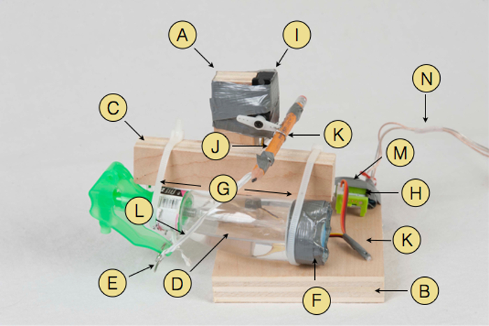
Figure 1: Photograph of the Nebulizer. Parts used: Wooden structure parts (A, B, C), spray bottle (D), bent steel rod (E), duct tape (F), plastic cable ties (G), 011 servo connector part (H), servo motor (I), pencil (J) held by bent paper clip (K), plastic covered wire twist ties (L) w1 wire connector (M) and speaker cable (N) leading to the battery. This figure is based on research originally published in reference19. Please click here to view a larger version of this figure.
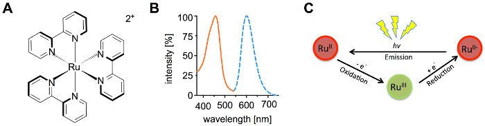
Figure 2. Properties of [Ru(bpy)3]2+. Structure (A) and excitation and emission spectra (B) of [Ru(bpy)3]2+. The oxidation/reduction based chemiluminescent catalytic cycle (C). This figure is based on research originally published in reference19. Please click here to view a larger version of this figure.
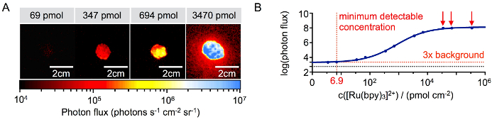
Figure 3: Detection Threshold of [Ru(bpy)3]2+. Representative signal intensities at different concentrations of [Ru(bpy)3]2+ on a microscope slide (A). Imaging signal quantification with detection threshold (red dotted line) and background (black dotted line) (B). This figure is based on research originally published in reference19. Please click here to view a larger version of this figure.
Figure 4: Chemiluminescence Imaging. Schematic drawing of a mouse and a nebulizer positioned in the bioluminescence reader (A) and schematic drawing (B) of the nebulizer spraying on a mouse. This figure is based on research originally published in reference19. Please click here to view a larger version of this figure.
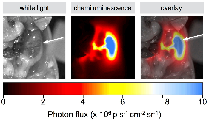
Figure 5: Detection of [Ru(bpy)3]2+ after Systemic Administration. White light, chemiluminescence, and overlay (from left to right). Images of a mouse body cavity that was injected with 33 nmol of [Ru(bpy)3]2+ and sprayed with (NH4)2Ce(NO3)6. The white arrow points towards the right kidney. This figure is based on research originally published in reference19. Please click here to view a larger version of this figure.
Figure 6: Detection of [Ru(bpy)3]2+ after Subdermal Administration. Popliteal lymph node imaging showing white light, chemiluminescence, and composite pictures for mice injected with [Ru(bpy)3]2+ (top) and PBS (bottom) in the hind limbs; 80 nmol in 10 µL of PBS, imaged 15 min after injection (A). White light and composite images for [Ru(bpy)3]2+ (top) and PBS (bottom)-treated excised popliteal lymph nodes (B). Quantification of chemiluminescent signals for PBS and [Ru(bpy)3]2+-treated lymph nodes (C).The data represents the mean ± SD. This figure is based on research originally published inreference19. Please click here to view a larger version of this figure.
Here, we have presented a technology that is capable of optically delineating tissue via the emission of photons created by a chemiluminescent reporter. In contrast to other, more established, technologies4,5,6,7,8,9, this chemiluminescent reporter system employs an imaging probe that is non-radioactive and facilitates detection at very high sensitivity levels. Perhaps even more importantly, chemiluminescence imaging does not require an incident light source (as in optical fluorescence imaging)20, a trait that minimizes autofluorescence and drastically reduces background signals.
The ruthenium reporter [Ru(bpy)3]Cl2 has an in vivo toxicity tolerable for imaging purposes (intraperitoneal mouse LD50: 20 mg/kg)21, is water soluble (up to 8 mM), and is stable in the bloodstream. The physicochemical properties of the metal complex are well-characterized and have already been investigated for the photodynamic therapy of cancer22,23. The oxidizing agent (NH4)2Ce(NO3)6 has been reported to have very low toxicity (oral rat LD50: 1600-3200 mg/kg)24 and is soluble in water at concentrations of up to 2.57 M at 20 °C25. In this article, a visual demonstration as well as text-based guidance for the construction of a remotely-operated nebulizing device are presented. In addition, we provide robust protocols for performing chemiluminescence imaging in a standard bioluminescence imaging device. We illustrate the use of [Ru(bpy)3]Cl2 for the visualization of tissues after both intravenous and subdermal injections in mice.
However, as with any other nascent imaging technology, there is room for improvement of our protocols. We believe that this proof-of-principle study could spur the development of multiple chemiluminescence applications for living systems. The following points could be addressed to further improve the technology and expand its scope.
A smaller second generation of remotely triggered spraying devices would allow the sample to be closer to the camera, hence improving spatial resolution. Improved optical equipment might further improve the detection limits of the method. The protocol could also be extended to imaging live animals. Exact control of the torque (by current and voltage) would allow a more exact control of the volume of reagent released with each spray. It is important to keep the nebulizer well-maintained. Not rinsing the nebulizer may destroy the nozzle. A fresh battery is crucial for the proper performance of the nebulizer. However, all the materials used for the nebulizer are inexpensive and readily commercially available. Following established synthetic protocols, the [Ru(bpy)3]2+ complex can easily be modified with various linkers, including maleimides26, amines27, and NHS esters28,29. This would enable bioconjugation to small molecules, peptides, or antibodies, and would thus facilitate specific molecular targeting30,31,32,33. Ultimately, targeted probe delivery could enable surgeons to identify small lesions and to accurately delineate surgical margins in the operating room with very high specificity. Also, the encapsulation of the highly water-soluble [Ru(bpy)3]2+ in nanomaterials-both targeted and untargeted-may also allow for the visualization of lesions while they are being surgically removed34,35,36. Finally, modifying the coordination sphere of the metal complex reporter and/or changing the transition metal center itself represent attractive routes to modulate and fine-tune the emission wavelengths within the visible and NIR ranges37,38.
Intraoperative chemiluminescence imaging needs a chemiluminescent reporter and, in our case, an oxidant, which can only be used within the limits of their toxicity and solubility. Tissue membranes can represent a barrier for the diffusion of the oxidant into the tissue, and hence, the signal generation. Since the chemiluminescent reporter is only generating one photon per cycle, the generated signal is rather weak. The ambient light in the operating room will therefore have to be prevented from entering the camera while the technique is in use. This might render ICI particularly interesting for the development of laparoscopic applications, where ambient light is naturally excluded.
We hope that this method may turn into a valuable tool for surgeons in the operating room. The absence of radioactivity is beneficial to the patient and operating team alike and makes fewer safety precautions necessary, potentially rendering this technique into a more attractive alternative.
The smooth operation of the nebulizer and its positioning play a crucial role for obtaining good results. Suboptimal angles and areas may contribute to signal variance. The control cable must be put through the door with care, and enough cable has to remain inside the bioluminescence reader so that it is not cramped or torn off.
Ultimately, chemiluminescence imaging is an extremely attractive new approach to molecular imaging. It is based on a foundation of well-established chemistry, employs inexpensive and readily available materials, and eschews both radiation and excitation light sources. As a result, we are both hopeful and confident that in the future, chemiluminescence imaging could have a profound effect on the surgical treatment of disease.
The authors have nothing to disclose.
The authors thank Prof. Jan Grimm and Mr. Travis Shaffer for their helpful discussions and Mr. David Gregory for editing the manuscript. Technical services provided by the MSK Animal Imaging Core Facility, supported in part by NIH Cancer Center Support Grant P30CA008748-48, are gratefully acknowledged. The authors thank the NIH (K25 EB016673 and R21 CA191679, T.R. and 4R00CA178205-02, B.M.Z.), the MSK Center for Molecular Imaging and Nanotechnology (T.R.), the Tow Foundation (B.C.), and the National Science Foundation Integrative Graduate Education and Research Traineeship (IGERT 0965983 at Hunter College for B.C. and T.M.S.) for their generous support. The research reported in this publication was supported by funding from the King Abdullah University of Science and Technology.
Wood part A (12.5×2.5×1.8 cm) | Woodcraft | 131404 | Cut from a 3/4” x 24” x 30” birch plywood sheet |
Wood part B (12.7×10.7×1.8cm) | Woodcraft | 131404 | Cut from a 3/4” x 24” x 30” birch plywood sheet |
Wood part C (11×2.5×1.8cm) | Woodcraft | 131404 | Cut from a 3/4” x 24” x 30” birch plywood sheet |
Screws (4×25 mm) | Screwfix | 79939 | |
Harmon Face Values 3oz mini sprayer | Bed, Bath and Beyond | ||
stainless steel rod (10 cm of 1/16” steel) | Metals Depot Int. Inc. | 2192 | |
Pencil Classic HB | Papermate | 58592 | |
Paper clip | Office Depot | 221720 | |
speaker cable | RCA Inc. | AH1650SN | |
Energizer 9V alkaline battery | Energizer Holdings Inc. | EN22 | |
Hitech HS-82MG Micro Servo Motor, 3.4kg/cm output torque @ 6V | Hitech RCD USA Inc. | 32082S | |
Name | Company | Catalog Number | Comments |
28 cm plastic cable ties | General Electric Inc. | 50725 | |
Duct tape | 3M Inc. | 3939 | |
littleBits w1 wire | littleBits Inc. | w1 wire | |
littleBits p1 power | littleBits Inc. | p1 power | |
littleBits i2 toggle switch | littleBits Inc. | i2 toggle switch | |
littleBits 011 servo | littleBits Inc. | 011 servo | |
20 cm plastic covered wire twist ties | Four Star Plastics | 71TIE8000 | |
Tris(2,2′-bipyridyl)dichlororuthenium(II) hexahydrate | Sigma-Aldrich Inc. | 224758 | |
Ammonium cerium(IV) nitrate | Sigma-Aldrich Inc. | 22249 | |
Isofluorane | Baxter Healthcare | 1001936060 | |
PBS | Sigma-Aldrich | PBS1 | |
Ethanol | Sigma-Aldrich | 2854 | |
Triethylamine | Sigma-Aldrich Inc. | T0886 | |
Water | Water was purified using a Milipore Mili-Q (R ≥ 18 MΩ) | ||
Female nude (outbred) mice | Jackson Laboratories | 1929 | age 5 – 6 weeks |
Strain C57BL/6J | |||
NU/J male mice at | Jackson Laboratories | 2019 | age 6 – 8 weeks |
IVIS 200 bioluminescence reader | Caliper Live Science | ||
Live Image 4.2 software | Perkin-Elmer | 128165 | |
Microscope slides | ThermoScientific | 4951PLUS4 |