We report a method for mesoscopic reconstruction of the whole mouse heart by combining new advancements in tissue transformation and staining with the development of an axially scanned light-sheet microscope.
Both genetic and non-genetic cardiac diseases can cause severe remodeling processes in the heart. Structural remodeling, such as collagen deposition (fibrosis) and cellular misalignment, can affect electrical conduction, introduce electromechanical dysfunctions and, eventually, lead to arrhythmia. Current predictive models of these functional alterations are based on non-integrated and low-resolution structural information. Placing this framework on a different order of magnitude is challenging due to the inefficacy of standard imaging methods in performing high-resolution imaging in massive tissue. In this work, we describe a methodological framework that allows imaging of whole mouse hearts with micrometric resolution. The achievement of this goal has required a technological effort where advances in tissue transformation and imaging methods have been combined. First, we describe an optimized CLARITY protocol capable of transforming an intact heart into a nanoporous, hydrogel-hybridized, lipid-free form that allows high transparency and deep staining. Then, a fluorescence light-sheet microscope able to rapidly acquire images of a mesoscopic field of view (mm-scale) with the micron-scale resolution is described. Following the mesoSPIM project, the conceived microscope allows the reconstruction of the whole mouse heart with micrometric resolution in a single tomographic scan. We believe that this methodological framework will allow clarifying the involvement of the cytoarchitecture disarray in the electrical dysfunctions and pave the way for a comprehensive model that considers both the functional and structural data, thus enabling a unified investigation of the structural causes that lead to electrical and mechanical alterations after the tissue remodeling.
Structural remodeling associated with cardiac diseases can affect electrical conduction and introduce electromechanical dysfunctions of the organ1,2. Current approaches used to predict functional alterations commonly employ MRI and DT-MRI to obtain an overall reconstruction of fibrosis deposition, vascular tree, and fiber distribution of the heart, and they are used to model preferential action potential propagation (APP) paths across the organ3,4. These strategies can provide a beautiful overview of the heart organization. However, their spatial resolution is insufficient to investigate the impact of structural remodeling on cardiac function at the cellular level.
Placing this framework at a different order of magnitude, where single cells can play individual roles on action potential propagation, is challenging. The main limitation is the inefficiency of standard imaging methods to perform high-resolution imaging (micrometric resolution) in massive (centimeter-sized) tissues. In fact, imaging biological tissues in 3D at high resolution is very complicated due to tissue opaqueness. The most common approach to perform 3D reconstructions in entire organs is to prepare thin sections. However, precise sectioning, assembling, and imaging require significant effort and time. An alternative approach that does not demand cutting the sample is to generate a transparent tissue. During the last years, several methodologies for clarifying tissues have been proposed5,6,7,8. The challenge to produce massive, transparent, and fluorescently-labeled tissues has been recently achieved by developing true tissue transformation approaches (CLARITY9, SHIELD10). In particular, the CLARITY method is based on the transformation of an intact tissue into a nanoporous, hydrogel-hybridized, lipid-free form that enables to confer high transparency by the selective removal of membrane lipid bilayers. Notably, this method has been found successful also in cardiac preparation11,12,13,14. However, since the heart is too fragile to be suitable for an active clearing, it must be cleared using the passive approach, which requires a long time to confer complete transparency.
In combination with advanced imaging techniques like light-sheet microscopy, CLARITY has the potential to image 3D massive heart tissues at micrometric resolution. In light-sheet microscopy, the illumination of the sample is performed with a thin sheet of light confined in the focal plane of the detection objective. The fluorescence emission is collected along an axis perpendicular to the illumination plane15. The detection architecture is similar to widefield microscopy, making the acquisition much faster than laser scanning microscopes. Moving the sample through the light sheet permits obtaining a complete tomography of big specimens, up to centimeter-sized samples. However, due to the intrinsic properties of the Gaussian beam, it is possible to obtain a very thin (of the order of a few microns) light-sheet only for a limited spatial extension, thus drastically limiting the field of view (FoV). Recently, a novel excitation scheme has been introduced to overcome this limitation and applied for brain imaging, allowing 3d reconstructions with isotropic resolution16.
In this paper, a passive clearing approach is presented, enabling a significant reduction of the clearing timing needed by the CLARITY protocol. The methodological framework described here allows reconstructing a whole mouse heart with micrometric resolution in a single tomographic scan with an acquisition time in the order of minutes.
All animal handling and procedures were performed in accordance with the guidelines from Directive 2010/63/EU of the European Parliament on the protection of animals used for scientific purposes and conformed to the principles and regulations of the Italian Ministry of Health. The experimental protocol was approved by the Italian Ministry of Health (protocol number 647/2015-PR). All the animals were provided by ENVIGO, Italy. For these experiments, 5 male C57BL/6J mice of 6 months of age were used.
1. Solution preparation
2. Heart isolation
3. Heart clearing
4. Cellular membrane staining
5. Heart mounting and acquisition
NOTE: All the components of the optical system are listed in detail in the Table of Materials.
The developed passive clearing setup allows to obtain a cleared adult mouse heart (with a dimension of the order 10 mm x 6 mm x 6 mm) in about 3 months. All the components of the setup are mounted as shown in Figure 1. The negligible temperature gradient between each clearing chamber (of the order of 3°C) allows maintaining the temperature in a proper range across all chambers.
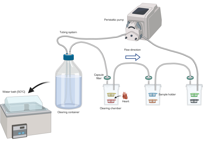
Figure 1: Schematic of the passive clearing setup. The clearing solution (after being filtered) circulates in succession through the sample chambers with the help of the peristaltic pump. The maintenance of the solution container in a water bath set at 50 °C allows the solution temperature to be between 37-45 °C within the chambers. Image created with Biorender.com. Please click here to view a larger version of this figure.
Figure 2 shows the result of the clearing process of an entire heart. As already reported by Costantini et al.16, the combination of the CLARITY methodology with TDE as RI-medium does not significantly change the sample's final volume nor leads to anisotropic deformation of the specimen.
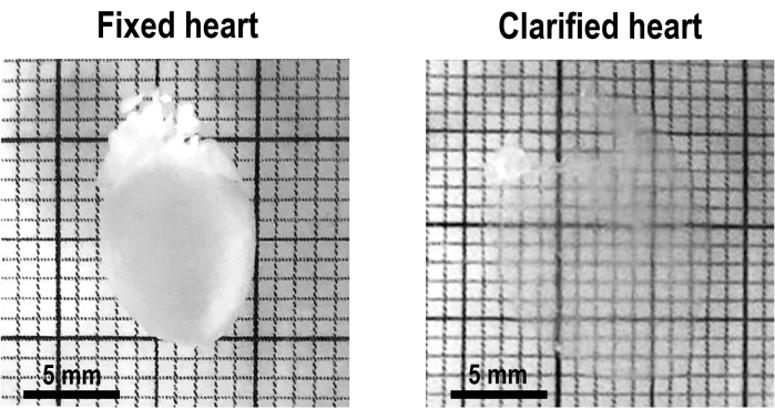
Figure 2: Representative image of a heart before (on the left) and after (on the right) the CLARITY protocol. The hearts become fully transparent and slightly oversized. Please click here to view a larger version of this figure.
Once the heart was cleared, cellular membranes were stained with an Alexa Fluor 633-conjugated WGA to perform the cytoarchitecture reconstruction of the entire organ. The custom-made fluorescence light-sheet microscope (Figure 3) was able to ensure 3D micron-scale resolution across the entire FoV.
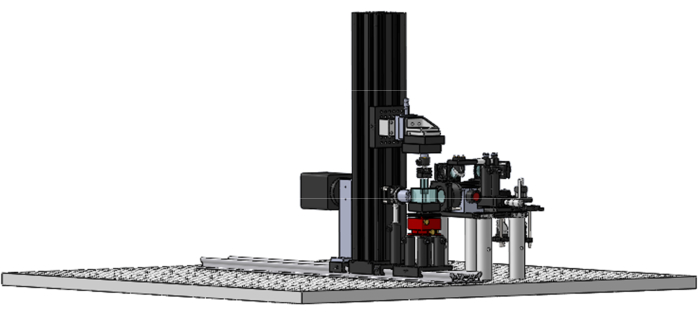
Figure 3: MesoSPIM. CAD rendering of the custom-made fluorescence light-sheet microscope. Please click here to view a larger version of this figure.
Considering the numerical aperture (NA = 0.1) of the detection optics, the radial (XY) Point Spread Function (PSF) of the system can be estimated in the order of 4-5 µm. On the other hand, the excitation optics produce a light-sheet with a minimum waist of about 6 µm (Full width half maximum, FWHM) that diverges up to 175 µm at the edge of the FoV (Figure 4A–C). The synchronization of the camera rolling shutter with the axial scan of the laser beam ensured to collect the emission signal only in the sample portion excited with the waist of the light-sheet, resulting in an average FWHM of about 6.7 µm along the entire FoV (Figure 4B–D).
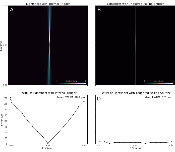
Figure 4: Light-sheet generation and characterization. (A) An excitation light-sheet generated with a laser source of 638 nm is focused on the center of the Field of View (FoV) and acquired with a pixel size of 3.25 µm and an Exposure Time of 10 ms. Light intensity is normalized and reported with a colormap. The Full Width Half Maximum (FWHM) of the light intensity profile is evaluated in 15 different positions along the FoV. Results are shown in C. (B) Image of the excitation light-sheet generated by the synchronization between the camera rolling shutter operating at 1.92 Hz and the light beam position driven by the tunable lens. The FWHM of the light intensity profile is evaluated along the FoV and results are shown in D. Please click here to view a larger version of this figure.
The Z-PSF of the microscope was also estimated by a tomographic reconstruction of the fluorescent nanosphere (Figure 5). An FWHM of 6.4 µm can be estimated by the fit, in good agreement with the previous assessment.
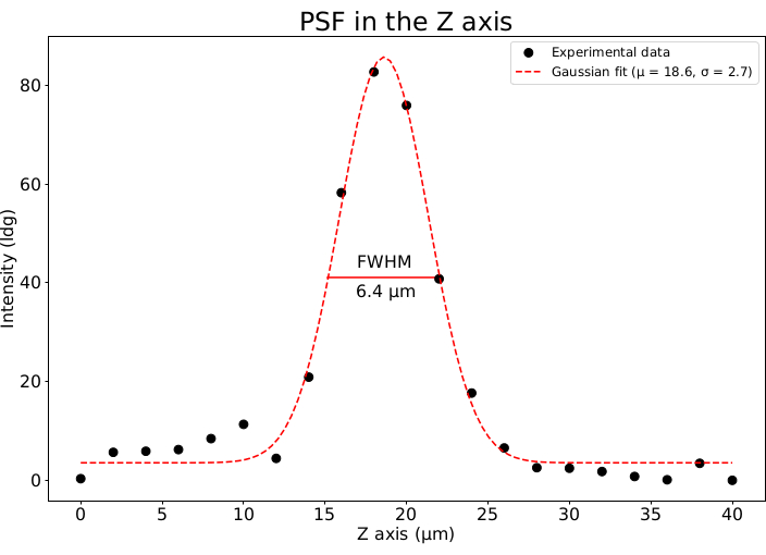
Figure 5: Point Spread Function in the Z-axis. The Point Spread Function (PSF) of the optical system is estimated by imaging fluorescent sub-micron-scale nanospheres (excited with a light sheet with a wavelength of 638 nm) with a pixel size of 3.25 µm × 3.25 µm × 2.0 µm. PSF intensity profile along the optical axis (Z) is represented as black dots. PSF profile is fitted with a Gaussian function with µ = 18.6 µm and σ = 2.7 µm. The FWHM of the PSF estimated by the fit is 6.4 µm. Please click here to view a larger version of this figure.
Owing to the high transparency of the tissue, it was possible to illuminate the whole heart without significant distortion of the axially scanned light-sheet at an excitation wavelength of 638 nm. The fluorescence signal was collected by the sCMOS sensor operating at 500 ms of exposure time and a frame rate of 1.92 Hz. Based on previous quantification, the tomographic acquisition was performed using a Z-scan velocity of 6 µm/s, and assuming a frame rate of 1.92 Hz, one frame every 3.12 µm was acquired, oversampling the system Z-PSF by about two times. Two representative frames (on the coronal and transverse planes) of the left ventricle chamber are shown in Figure 6. This result confirms the potentiality of the system to resolve single cellular membranes in three dimensions with a sufficient Signal/Noise ratio in the entire organ (Figure 6).
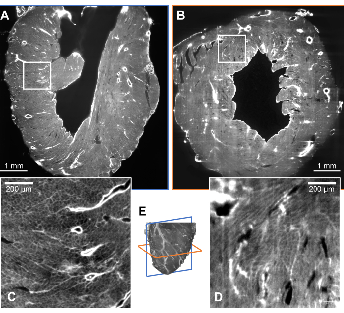
Figure 6: Mouse heart tissue reconstruction. The clarified heart was stained with WGA conjugated to Alexa Fluor 633 and excited by a laser source with a wavelength of 638 nm. (A) Coronal and (B) transverse representative sections. (C–D) Tissue transformation produces high tissue transparency, allowing to resolve small structures in the wall depth. The optical system shows an axial resolution sufficient to resolve micrometric structures (panel. D). (E) 3D low-resolution heart rendering. Please click here to view a larger version of this figure.
In this work, a successful approach to clear, stain, and image a whole mouse heart at high resolution was introduced. First, a tissue transformation protocol (CLARITY) was optimized and performed, slightly modified for its application on the cardiac tissue. Indeed, to obtain an efficient reconstruction in 3D of a whole heart, it is essential to prevent the phenomenon of light scattering. The CLARITY methodology allows us to obtain a highly transparent intact heart, but it requires long incubation times when performed passively (about 5 months). With respect to the brain, the cardiac tissue is not suitable for an active clearing, which takes advantage of an electric field. Even at low voltages, the electric field leads to damages and tissue breakages. Here, a passive clearing approach was optimized to obtain a completely cleared heart in about 3 months. After isolating and cannulating the heart through the proximal aorta, the CLARITY methodology was performed as described in section 3 of the protocol. To speed up the procedure, a homemade passive clearing setup was arranged (Figure 1), which ended up decreasing the timing of tissue clearing by about 40%. The setup is composed of a container for the clearing solution, a water bath, a peristaltic pump, several chambers containing different sample holders, capsule filters for each chamber, and a tubing system for the recirculation of the solution. The pump extracts and circulates the solution from the container in succession through each of the chambers, where the samples are held for clearing. Before entering the chambers, the solution flows through a capsule filter to trap the lipids flushed away from tissues during the clearing. The optimal temperature for the clearing solution, between 37-45 °C, is maintained within the chambers during the recirculation by keeping the solution container in a water bath at 50 °C. It is advised to change the clearing solution in the container once a week during the procedure. All components used are listed in detail in the Table of Materials. The optimized solution presented here allows us to obtain a whole passively cleared mouse heart in a significantly shorter time with respect to the standard passive clearing technique, thus reducing the required experimental time without damaging the organ. The staining approach was also optimized for homogeneous labeling of the cellular membranes and endothelium, using a fluorescent lectin (WGA – Alexa Fluor 633).
The heart cytoarchitecture has been reconstructed by developing a dedicated mesoSPIM that axially sweeps the light-sheet across the sample (https://mesospim.org). The custom-made fluorescence light-sheet microscope (Figure 3) was able to rapidly acquire images of a mesoscopic FoV (of the order of millimeters) with micrometric resolution. In this way, single cardiomyocytes can be resolved and mapped into a 3D reconstruction of the entire organ. The microscope illuminates the cleared sample with a light-sheet, dynamically generated by scanning a laser beam at 638 nm using a galvanometric mirror. A sCMOS camera characterizes the detection arm in a 2x magnification scheme which enables it to acquire the entire FoV in a single scan. The fluorescence signal was selected by placing a long-pass filter after the objective. The camera was set to work in rolling shutter mode: at any time, the lines of active camera pixels (i.e., exposed to the image) are synchronized with the in-plane shift of the focal band of the light-sheet, performed by an electrically tunable lens. This approach maximized the optical sectioning capability in the whole FoV by only acquiring images in the thinnest part of the focused light-sheet. This solution differs from conventional configurations, where acquisition involves the entire range of focal depth of the light-sheet, preventing peak optical sectioning resolution in large part of the FoV. An integrated sample stage supports cuvettes, thereby optimizing positioning and enabling axial movement of the sample during the imaging process. In this way, tomographic reconstructions are possible by acquiring consecutive internal sections. The images obtained have a mesoscopic FoV and a micrometric resolution, while the acquisition time required for a whole mouse heart is ~ 15 min. The synchronization between the camera rolling shutter and the excitation light beam sweeping the FoV allows acquiring the entire image plane with a high spatial resolution (Figure 4). This allows direct reconstruction of the sample in a single tomographic acquisition, without the necessity of sample radial displacement and multi-adjacent-stacks-based imaging. Notably, the microscope allowed the reconstruction of the entire organ of about (10 mm x 6 mm x 6 mm) in a single imaging session, with a near-isotropic voxel size and a sufficient signal-to-background ratio to resolve single cells across the whole organ potentially.
It is noteworthy that the proposed protocol presents some critical steps that must be performed carefully to achieve good results. In particular, the cannulation of the heart through the proximal aorta can be quite difficult, but it is an essential step to wash and fix the organ properly. Judd et al.17, showed how to perform this step effectively. Moreover, the degassing procedure needed by the CLARITY protocol is quite complex too, but it is essential for tissue preservation; if this step is not performed properly, the tissue could encounter damages and decay during the incubation in clearing solution.
Furthermore, although the presented experimental workflow is suitable for small fluorescent probes, the use of immunohistochemistry does not always provide good efficiency in the staining due to the higher molecular weight of the antibodies. Each immunostaining protocol requires proper optimization, and different approaches have been conceived to improve the antibody penetration, for example, tissue expansion18 and/or variations in pH and ionic strength19.
The mesoSPIM setup also presents two main limitations: i) the light-sheet preservation across the sample is strongly dependent on the tissue transparency, and ii) the dimension of the camera sensor limits the FoV. Guaranteeing a perfect refractive index matching inside the entire heart is very challenging, and small variations on the refractive index can produce light scattering, leading to degradation of the image quality. In this respect, a dual-side illumination scheme can be introduced. Two excitation arms can generate two distinct and aligned dynamic light sheets with maximally focused illumination by alternating the illumination from one side to the other of the specimen. Also, the FoV can be improved by using a new generation high-resolution back-illuminated sCMOS with very large sensors in combination with high numerical aperture telecentric lenses with low field distortion. This implementation would allow us to reconstruct bigger organs or expanded tissues maintaining the same optical section capability and thus producing micron-scale 3D images of centimeter-sized cleared samples.
Although the presented protocol still requires a long time for sample preparation and a high level of transparency to obtain a reliable cytoarchitecture reconstruction of the entire organ, the main significance of the approach resides in the improvements of the clearing protocol and the capability to perform mesoscopic reconstruction in a single scan at micrometric resolution. In the future, these advances can be combined with a multi-staining protocol to achieve whole-organ reconstruction integrating different biological structures.
The authors have nothing to disclose.
This project has received fundings from the European Union’s Horizon 2020 research and innovation programme under grant agreement No 952166 (REPAIR), MUR under the FISR program, project FISR2019_00320 and Regione Toscana, Bando Ricerca Salute 2018, PERCARE project.
2-2’ Thiodiethanol | Sigma-Aldrich | 166782 | |
Acrylamide | Bio-Rad | 61-0140 | |
AV-044 Initiator | Wako Chemicals | AVP5874 | |
Bis-Acrylamide | Bio-Rad | 161-042 | |
Boric Acid | Sigma-Aldrich | B7901 | |
Camera | Hamamatsu | Orca flash 4.0 v3 | |
Camera software | Hamamatsu | HC Image | |
Collimating lens | Thorlabs | AC254-050-A-ML | |
Detection arm | Integrated optics | 0638L-15A-NI-PT-NF | |
Excitation lens | Nikon | 91863 | |
Exteraìnal quartz cuvette | Portmann Instruments | UQ-753 | |
Fold mirrors | Thorlabs | BBE1-E02 | |
Galvanometric mirror | Thorlabs | GVS211/M | |
Glucose | Sigma-Aldrich | G8270 | |
HCImage Live | Hamamatsu | 4.6.1.19 | |
HEPES | Sigma-Aldrich | H3375 | |
Internal quartz cuvette | Portmann Instruments | UQ-204 | |
KCl | Sigma-Aldrich | P4504 | |
Laser source | Integrated Optics | 0638L-15A-NI-PT-NF | |
Long-pass filter | Thorlabs | FELH0650 | |
Magnetic base | Thorlabs | KB25/M | |
MgCl2 | Chem-Lab | CI-1316-0250 | |
Motorized traslator | Physisk Instrument | M-122.2DD | |
NaCl | Sigma-Aldrich | 59888 | |
Objective | Thorlabs | TL2X-SAP | |
Paraformaldehyde | Agar Scientific | R1018 | |
Phosphate Buffer Solution | Sigma-Aldrich | P4417 | |
Polycap AS | Whatman | 2606T | |
Relay lens | Qioptiq | G063200000 | |
Sodium Dodecyl Sulfate | Sigma-Aldrich | L3771 | |
Tube lens | Thorlabs | ACT508-200-A-ML | |
Tunable lens | Optotune | EL-16-40-TC-VIS-5D-1-C | |
Vacuum pump | KNF Neuberger Inc | N86KT.18 | |
Water bath | Memmert | WTB |