Summary

أدوات الموائع الدقيقة للتحقيق في التفاعلات الفطرية الميكروبية على المستوى الخلوي

Published: June 23, 2022
doi:

Summary

نظرا لعتامة التربة ، لا يمكن بسهولة تصور التفاعلات بين الميكروبات المكونة لها بدقة خلوية. هنا ، يتم تقديم أداتين للسوائل الدقيقة ، والتي توفر فرصا جديدة للتحقيق في التفاعلات الفطرية الميكروبية. الأجهزة متعددة الاستخدامات وسهلة الاستخدام ، مما يتيح التحكم الزماني المكاني العالي والتصوير عالي الدقة على المستوى الخلوي.

Abstract

الفطريات الخيطية هي سكان ناجحون في التربة وتلعب دورا رئيسيا في النظم الإيكولوجية للتربة ، مثل تحلل المواد العضوية وغير العضوية ، وكذلك تنظيم مستويات المغذيات. هناك يجدون أيضا العديد من الفرص للتفاعل مع مجموعة متنوعة من الميكروبات الأخرى مثل البكتيريا أو الفطريات الأخرى. ومع ذلك ، فإن دراسة التفاعلات الفطرية على المستوى الخلوي يمكن أن تكون صعبة بسبب طبيعة التربة الشبيهة بالصندوق الأسود. ويجري تطوير أدوات جديدة للموائع الدقيقة لدراسة التفاعلات الفطرية؛ يتم تسليط الضوء على منصتين مصممتين لدراسة التفاعلات البكتيرية الفطرية والفطرية والفطرية. داخل هذه القنوات الدقيقة ، يمكن مراقبة التفاعلات الفطرية الميكروبية في بيئات فيزيائية كيميائية خاضعة للرقابة بدقة زمنية ومكانية أعلى مما كان ممكنا في السابق. وقد أسفر تطبيق هذه الأدوات عن العديد من الرؤى البيولوجية الجديدة، مثل ملاحظة الارتباط القطبي البكتيري بالهيفاي أو الكشف عن العداوات الفطرية الفطرية غير المميزة. ومن السمات الرئيسية لهذه المنهجيات سهولة استخدام هذه الأداة من قبل غير الخبراء، مما ينتج عنه تكنولوجيات قابلة للترجمة بدرجة عالية لاستخدامها في مختبرات علم الأحياء الدقيقة.

Introduction

التربة هي بيئة متنوعة بشكل استثنائي تحتوي على وفرة من الكائنات الحية الدقيقة التي لها دور أساسي في دورات الكربون والفوسفور 1,2. الفطريات الخيطية هي عنصر رئيسي في العديد من النظم الإيكولوجية كمتحللة للمواد العضوية وغير العضوية ويمكن أن تعزز تغذية النباتات من خلال تشكيل علاقات تكافلية 3,4. داخل التربة ، تتفاعل الفطريات ديناميكيا مع العديد من الميكروبات مثل الفطريات الأخرى5 والبكتيريا6 والفيروسات7 والديدان الخيطية8. هذه التفاعلات لها عواقب وخيمة على صحة التربة والنبات. ومع ذلك، وبسبب الافتقار إلى النظم التجريبية المناسبة القادرة على تصوير الكائنات الحية الدقيقة المتفاعلة بدقة عالية، لا يزال العديد منها غير محدد.

وللتحقيقات المتعلقة بالتفاعلات البكتيرية الفطرية والتفاعلات الفطرية والفطرية تطبيقات قيمة في طائفة من المجالات، بما في ذلك مضادات الميكروبات في الطب وعوامل المكافحة البيولوجية في الزراعة. على سبيل المثال ، ينتج الفطر Coprinopsis cinerea الببتيد copsin، الذي ثبت أنه يظهر نشاطا مضادا للبكتيريا ضد العامل الممرض البشري Listeria monocytogenes9. وبالمثل ، يستخدم المركب المشتق من الفطريات ، griseofulvin ، على نطاق واسع كعلاج للعدوى الفطرية البشرية ، بالإضافة إلى أنه قادر على تثبيط نمو الفطريات المسببة للأمراض النباتية Alternaria solani10,11. كما ثبت أن العديد من سلالات بكتيريا Bacillus subtilis التي تعيش في التربة هي عوامل فعالة للمكافحة الحيوية للنبات الفطري الممرض Rhizoctonia solani12,13. ومع ذلك، ونظرا للقيود المرتبطة بالمنهجيات التقليدية، فإن مؤسسات التمويل الدولية ومؤسسات التمويل الأجنبي غير مفهومة بشكل جيد على مستوى الخلايا المفردة.

عادة ما تستكشف الدراسات التقليدية BFIs و FFIs على النطاق الكلي باستخدام ألواح أجار مع نوعين أو أكثر في المواجهة. يتم تقييم تفاعلها عن طريق قياس معدلات النمو وإنتاج الأيض للأنواع المواجهة14،15،16 ؛ ومع ذلك ، يتم حل هذه المنهجية فقط على مستوى المستعمرة. لدراسة التفاعلات على المستوى الخلوي ، يمكن زراعة اللقاحات البكتيرية والفطرية على شرائح المجهر الزجاجية المغلفة بالأجار والتي يتم تصويرها بعد ذلك تحت المجهر17. ومع ذلك ، قد يكون من الصعب اتباع hypha واحد باستخدام شرائح المجهر بسبب عدم وجود حبس ، مما يعني أنه من الصعب الحصول على صور الفاصل الزمني. علاوة على ذلك ، فإن فرصة حصر الكائنات الحية الدقيقة الأخرى مكانيا داخل مناطق محددة من الميسيليوم الفطري أو إنشاء بيئات كيميائية محددة يمكن إزعاجها ، على سبيل المثال ، غير ممكنة في مثل هذه الإعدادات. تضيف طبيعة “الصندوق الأسود” للتربة أيضا إلى تعقيد دراسة التفاعلات الفطرية الميكروبية على مستوى الخلايا المفردة18. من خلال مراقبة الأنواع المتفاعلة بعيدا عن التنوع المذهل لميكروبيوم التربة ، يمكن تقييم الطريقة الدقيقة التي يتفاعل بها الأعضاء الأفراد. وبالتالي ، هناك حاجة مستمرة إلى منصات متعددة الاستخدامات تمكن من التصوير عالي الدقة وخلية واحدة ل BFIs و FFIs.

توفر تقنيات الموائع الدقيقة ، ما يسمى بأنظمة المختبر على رقاقة ، منصة مثالية لدراسة BFIs و FFIs على مستوى الخلايا المفردة. تم اعتماد مجال الموائع الدقيقة ، الناشئة عن التقنيات المطورة للتحليل الكيميائي والإلكترونيات الدقيقة ، من قبل العلوم البيولوجية19. تنظم تقنيات الموائع الدقيقة كميات صغيرة من السوائل داخل شبكة مخصصة من القنوات المصغرة ، لها بعد واحد على الأقل على مقياس الميكرومتر ، ويتوسع استخدامها في الأبحاث البيولوجية20. على وجه الخصوص ، تم تطوير أجهزة الموائع الدقيقة لدراسة نمو الفطريات الخيطية21،22،23،24،25،26،27،28،29،30. إحدى فوائد استخدام هذه التكنولوجيا هي أن حبس hyphae وتوزيع العناصر الغذائية داخل القنوات الدقيقة يشبه إلى حد كبير بنية بيئة التربة أكثر من طرق الأجار التقليدية31. في الآونة الأخيرة ، تم استخدام منصات الموائع الدقيقة للتحقيق في التفاعلات بين العدلات البشرية ومسببات الأمراض الفطرية32 ، والبكتيريا وجذور النباتات 33 ، وكذلك الفطريات والديدان الخيطية34,35.

واحدة من المزايا العديدة لاستخدام الموائع الدقيقة لدراسة التفاعلات الميكروبية تشمل التحكم المحدد في بيئة القنوات الدقيقة. على سبيل المثال ، يمكن استغلال أنظمة التدفق الرقائقي لتوليد تدرجات تركيز محددة ، وهو أمر مفيد بشكل خاص عند فحص التاكسي الكيميائي البكتيري36. ميزة أخرى هي أن الطبيعة الشفافة ل poly (dimethylsiloxane) (PDMS) ، وهو بوليمر مطاطي غير مكلف ومتوافق حيويا يستخدم عادة في تصنيع أجهزة الموائع الدقيقة ، يسهل التصوير عالي الدقة للخلايا المفردة باستخدام المجهر الساطع والفلوري37. وبالمثل ، فإن حبس الميكروبات داخل القنوات الدقيقة يعني أنه يمكن إجراء تجارب الفاصل الزمني التي تتعقب الخلايا المفردة ، مما يسمح بتسجيل الاستجابات الخلوية الفردية وتحديدها كميا37. وأخيرا، بما أنه يمكن تصميم أجهزة الموائع الدقيقة لتكون سهلة الاستخدام، يمكن استخدامها بسهولة من قبل غير الخبراء38.

ومن المهم تعزيز المعرفة بالتفاعلات بين الكائنات الحية الدقيقة التي تعيش في التربة لتحسين ممارسات الإدارة المستدامة للنظم الإيكولوجية التي تحافظ على التنوع البيولوجي وللتخفيف من أثر تغير المناخ على البيئات الأرضية39. وبالتالي ، فإن تطوير أدوات جديدة للموائع الدقيقة أمر أساسي لتوسيع فهم الفطريات وتفاعلاتها على المستوى الخلوي. وسيركز البروتوكول هنا على جهازين من أجهزة الموائع الدقيقة المنتجة لدراسة BFIs40 و FFIs41 كما هو موضح في الشكل 1.

Figure 1
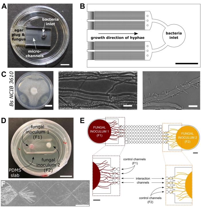
الشكل 1: التمثيل البصري والتخطيطي لأجهزة التفاعل البكتيري الفطري (BFI) والتفاعل الفطري الفطري (FFI). (أ) صورة لجهاز BFI. يتم وضع قابس mycelial عند مدخل أحد طرفي القنوات الدقيقة للسماح بنمو hyphal في الجهاز. المدخل البكتيري في الطرف المعاكس. شريط المقياس = 5 مم (B) نظرة عامة تخطيطية على جهاز BFI ، تصور موضع المداخل البكتيرية واتجاه نمو الواصلة من خلال القنوات الدقيقة التفاعلية. يبلغ عمق القنوات 10 ميكرومتر وعرضها 100 ميكرومتر وطولها 7 مم ، مع 28 قناة مراقبة في المجموع. (ج) فحص المواجهة على صفيحة أجار بين Coprinopsis cinerea و Bacillus subtilis NCIB 3610 ، شريط المقياس = 20 مم (يسار). صور مجهرية تظهر التفاعل بين C. cinerea و B. subtilis NCIB 3610 داخل القناة الدقيقة (الوسطى واليمنى) ، أي التعلق القطبي للبكتيريا ب hyphae الفطرية. شريط المقياس = 25 ميكرومتر (وسط) و 10 ميكرومتر (يمين). (د) صورة لجهاز FFI مرتبط بطبق بتري ذو قاع زجاجي ، ملقح مزدوج بسدادات mycelial. شريط المقياس = 1 سم (E) نظرة عامة تخطيطية لجهاز FFI. يتم إدخال اثنين من سدادات التطعيم الفطرية في المداخل في أي من طرفي الجهاز ، مما يسمح بالاستكشاف الواصلي للقنوات الدقيقة. ترتبط قنوات التحكم بمدخل فطري واحد فقط ولها قناة مسدودة ، مما يمنع التفاعلات بين فطريات الاختبار. تربط قنوات التفاعل كلا من المداخل الفطرية وتسمح بالتفاعلات الواصلة بين مواضيع الاختبار داخل القناة الدقيقة. تتكون كل قناة تفاعل من 18 قسما على شكل ماس ، يبلغ طولها الإجمالي 8.8 مم (490 × 430 ميكرومتر لكل ماسة) ، وعمقها 10 ميكرومتر ، ولها منطقة ربط بين كل ماسة تبلغ 20 ميكرومتر. يتم تكرار أنواع القنوات ، أشرطة المقياس = 1 مم. (F) منطقة التفاعل بين جبهتين واصليتين تقتربان ، تنمو من طرفي نقيض من قناة التفاعل المترابطة. صورة مجهرية لتباين الطور ، شريط مقياس = 250 ميكرومتر. تم تعديل اللوحات الواردة في هذا الشكل من Stanley et al., 2014 (A-C)40 and Gimeno et al., 2021 (D-F)41. يرجى النقر هنا لعرض نسخة أكبر من هذا الرقم.

Protocol

ملاحظة: يرد ملخص للإجراءات المبينة في هذا البروتوكول بصورة مرئية في الشكل 2. الشكل 2: تمثيل تخطيطي للمنهجية المقدمة يتألف من خمسة أقسام رئيسية مفصلة في هذا البروتوكول.<…

Representative Results

يتم عرض النتائج التمثيلية من أجهزة BFI40 و FFI41 النموذجية. يمكن بسهولة الحصول على قياسات معدل نمو Hyphal باستخدام هذه الأجهزة مع تقنيات الفحص المجهري الأساسية. يوضح الشكل 3A-B التفاعلات البكتيرية الفطرية بين C. cinerea hyphae و B. subtilis …

Discussion

تقدم هذه المقالة بروتوكولا لدراسة التفاعلات الفطرية الميكروبية باستخدام الموائع الدقيقة للقناة. يهدف المؤلفون إلى إظهار تنوع هذه الأجهزة وتشجيع التكيف لتناسب اهتمامات الباحث. باستخدام أجهزة BFI و FFI النموذجية ، يمكن دراسة التفاعلات الفطرية الميكروبية بمزيد من التفصيل مما كان يمكن الوصول…

Declarações

The authors have nothing to disclose.

Acknowledgements

نحن نقر بالدعم المالي المقدم من قسم الهندسة الحيوية في إمبريال كوليدج لندن و The Leverhulme Trust (مرجع منحة البحث: RPG-2020-352).

Materials

Agar Difco Laboratories 214010 Used to solidify culture medium for bacterial and fungal cultivation within Petri dishes
Aluminum foil Fisher Scientific Ltd 11759408
AutoCAD 2021 Autodesk, USA
Autoclave (VX-75) Systec
Centrifuge (5810R) Eppendorf
Chlorotrimethysilane Merck Life Sciences 386529 CAUTION: Chlorotrimethylsilane is a hazardous substance. Wear appropriate PPE and handle with care. Avoid contact with skin and eyes and prevent inhalation. Keep away from sources of ignition and use in a well-ventilated area.
Cork borer SLS COR1000
Developer solution (mr-Dev 600) Microresist Technologies CAUTION: mr-Dev 600 developer solution is flammable
Erlenmeyer flasks VWR 214-1108 e.g. 200 mL; choose size to suit your exact needs
Ethanol (70% v/v)  Fisher Scientific Ltd E/0650DF/15 Diluted from 99.8% (Analytical Reagent Grade)
Fiji ImageJ Exemplar software package for imaging processing
Filtered, compressed air Available as standard in most labs. Altervatively, an oil-free compressor with air regulator can be used.
Flat-headed wafer tweezers SLS INS5026
Forceps Fisher Scientific Ltd 10008051 Bent, sharp
Glass bottom petri dish World Precision Instruments FD35-100 35 mm
Glass bottom petri dish World Precision Instruments FD5040-100 50 mm
Glass crystallisation dishes VWR 216-1865 Used for washing of PDMS slabs
Glass crystallisation dishes VWR 216-1866 Used in the development of master moulds
Glass media bottles Fisher Scientific Ltd 15456113 e.g. 250 mL; choose size to suit your exact needs
Glass syringe (Hamilton) Fisher Scientific Ltd 10625251 Used for dispensing chlorotrimethylsilane
Hot plate (HP 160 III BM) SAWATEC
Inoculation loop VWR COPA175CS01
Isopropyl alcohol Sigma-Aldrich W292907
Laminar flow hood Air Science (PCR) Exemplar laminar flow hood used for device fabrication
LB medium Fisher Scientific Ltd BP9723-500 Exemplar nutrient broth for bacterial overnight culture
Light emitting diode light engine (LedHUB) Omicron-Laserage Laserprodukte GmbH Exemplar light source that can be used for imaging fungal-microbial interactions (fluorescence)
MA6 Ultraviolet mask aligner Suss Microtec
Malt extract VWR 84618 Used to make exemplar fungal culture medium (Malt extract agar)
Mask Writer Applied Materials 4700DP Example of a mask writer which can be used to print photo-mask for photolithography
Master mould plastic mount 3D-printed bespoke holder manufactured in-house
Microbiological safety cabinet  (BioMat2) Contained Air Solutions Exemplar MSC used for microbial culture and device inoculation
Milli-Q purified water Available as standard in biology labs. 
NaOH Fisher Scientific Ltd BP359-500
NIS-Elements Advanced Research imaging software Nikon Exemplar software package for image acquisition
NIS-Elements Free Viewer Nikon Exemplar software package for viewing acquired images
Oven (Binder BD115) Fisher Scientific Ltd 15602126 Used for curing poly(dimethylsiloxane)(PDMS)
Oven (CLO-2AH-S) KOYO Used for preparing silicon wafers
Parafilm Bemis HS234526B transparent film
Petri dishes, square sterile Fisher Scientific Ltd 11708573 120.5 mm
Petri dishes, sterile Fisher Scientific Ltd 15370366 90 mm 
Photolithography mask Micro Lithography Services Ltd. UK
Plasma cleaner (Zepto) Diener Electronic 100012601
Plastic cup Semadeni 8323
Plastic spatula Semadeni 3340
Portable precision balance (OHAUS Scout) Fisher Scientific Ltd 15519631 Used for weighing PDMS, media components etc.
Precision cutter Syneo HS1251135P1183 Cutting edge diameter: 3.18 mm
Precision cutter  Syneo HS1871730P1183S Cutting edge diameter: 4.75 mm
Profilometer  Bruker Dektak XT-stylus
Razor blades Häberle Labortechnik 9156110
Refridgerator Haden 4-6 °C
Retiga R1 CCD camera Qimaging Exemplar camera that can be used for imaging fungal-microbial interactions
Scotch magic tape Office Depot 3969954 19 mm invisible tape; clear tape
Shaking incubator (Cole-Parmer SI500) Fisher Scientific Ltd 10257954
Silicon wafer Inseto 100 mm
Soda lime glass plate Inseto 125 mm x 125 mm x 2 mm. Used to hold photolithography mask in mask aligner
Sodium chloride Sigma-Aldrich S7653
Spincoater  SAWATEC SM-180-BM
SU-8 2010 photoresist MicroChem CAUTION: SU-8 photoresist is hazardous, take care when handling and prevent inhalation and contact with skin. Flammable, potentially carcinogenic and toxic to the environment. 
Sylgard 184 elastomer kit VWR 634165S Used for the preparation of poly(dimethylsiloxane)(PDMS) devices
Temperature controlled incubator Okolab Exemplar incubator that can be used for imaging fungal-microbial interactions
Ti2-E inverted epifluorescence microscope Nikon MEA54000 Exemplar microscope that can be used for imaging fungal-microbial interactions
Ultrasonic cleaner S-Line Fisher Scientific Ltd FB15050
Vacuum desiccator Fisher Scientific Ltd 10528861 Silianisation and PDMS degassing should be conducted in separate desiccators
x10/0.3 NA CFI Plan Fluor DL objective lens Nikon MRH20105 Exemplar objective lens that can be used for imaging fungal-microbial interactions
x20/0.45 NA CFI Plan Fluor DL objective lens Nikon MRH48230 Exemplar objective lens that can be used for imaging fungal-microbial interactions

Referências

  1. Zhu, Y. -. G., Miller, R. M. Carbon cycling by arbuscular mycorrhizal fungi in soil-plant systems. Trends in Plant Science. 8 (9), 407-409 (2003).
  2. Dai, Z., et al. Long-term nutrient inputs shift soil microbial functional profiles of phosphorus cycling in diverse agroecosystems. The ISME Journal. 14 (3), 757-770 (2020).
  3. Op De Beeck, M., et al. Regulation of fungal decomposition at single-cell level. The ISME Journal. 14 (4), 896-905 (2020).
  4. Bender, S. F., et al. Symbiotic relationships between soil fungi and plants reduce N2O emissions from soil. The ISME Journal. 8 (6), 1336-1345 (2014).
  5. Dullah, S., et al. Melanin production and laccase mediated oxidative stress alleviation during fungal-fungal interaction among basidiomycete fungi. IMA Fungus. 12 (1), 33 (2021).
  6. Deveau, A., et al. Bacterial-fungal interactions: ecology, mechanisms and challenges. FEMS Microbiology Reviews. 42 (3), 335-352 (2018).
  7. Bian, R., et al. Facilitative and synergistic interactions between fungal and plant viruses. Proceedings of the National Academy of Sciences of the United States of America. 117 (7), 3779-3788 (2020).
  8. Jiang, X., Xiang, M., Liu, X. Nematode-trapping fungi. Microbiology Spectrum. 5 (1), (2017).
  9. Essig, A., et al. a novel peptide-based fungal antibiotic interfering with the peptidoglycan synthesis. Journal of Biological Chemistry. 289 (50), 34953-34964 (2014).
  10. Tang, H. -. Y., Zhang, Q., Li, H., Gao, J. -. M. Antimicrobial and allelopathic metabolites produced by Penicillium brasilianum. Natural Product Research. 29 (4), 345-348 (2015).
  11. Bai, Y. -. B., et al. Antifungal activity of griseofulvin derivatives against phytopathogenic fungi In vitro and In vivo and three-dimensional quantitative structure-activity relationship analysis. Journal of Agricultural and Food Chemistry. 67 (22), 6125-6132 (2019).
  12. Solanki, M. K., et al. Characterization of antagonistic-potential of two Bacillus strains and their biocontrol activity against Rhizoctonia solani in tomato. Journal of Basic Microbiology. 55 (1), 82-90 (2015).
  13. Jamali, H., Sharma, A., Srivastava, A. K. Biocontrol potential of Bacillus subtilis RH5 against sheath blight of rice caused by Rhizoctonia solani. Journal of Basic Microbiology. 60 (3), 268-280 (2020).
  14. Válková, H., Novotný, &. #. 2. 6. 8. ;., Malachová, K., Šlosarčíková, P., Fojtík, J. Effect of bacteria on the degradation ability of Pleurotus ostreatus. Science of The Total Environment. 584-585, 1114-1120 (2017).
  15. Leyva-Rojas, J. A., Coy-Barrera, E., Hampp, R. Interaction with soil bacteria affects the growth and amino acid content of Piriformospora indica. Molecules. 25 (3), 572 (2020).
  16. Dullah, S., et al. Fungal interactions induce changes in hyphal morphology and enzyme production. Mycology. 12 (4), 279-295 (2021).
  17. Marfetán, J. A., Romero, A. I., Folgarait, P. J. Pathogenic interaction between Escovopsis weberi and Leucoagaricus sp.: mechanisms involved and virulence levels. Fungal Ecology. 17, 52-61 (2015).
  18. Cortois, R., De Deyn, G. B. The curse of the black box. Plant and Soil. 350 (1), 27-33 (2012).
  19. Whitesides, G. M. The origins and the future of microfluidics. Nature. 442 (7101), 368-373 (2006).
  20. Sackmann, E. K., Fulton, A. L., Beebe, D. J. The present and future role of microfluidics in biomedical research. Nature. 507 (7491), 181-189 (2014).
  21. Hanson, K. L., et al. Fungi use efficient algorithms for the exploration of microfluidic networks. Small. 2 (10), 1212-1220 (2006).
  22. Held, M., Edwards, C., Nicolau, D. V. Probing the growth dynamics of Neurospora crassa with microfluidic structures. Fungal Biology. 115 (6), 493-505 (2011).
  23. Thomson, D. D., et al. Contact-induced apical asymmetry drives the thigmotropic responses of Candida albicans hyphae. Cellular Microbiology. 17 (3), 342-354 (2015).
  24. Lee, K. K., Labiscsak, L., Ahn, C. H., Hong, C. I. Spiral-based microfluidic device for long-term time course imaging of Neurospora crassa with single nucleus resolution. Fungal Genetics and Biology. 94, 11-14 (2016).
  25. Asenova, E., Lin, H. Y., Fu, E., Nicolau, D. V., Nicolau, D. V. Optimal fungal space searching algorithms. IEEE Transactions on NanoBioscience. 15 (7), 613-618 (2016).
  26. Soufan, R., et al. Pore-scale monitoring of the effect of microarchitecture on fungal growth in a two-dimensional soil-like micromodel. Frontiers in Environmental Science. 6, (2018).
  27. Uehling, J. K., et al. Microfluidics and metabolomics reveal symbiotic bacterial-fungal interactions between Mortierella elongata and Burkholderia include metabolite exchange. Frontiers in Microbiology. 10, 2163 (2019).
  28. Millet, L. J., et al. Increasing access to microfluidics for studying fungi and other branched biological structures. Fungal Biology and Biotechnology. 6 (8), 1-14 (2019).
  29. Baranger, C., Fayeulle, A., Le Goff, A. Microfluidic monitoring of the growth of individual hyphae in confined environments. Royal Society Open Science. 7 (8), 191535 (2020).
  30. Aleklett, K., Ohlsson, P., Bengtsson, M., Hammer, E. C. Fungal foraging behaviour and hyphal space exploration in micro-structured Soil Chips. The ISME Journal. 15 (6), 1782-1793 (2021).
  31. Aleklett, K., et al. Build your own soil: exploring microfluidics to create microbial habitat structures. The ISME Journal. 12 (2), 312-319 (2018).
  32. Ellett, F., Jorgensen, J., Frydman, G. H., Jones, C. N., Irimia, D. Neutrophil interactions stimulate evasive hyphal branching by Aspergillus fumigatus. PLOS Pathogens. 13 (1), 1006154 (2017).
  33. Massalha, H., Korenblum, E., Malitsky, S., Shapiro, O. H., Aharoni, A. Live imaging of root-bacteria interactions in a microfluidics setup. Proceedings of the National Academy of Sciences of the United States of America. 114 (17), 4549-4554 (2017).
  34. Schmieder, S. S., et al. Bidirectional propagation of signals and nutrients in fungal networks via specialized hyphae. Current Biology. 29 (2), 217-228 (2019).
  35. Tayyrov, A., Stanley, C. E., Azevedo, S., Künzler, M. Combining microfluidics and RNA-sequencing to assess the inducible defensome of a mushroom against nematodes. BMC Genomics. 20 (1), 243 (2019).
  36. Stanley, C. E., Grossmann, G., Casadevall i Solvas, X., deMello, A. J. Soil-on-a-Chip: microfluidic platforms for environmental organismal studies. Lab on a Chip. 16 (2), 228-241 (2016).
  37. Stanley, C. E., vander Heijden, M. G. A. Microbiome-on-a-Chip: new frontiers in plant-microbiota research. Trends in Microbiology. 25 (8), 610-613 (2017).
  38. Ortseifen, V., Viefhues, M., Wobbe, L., Grünberger, A. Microfluidics for biotechnology: bridging gaps to foster microfluidic applications. Frontiers in Bioengineering & Biotechnology. 8, 589074 (2020).
  39. Jansson, J. K., Hofmockel, K. S. The soil microbiome-from metagenomics to metaphenomics. Current Opinion in Microbiology. 43, 162-168 (2018).
  40. Stanley, C. E., et al. Probing bacterial-fungal interactions at the single cell level. Integrative Biology (Camb). 6 (10), 935-945 (2014).
  41. Gimeno, A., et al. A versatile microfluidic platform measures hyphal interactions between Fusarium graminearum and Clonostachys rosea in real-time. Communications Biology. 4 (1), 262 (2021).
  42. Duffy, D. C., McDonald, J. C., Schueller, O. J. A., Whitesides, G. M. Rapid prototyping of microfluidic systems in poly(dimethylsiloxane). Analytical Chemistry. 70 (23), 4974-4984 (1998).
  43. Stanley, C. E., et al. Fabrication and use of the dual-flow-RootChip for the imaging of Arabidopsis roots in asymmetric microenvironments. Bio-protocol. 8 (18), 3010 (2018).
  44. Choi, C. -. H., Lee, H., Weitz, D. A. Rapid patterning of PDMS microfluidic device wettability using syringe-vacuum-induced segmented flow in nonplanar geometry. ACS Applied Materials & Interfaces. 10 (4), 3170-3174 (2018).
  45. Sanders, E. R. Aseptic laboratory techniques: plating methods. Journal of Visualized Experiments. (63), e3064 (2012).
  46. Schindelin, J., et al. Fiji: an open-source platform for biological-image analysis. Nature Methods. 9 (7), 676-682 (2012).
  47. Harting, R., et al. Pseudomonas strains induce transcriptional and morphological changes and reduce root colonization of Verticillium spp. Frontiers in Microbiology. 12, 652468 (2021).
  48. Boenisch, M. J. . Structural and molecular characterisation of the penetration process of Fusarium graminearum during Fusarium head blight infection. , (2013).
  49. Eynck, C., Koopmann, B., Grunewaldt-Stoecker, G., Karlovsky, P., von Tiedemann, A. Differential interactions of Verticillium longisporum and V. dahliae with Brassica napus detected with molecular and histological techniques. European Journal of Plant Pathology. 118 (3), 259-274 (2007).
  50. Ghanem, N., Stanley, C. E., Harms, H., Chatzinotas, A., Wick, L. Y. Mycelial effects on phage retention during transport in a microfluidic platform. Environmental Science & Technology. 53 (20), 11755-11763 (2019).
  51. Alrifaiy, A., Lindahl, O. A., Ramser, K. Polymer-based microfluidic devices for pharmacy, biology and tissue engineering. Polymers. 4 (3), 1349-1398 (2012).
  52. Duncombe, T. A., Tentori, A. M., Herr, A. E. Microfluidics: reframing biological enquiry. Nature Reviews Molecular Cell Biology. 16 (9), 554-567 (2015).
  53. Hoelzle, D., et al. Microfluidic device design, fabrication, and testing protocols. Protocol Exchange. , (2015).

Play Video

Citar este artigo
Masters-Clark, E., Clark, A. J., Stanley, C. E. Microfluidic Tools for Probing Fungal-Microbial Interactions at the Cellular Level. J. Vis. Exp. (184), e63917, doi:10.3791/63917 (2022).

View Video