In this video article, we describe the in vitro synthesis of modified mRNA for induction of protein expression in cells.
The exogenous delivery of coding synthetic messenger RNA (mRNA) for induction of protein synthesis in desired cells has enormous potential in the fields of regenerative medicine, basic cell biology, treatment of diseases, and reprogramming of cells. Here, we describe a step by step protocol for generation of modified mRNA with reduced immune activation potential and increased stability, quality control of produced mRNA, transfection of cells with mRNA and verification of the induced protein expression by flow cytometry. Up to 3 days after a single transfection with eGFP mRNA, the transfected HEK293 cells produce eGFP. In this video article, the synthesis of eGFP mRNA is described as an example. However, the procedure can be applied for production of other desired mRNA. Using the synthetic modified mRNA, cells can be induced to transiently express the desired proteins, which they normally would not express.
In cells, the transcription of messenger RNA (mRNA) and the following translation of mRNA into desired proteins ensure the proper functioning of cells. Hereditary or acquired genetic disorders can lead to insufficient and dysfunctional synthesis of proteins and cause severe diseases. Thus, a new therapeutic approach to induce the production of missing or defective proteins is the exogenous delivery of synthetic modified mRNA into cells, which codes for the desired protein. Thereby, cells are triggered to synthesize functional proteins, which they normally cannot produce or would naturally not need. Using this approach, genetic disorders can be corrected by introduction of mRNA that codes for the defective or missing protein 1. The mRNA therapy can also be used for vaccination to synthetize protein antigens, which are expressed by tumor cells or pathogens. Thereby, host immune system can be activated to effectively eliminate tumor cells or prevent infections 2,3. Furthermore, in recent years, mRNA was successfully used to generate induced pluripotent stem cells (iPSCs). For this purpose, fibroblasts were transfected with mRNAs to induce the expression of reprogramming factors 4-6 and to convert them in iPSCs with an enormous potential in regenerative medicine.
Previously, the use of conventional mRNA was associated with low stability and strong immunogenicity. Thus, clinical applications of conventional mRNAs were limited. However, the replacement of cytidine and uridine by 5-methylcytidine and pseudouridine within the mRNA molecule by Kariko and colleagues rendered mRNA molecules stable in biological fluids and dramatically reduced immune activation 7-10, which now allows the clinical applicability of modified mRNAs.
Using synthetically produced modified mRNAs, desired gene transcripts can be temporarily delivered in vivo 11 or in vitro to induce protein expression. The introduced mRNA is translated under physiological conditions by the cellular translation machinery. Due to lack of integration into the host cell genome compared to viral gene therapy vectors, the risk of oncogenesis is prevented 12,13. Thus, therapy using modified synthetic mRNA will get better clinical acceptance in the future.
Here, we describe a detailed protocol for production of modified mRNA (Figure 1), transfection of cells with mRNA and the evaluation of protein expression in transfected cells.
1. Augmentation of Plasmids Containing Required Coding DNA Sequences (CDS)
2. Amplification of Plasmid Inserts and Adding of Poly T-tail by Polymerase Chain Reaction (PCR)
3. Control Step: Quality of PCR Product
4. In Vitro Transcription (IVT)
5. Treatment of Purified mRNA with Antarctic Phosphatase
6. Control Step: Quality of Synthesized Modified mRNA
7. Preparing of Cells for Transfection
8. Performing mRNA Transfection of Cells
9. Flow Cytometric Analyses of eGFP Expression in Cells
10. Measurement of Protein Expression Over Time
Using a pcDNA 3.3 plasmid containing the CDS of eGFP, the synthesis of modified eGFP mRNA was established (Figure 1). After insert amplification by PCR and poly T-tailing, a clear band with a length of approximately 1,100 bp is detected (Figure 2). Increasing the IVT time augmented the yield of mRNA (Figure 3). After the IVT, a clear mRNA band with a length of approximately 1,100 bp was detected, which corresponds to the length of eGFP mRNA to be produced (Figure 4).
The functionality of the generated eGFP mRNA was tested by transfection of HEK293 cells. For this purpose, transfection complexes (lipoplexes) were generated using a cationic lipid transfection reagent. The transfection was performed with 2 x 10⁵ cells per well of 12-well plate. The production of eGFP in the cells was detected 24 hr after transfection using fluorescence microscopy (Figure 5) and flow cytometry (Figure 6).
HEK293 cells were transfected with eGFP mRNA. eGFP expression was determined 1, 2, and 3 days after transfection to evaluate the duration of protein expression in the cells (Figure 7). After 24 hr, the protein expression was highest in the cells. The amount was reduced 1.6-fold every next day. Even after 3 days, the cells contained eGFP.
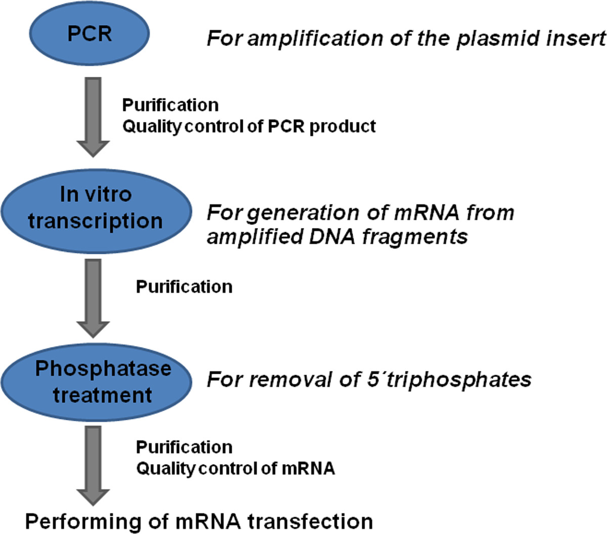
Figure 1: Overview of the modified mRNA production process. Coding DNA sequences (CDS) with known flanking sequences are amplified by PCR using specific primers. The PCR product is purified and the quality of the generated DNA is determined. The mRNA is generated from DNA product using the in vitro transcription process. The product is purified and treated with phosphatase to remove 5'-triphosphates. After the additional purification and quality control of generated mRNA, the mRNA transfections can be performed.
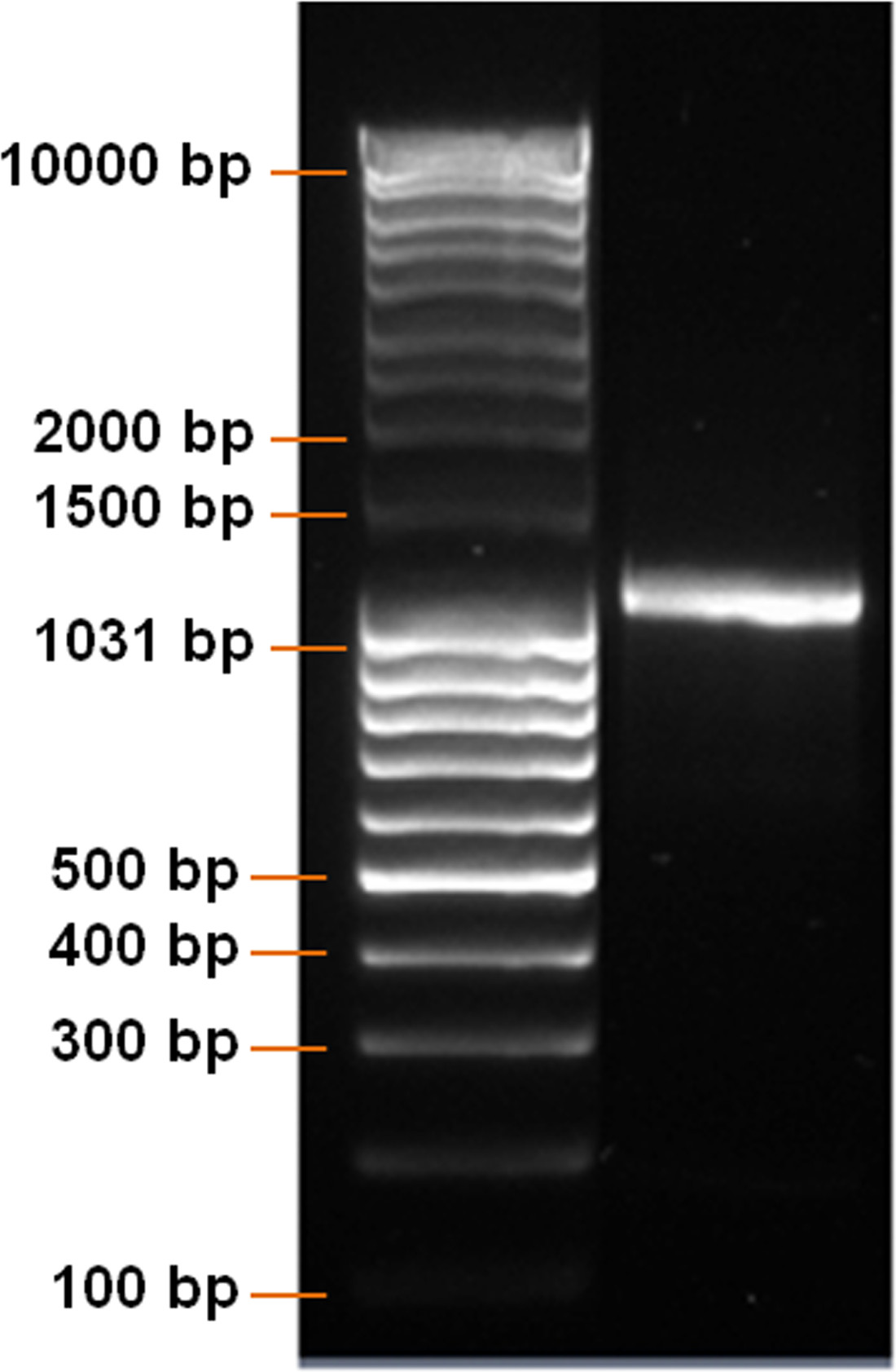
Figure 2: Analysis of DNA product after PCR. DNA ladder and PCR product were run on a 1% agarose gel. A clear DNA band with a length of approximately 1,100 bp should be detected.
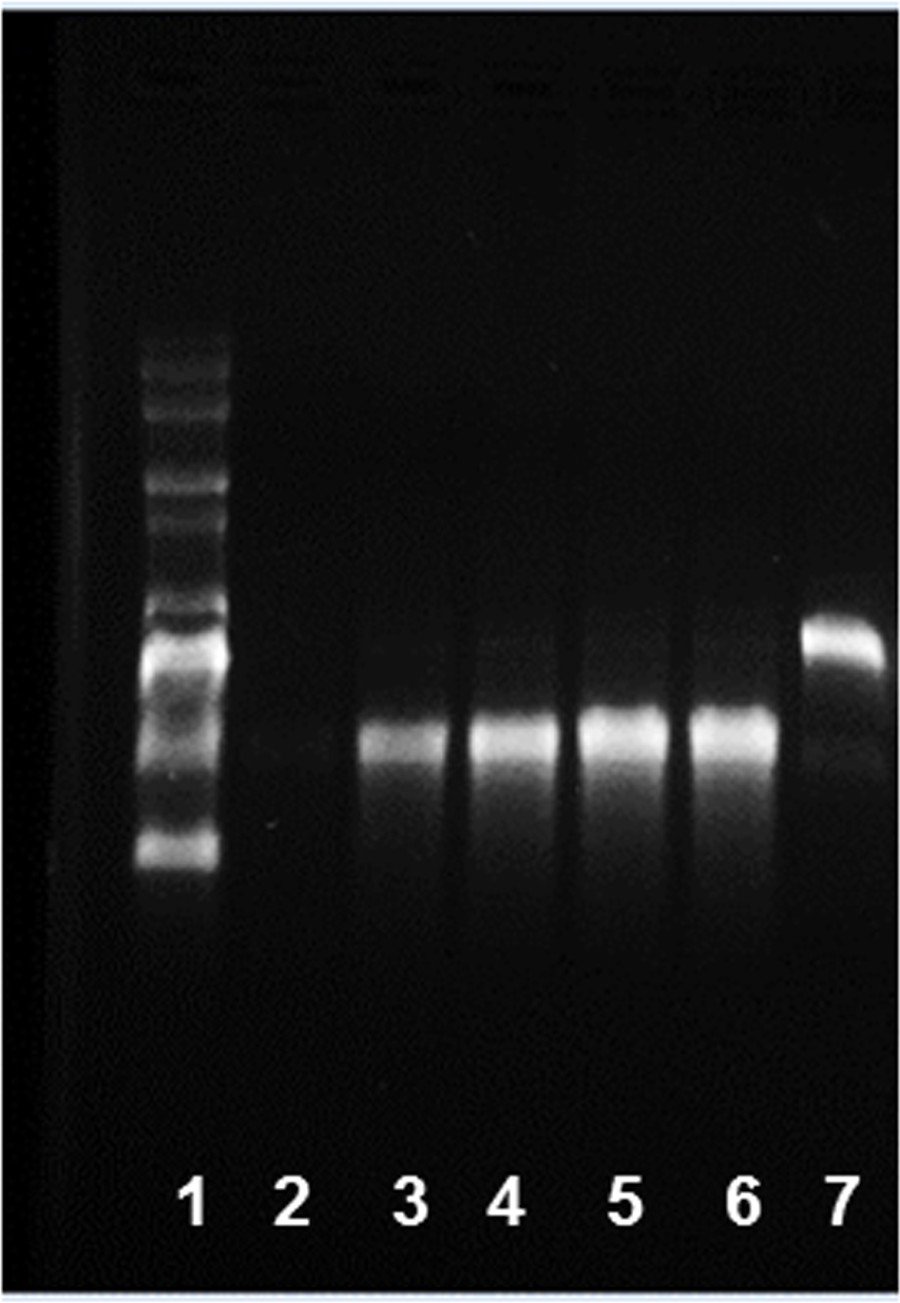
Figure 3: Kinetics of in vitro transcription for generation of eGFP mRNA. 1) RNA ladder and IVT products after 2) 0 min, 3) 10 min, 4) 30 min, 5) 180 min, 6) 360 min, and 7) DNA template alone were run on a 1% agarose gel.
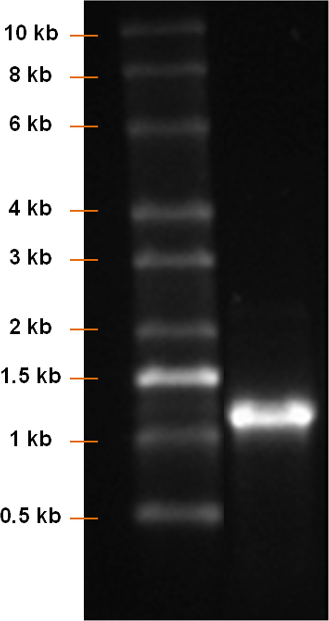
Figure 4: Analysis of mRNA product after IVT. RNA ladder and IVT product were run on a 1% agarose gel. A clear mRNA band with a length of approximately 1,100 bp should be detected.
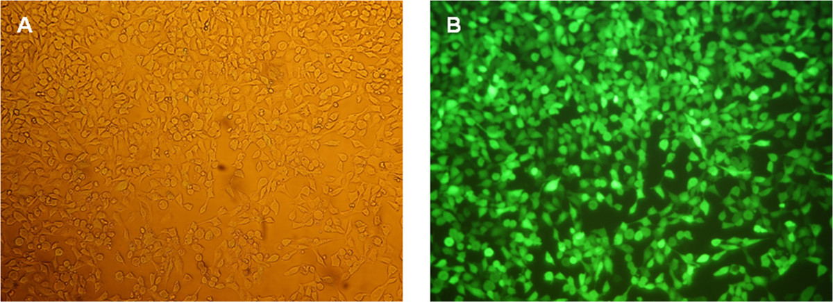
Figure 5: Fluorescence microscopic analyses of HEK293 cells 24 hr after eGFP mRNA transfection. (A) Phase contrast image of the cells at a magnification of 100X. (B) Fluorescence images of the cells at a magnification of 100X.
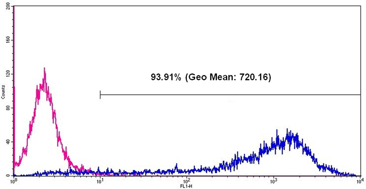
Figure 6: Flow cytometric analyses of eGFP expression in HEK293 cells 24 hr after eGFP mRNA transfection. The pink line represents cells without mRNA transfection and the blue line represents eGFP positive cells after eGFP mRNA transfection. After eGFP mRNA transfection, 93.91% of all measured cells are positive and the Geo Mean (geometric mean of fluorescence intensity) is 720.16.
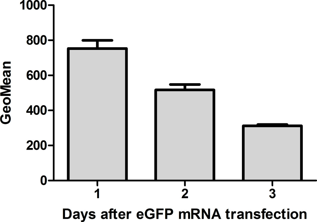
Figure 7: Flow cytometric analyses of eGFP expression in HEK293 cells 1, 2, and 3 days after eGFP mRNA transfection. The protein expression is highest 24 hr after mRNA transfection. Thereafter, the amount is reduced 1.6-fold every day (n = 3).
Table 1: Composition of PCR mixture.
Component | Final concentration | Amount (µl) |
Forward Primer | 0.7 µM | 7 |
Reverse Primer | 0.7 µM | 7 |
5x Q-Solution | 1x | 20 |
5x HotStar HiFidelity PCR Buffer | 1x | 20 |
Plasmid DNA | 50 ng / 100µl | Variable |
HotStar HiFidelity DNA Polymerase (2.5 U/µl) | 2.5 U | 1 |
Nuclease-free water | Variable | |
Total volume | 100 |
Table 1: Composition of PCR mixture.
Cycle Number | Time | Temperature (°C) | |
Initial denaturation step | 1 | 5 min | 95 |
3-step cycling | 2-25 | ||
· Denaturation | 45 sec | 95 | |
· Annealing | 1 min | 55 | |
· Extension | 1 min | 72 | |
Final extension step | 26 | 10 min | 72 |
End of PCR cycling | Indefinite | 4 |
Table 2: PCR cycling protocol.
Component | Stock concentration (mM) | Final concentration (mM) | Volume (µl) |
ATP (from MEGAscript T7 Kit) | 75 | 7.5 | 4 |
GTP (from MEGAscript T7 Kit) | 75 | 1.875 | 1 |
Me-CTP (from Trilink) | 100 | 7.5 | 3 |
Pseudo-UTP (from Trilink) | 100 | 7.5 | 3 |
3´-O-Me-m7G(5´)ppp(5´)G RNA cap structure analog | 10 | 2.5 | 10 |
Total volume | 23 |
Table 3: Composition of NTP/cap analog mixture.
Component | Final concentration | Amount (µl) |
Nuclease-free water | Variable | |
RNase Inhibitor | 40 U | 1 |
NTP/cap analog mixture (from step 4.3) | 23 | |
PCR product | 1 µg | Variable |
10x reaction buffer | 1x | 4 |
10x T7 RNA polymerase enzyme mix | 1x | 4 |
Total volume | 40 |
Table 4: Composition of in vitro transcription (IVT) reaction mixture.
Component | Amount (µl) |
Formamide | 3.3 |
37% formaldehyde | 1 |
MEN buffer (10x) | 1 |
6x loading buffer (supplied with the peqGOLD Range Mix DNA-Ladder) | 1.7 |
Total volume | 7 |
Table 5: Preparation of loading buffer for RNA gel electrophoresis.
Cell Culture Medium and Buffer | |
HEK-293 cell culture medium | Add 25 ml of FCS, 2.5 ml of penicillin/streptomycin, 2.5 ml of L-glutamine in 220 ml DMEM high glucose. Store the medium at 4 °C and use it within 2 weeks. |
TBE buffer (10x) | Dissolve 0.9 M Tris base, 0.9 M boric acid and 20 mM EDTA in 1 L water (Ampuwa). The pH of the buffer is 8. |
MEN buffer (10x) | Dissolve 200 mM MOPS, 50 mM NaOAc, 10 mM EDTA in 1 L water (Ampuwa). Adjust the pH value with NaOH to 7. |
Table 6: Cell culture medium and buffers.
The mRNA therapy has tremendous potential in the field of regenerative medicine, treatment of diseases and vaccination. In this video, we demonstrate the production of a stabilized, modified mRNA for induction of protein expression in cells. Using this protocol, other desired mRNA can be generated. The in vitro synthesis of modified mRNA allows the transfection of cells with desired mRNAs to induce expression of target proteins. Thereby, the desired protein is expressed transiently under physiological conditions until the exogenously delivered mRNA is completely degraded.
In this video, the expression of eGFP was demonstrated for 3 days after a single transfection of HEK293 cells with eGFP mRNA. mRNA molecules coding other proteins could lead to a shorter protein expression period. The expression of the protein decreases due to degradation of the exogenously delivered mRNA. Therefore, the limitation of this technique for some applications might be the transient induction of protein expression. Thus, to maintain protein expression in cells for a longer period, repeated delivery of mRNA is required. Although, mRNA transfection has the advantage of non-integration into the host genome, which prevents insertional mutagenesis and the development of cancer and leukemia compared to viral vectors, the in vivo transfection efficiency could be less than using viral vectors.
The required mRNA concentration and amount of transfection reagent should be optimized for each different cell type14, which are the target cells for exogenous delivery of mRNA to produce the missing protein.
Repeated freezing and thawing of mRNA should be avoided to maintain the stability of the produced mRNA. Therefore, working aliquots can be prepared. After PCR and IVT, only a single specific band should be detected. Otherwise, the number of PCR cycles, primer annealing temperature, and/or amount of plasmid DNA should be optimized to obtain the specific DNA product for IVT. Furthermore, the IVT time and the amount of DNA template for IVT can be optimized to obtain mRNA of a specific length in sufficient amounts.
The authors have nothing to disclose.
This project was funded by the European Social Funds in Baden-Wuerttemberg, Germany.
Name of Material/ Equipment | Company | Catalog Number | Comments/Description |
Cell culture | |||
DMEM, high glucose | PAA | E15-009 | |
FBS | Life Technologies | 10500 | |
Penicillin/Streptomycin | PAA | P11-010 | 100x |
L-glutamine | PAA | M11-004 | 200 mM |
DPBS without calcium and magnesium | PAA | E15-002 | |
0.04% Trypsin / 0.03% EDTA | Promocell | C-41020 | |
TNS | Promocell | C-41120 | Trypsin Neutralizing Solution, 0.05% trypsin inhibitor in 0.1% BSA |
HEK-293 cells | ATCC | CRL-1573 | |
Consumables | |||
Tissue culture plates, 12-well | Corning | 3512 | |
Cell culture flask (75 cm2) | Corning | 430641 | |
DNase-and RNase-free 1.5 ml sterile microcentrifuge tubes | Eppendorf | 0030 121.589 | Safe-Lock, Biopur |
15 ml conical tubes | greiner bio-one | 188271 | |
PCR clean and sterile epT.I.P.S. dualfilter pipette tips | Eppendorf | 10 µl: 022491202, 100 µl: 022491237, 1000 µl: 022491253 | |
Cryovial | greiner bio-one | 122279-128 | Cryo.s |
14 ml polypropylene round bottom tube for bacterial culture | BD Falcon | 352059 | |
Plasmid amplification and purification | |||
pcDNA 3.3_eGFP Plasmid | Addgene | 26822 | |
One Shot Top10 chemically component Escherichia coli | Invitrogen | C4040-10 | |
Sterile water (Ampuwa) | Fresenius Kabi | 1636071 | |
LB medium (Luria/Miller) | Carl Roth | X968.1 | Dissolve 25 g l-1 in sterile water. |
LB agar (Luria/Miller) | Carl Roth | X969.1 | Dissolve 40 g l-1 in sterile water. |
Ampicillin Ready Made Solution | Sigma Aldrich | A5354 | 100 mg/ml |
Glycerol | Sigma Aldrich | G2025 | |
QIAprep Spin Miniprep Kit | Qiagen | 27104 | |
mRNA production | |||
HotStar HiFidelity Polymerase Kit | Qiagen | 202602 | |
QIAquick PCR Purification Kit | Qiagen | 28106 | |
MEGAscript T7 Kit | Life Technologies | AM1334 | |
5-Methylcytidine-5´-triphosphate | Trilink | N1014 | 5-Methyl-CTP |
Pseudouridine-5´-triphosphate | Trilink | N1019 | Pseudo-UTP |
3´-O-Me-m7G(5´)ppp(5`)G RNA cap structure analog | New England Biolabs | S1411L | |
RiboLock RNase Inhibitor | Thermo Scientific | EO0381 | 40 U/µl |
TURBO DNase | Life Technologies | AM1334 | 2 U/µl (from MEGAscript T7 Kit) |
Antarctic phosphatase | New England Biolabs | MO289S | |
RNeasy mini kit | Qiagen | 74104 | |
RNaseZap solution | Life Technologies | AM9780 | |
Transfection | |||
Lipofectamine 2000 Transfection Reagent | Invitrogen | 11668-019 | Cationic lipid transfection reagent |
Opti-MEM I Reduced Serum Media | Invitrogen | 11058-021 | Improved Minimal Essential Medium (MEM) with reduced Fetal Bovine Serum (FBS) supplementation |
Gel electrophoresis | |||
Agarose | Sigma-Aldrich | A9539 | |
Gelred Nucleic Acid Gel Stain | Biotium | 41003 | 10,000x in water |
peqGOLD Range Mix DNA-Ladder | Peqlab | 25-2210 | |
0.5-10 kb RNA Ladder | Invitrogen | 15623-200 | |
1x TBE buffer | |||
Flow cytometry analyses | |||
CellFIX (1x) | BD Biosciences | 340181 | 10x concentrate |
Primer for insert amplification and poly (T) tail PCR | |||
Forward Primer (HPLC-grade) 10 µM | Ella Biotech | 5´-TTGGACCCTCGTACAGAAGCTAATACG-3´ | |
Reverse Primer (HPLC-grade) 10 µM | Ella Biotech | 5´- T120-CTTCCTACTCAGGCTTTATTCAAAGACCA-3´ | |
Equipment | |||
Cell incubator | Binder | CO2 (5%) and O2 (20%) | |
CASY cell counter | Schärfe System | ||
Sterile workbench | BDK Luft-und Reinraumtecknik GmbH | ||
Bacterial incubator | Incutec | ||
Water bath | |||
ScanDrop spectrophotometer | Analytic Jena | ||
PCR thermocycler | Eppendorf | ||
Microcentrifuge | Eppendorf | ||
Vortex | peqlab | ||
Thermomixer | Eppendorf | ||
Gel apparatus for electrophoresis | Bio-Rad | ||
Gel documentation system | Bio-Rad | ||
FACScan System | BD Biosciences | ||
Fluorescence microscope | Nikon | ||
Phase-contrast microscope | Zeiss |