Culturing Mouse Cardiac Valves in the Miniature Tissue Culture System
Summary
Here, we present an ex vivo flow model in which murine cardiac valves can be cultured allowing the study of the biology of the valve.
Abstract
Heart valve disease is a major burden in the Western world and no effective treatment is available. This is mainly due to a lack of knowledge of the molecular, cellular and mechanical mechanisms underlying the maintenance and/or loss of the valvular structure.
Current models used to study valvular biology include in vitro cultures of valvular endothelial and interstitial cells. Although, in vitro culturing models provide both cellular and molecular mechanisms, the mechanisms involved in the 3D-organization of the valve remain unclear. While in vivo models have provided insight into the molecular mechanisms underlying valvular development, insight into adult valvular biology is still elusive.
In order to be able to study the regulation of the valvular 3D-organization on tissue, cellular and molecular levels, we have developed the Miniature Tissue Culture System. In this ex vivo flow model the mitral or the aortic valve is cultured in its natural position in the heart. The natural configuration and composition of the leaflet are maintained allowing the most natural response of the valvular cells to stimuli. The valves remain viable and are responsive to changing environmental conditions. This MTCS may provide advantages on studying questions including but not limited to, how does the 3D organization affect valvular biology, what factors affect 3D organization of the valve, and which network of signaling pathways regulates the 3D organization of the valve.
Introduction
Heart valve disease is a major cause of morbidity and mortality in the Western world; its prevalence increases with age and it affects more than 10% of the population 75 years and older1. The valves of the systemic part of the heart, the aortic and mitral valves, are mostly affected. Heart valve disease is characterized by the loss of the highly organized structure of the valves, which results in the alteration of the mechanical properties2. The structural integrity is therefore critical for the function of the valve.
The leaflets of the valve are comprised of valvular interstitial cells (VIC), valvular endothelial cells (VEC), and extracellular matrix, which is highly organized in an layered pattern3,4. The VICs are responsible for the ECM synthesis, degradation and organization. Factors emanating from the bloodstream, ECs or residing in the ECM act on the VICs orchestrating its function. In addition, mechanical forces act on the leaflet during the cardiac cycle resulting in laminar or oscillatory shear stress, compressive or tensile stresses influencing the behavior of VICs5.
In order to understand how the structure of the valve is regulated, it must first be understood how VICs respond to the diverse set of stimuli experienced during the cardiac cycle. In vitro studies have been very informative about the characteristics and abilities of the valvular cells. The response of these cells in vitro, however, may not always accurately mimic the in vivo response6; for example, the response of VIC to stimuli is dependent on the presence of ECs and the ECM composition5. Furthermore, the response of the valvular cells to stimuli depends on their specific location in the leaflet7. In addition to biochemical stimuli, the behavior of the valvular cells is determined by mechanical forces acting on the valve8. Each region of the valve is subjected to its own specific set of hemodynamic stresses. Although current ex vivo models have shown that mechanical forces are important determinants of valvular structure5, the associated mechanisms are still unclear. While in vivo models have provided insight into the molecular mechanisms underlying valvular development9,10, insights into adult valvular biology is still elusive.
Therefore, an ex vivo flow model was developed in which the cardiac valves can be cultured in their natural position in the heart for an extended period of time11. This has the advantage that the valves remain in their natural configuration and the VICs experience the same environment as in vivo, making the VICs responses to stimuli as natural as possible. In addition, the culture of the valves in their natural position in the heart facilitates subjecting each valvular region to the relevant hemodynamic stresses. In this ex vivo model, i.e., the Miniature Tissue Culture System (MTCS), the valves can be subjected to different biochemical and hemodynamic stimuli allowing the investigation of their role in cardiac valve remodeling.
Protocol
This protocol follows the LUMC guidelines of the animal research ethics committee.
1. Preparation of Instruments, Culture Medium, and MTCS
Note: Perform all preparations in the laminar flow hood. The MTCS perfusion chamber, bubble trap and stand are described in Lieber et al., 201011.
- Disinfect the forceps and micro scissors with 70% ethanol. Prepare a 5 ml syringe with 21G needle with sterile Tyrode’s Buffer (130 mM NaCl, 5.4 mM KCl, 6.0 mM Hepes, 1.2 mM MgSO4, 1.2 mM KH2PO4, 20 mM Glucose, 1.5 mM CaCl2.2H2O, pH=7.2). Prepare a 5 ml syringe with 21G needle with sterile KCl solution (100 mM).
- Prepare 65 ml of medium per heart: DMEM supplemented with 10% Fetal Calf Serum, antibiotic/antimycotic (A/A; 100 units of penicillin,100 μg of streptomycin, and 0.25 ug of amphotericin B/ml), Insulin-Transferrin-Selenium (ITS; 10 μg/ml insulin, 5.5 μg/ml Transferrin, 6.7 ng/ml sodium selenite) and filter sterilize.
- Sterilize the perfusion chamber and bubble trap by spraying with 70% ethanol and dry with paper towel. Assemble the MTCS (Figure 1D), however, initially without the perfusion chamber. Position the bubble trap on the stand. Use a peristaltic roller pump with easy-load pump heads.
- Using silicon tubing make connections between the reservoir (50 ml tube) and the pump, the pump and the bubble trap, the bubble trap and reservoir (see Figure 1D). Make the tubing inside the incubator long enough to allow medium to reach gas equilibrium with the gas in the incubator through the gas permeable silicon tubing (1 m) and the tubing outside the incubator as short as possible.
- Fill the 50 ml tube with 70% ethanol, turn on the pump (flow speed around 1 ml/min to allow proper circulation) and let the ethanol circulate for 30 min to sterilize all tubing. Remove the ethanol by emptying the reservoir and pumping out the ethanol from the system.
- Fill the 50 ml tube with sterile distilled water, turn on the pump and let the water circulate for 30 min to get rid of remaining ethanol. Remove the water by emptying the reservoir and pumping out the water from the system.
- Fill the 50 ml tube with 45 ml of medium, put the stand in the incubator. Turn on the pump (flow speed around 1 ml/min) and circulate the medium to fill all tubing, remove all air bubbles and allow the medium to adapt to the gas composition in the incubator for at least 1 hr. Maintain the incubator at standard tissue-culture conditions (5% CO2 and 37 °C). Ensure that no bubbles are present in the tubing.
2. Isolation of Mouse Heart
- Inject the mouse with 500 units of Heparin. This will prevent the blood from clotting and allows the removal of blood from the heart. After 10 min, anesthetize the mouse in an induction chamber with 4% isoflurane using a precision vaporizer. The anesthesia is sufficient when the mouse is unresponsive to toe pinch.
- Transfer the mouse to a dissection board and maintain anesthesia by means of a facemask connected to a coaxial circuit. Disinfect the fur of the mouse with 70% alcohol. Using scissors open the abdominal cavity to expose the vena cava and diaphragm.
- To expose the heart, make lateral incisions starting from the last to the first ribs and reflect the thoracic cage over mouse’s head. Stop the anesthesia as mouse is not breathing anymore. Observe the heart beating.
- Insert the 21G needle attached to a 5 ml syringe containing sterile Tyrode’s Buffer into the inferior vena cava from the abdominal into the thoracic cavity (through diaphragm). Make sure that blood can exit from the vena cava caudal from the needle insertion. Perfuse the Tyrode’s Buffer gently and with constant pressure into the vena cava until the heart loses partly its red color. Note: The heart remains beating allowing the removal of the blood from the heart.
- Replace the needle with the 21G needle attached to a 5 ml syringe containing sterile KCl solution. Gently perfuse the KCl solution into the vena cava until the heart stops beating and remove the needle. Note: The KCl solution preserves the heart in the relaxed diastolic phase, which will allow easier insertion of the perfusion needles and perfusion of the coronary system.
- Slightly lift the heart using curved forceps and using scissors dissect the heart free from surrounding tissue but leave arteries and veins proximal to the heart intact and attached to the heart. Transfer the heart to a 15 ml tube containing ice-cold PBS supplemented with antibiotic/antimycotic (A/A). Store the heart in ice-cold PBS for up to 3 hr.
3. Cannulation of the Mouse Hearts in the Perfusion Chamber
- Perform all procedures in laminar flow hood. Transfer the isolated heart from the 15 ml tube to a 10 cm Petri dish and add PBS supplemented with A/A.
- Under a dissecting microscope, use the micro scissor and forceps to remove all non-cardiac tissue but preserve the ascending aorta to at least the bifurcation with the brachiocephalic artery and the pulmonary veins, 2 mm proximal to the heart. Cut off the tip of the apex of the heart to create access to the left ventricular lumen (typically 2 mm). Place the perfusion chamber in the flow hood under the dissecting microscope.
- Attach a 5 ml syringe with medium to the insert needle using the silicone tubing. Fill the perfusion chamber with 20 ml medium and put the heart on the rotation stage in between the 2 blunted needles in the perfusion chamber (Figure 1). Adjust the height of the rotation stage so the heart is positioned in front of the needles.
4. Ligation for Culturing the Mitral Valve (see Figure 1)
- Ligate the aorta with suture (silk 7.0). Ligate the left atrial appendage with suture. Insert the needle (nr. 1 in Figure 1) through the pulmonary vein into the left atrium and ligate with suture proximal to its entry in the left atrium. Make sure the needle is not too far into the left atrium as this would damage the mitral valve.
- Insert the needle (nr.2 in Figure 1) with linear motion into the left ventricle. Transfer the medium from the perfusion chamber to a 50 ml tube. Seal the needle to the myocardium with biocompatible glue.
- After the glue has dried, carefully inject medium into heart by connecting a medium-filled syringe to needle nr.1 and check if there is any leakage. Ensure that medium exits from needle nr.2 and the last remaining blood is perfused out of the heart.
5. Ligation for Culturing the Aortic Valve (see Figure 1)
- Insert the needle (nr.1 in Figure 1) in the aorta and ligate with suture. Make sure the needle is not too far into the aorta as this would damage the aortic valve. Insert the needle (nr.2 in Figure 1) with linear motion into the left ventricle. Transfer the medium from the perfusion chamber to a 50 ml tube. Seal the needle to the myocardium with biocompatible glue.
- After the glue has dried, carefully inject medium into the heart by connecting a medium-filled syringe to needle nr.2 and check if there is any leakage. Ensure that medium exits from needle nr.1 and the last remaining blood is perfused out of the heart.
6. Place Perfusion Chamber on Stand
- Fill up perfusion chamber with the 20 ml transferred medium. Place gasket and lid on the chamber and tighten with washer and screws.
- Remove the assembled MTCS from the incubator. Attach the perfusion chamber on the stand. Connect the tubing (see Figure 1D). Place the stand in the incubator (5% CO2 and 37 °C). Connect the tubing to the pump. Turn on the pump with flow speed around 600 μl/min.
- Make sure that in the reservoir and/or the bubble trap the level of the medium allows the visualizations of the presence of the flow by the falling of the incoming medium drops. After culture, the heart is removed from the system, fixed and processed for histological examination.
Representative Results
The aortic valve (Figure 2) or mitral valve11 can be cultured for at least 3 days. By culturing in the open position (which represents the systolic position for the aortic valve and the diastolic position for the mitral valve), valvular cells remain viable. No cell death is observed as determined by the absence of TUNEL-positive cells (Figure 2H,I) or cleaved caspase-3 expression (not shown and Lieber et al., 201011). The collagen distribution (as visualized by Masson’s Trichrome staining) is similar to the native condition when cultured in 1% serum (Figure 2D,E). The valves are responsive to changing culture conditions. Increasing the amount of serum to 10% results in thicker leaflets (Figure 2C). Furthermore, a clear collagen-free region is observed at the ventricular side of the leaflet near the attachment to the aortic wall (arrow in Figure 2F). Together these observations show that the cultured valves remain viable, resemble their native counterparts and are responsive to environmental changes.
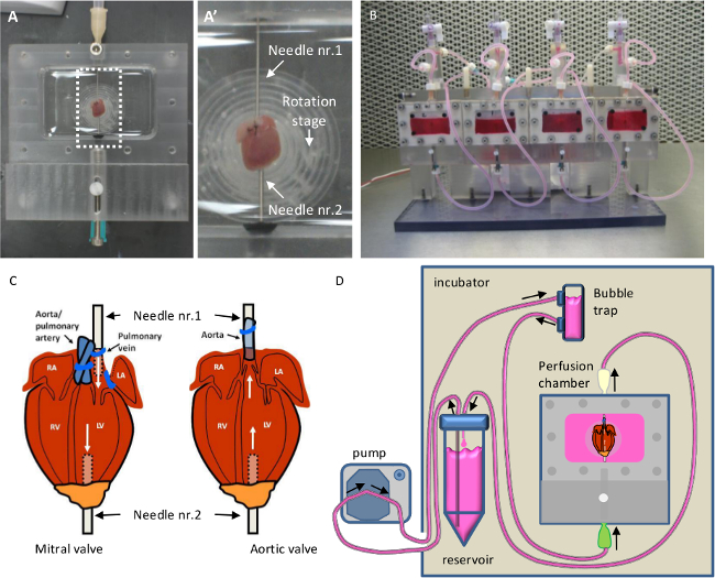
Figure 1. The Miniature Tissue Culture system. (A) In the perfusion chamber the heart is laying on the rotation stage and ligated with the blunted needles indicated with nr.1 and nr.2. (B) Perfusion chamber is mounted on the stand. (C) Schematic drawing of the heart when culturing the mitral valve (left) or aortic valve (right). (D) Schematic drawing of the setup of the ex vivo flow system for aortic valve culture. This figure has been partly modified from previous study11. Please click here to view a larger version of this figure.
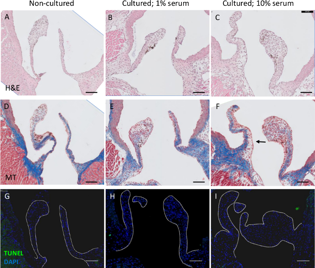
Figure 2. Histological analysis of cultured valves from 2 months old mice. (A-C) heamatoxylin and eosin (H&E) (D-F) Masson’s Trichrome staining and G-I) TUNEL staining of a non-cultured aortic valve (A,D,G), an aortic valve cultured in the presence of 1% serum (B,E,H) or 10% serum (C,F,I) for 3 days in the MTCS. The valve cultured with 1% serum appears similar to the non-cultured valve, whereas the valve cultured with 10% serum is thicker (C) and has an altered ECM pattern (F). No apoptosis is observed in the valves (G-I). Scale bar is 100 um. Please click here to view a larger version of this figure.
Discussion
Critical steps in culturing the cardiac mouse valves include making the time between excision of the heart from the mouse and the ligation in the perfusion chamber as short as possible to ensure viability and ligation of the needles perpendicular to the valves to ensure proper direction of the flow. Additionally, checking the flow after ligation in the perfusion chamber without medium ensures proper insertion and ligation of needles. It is critical to maintain a sterile culture and prevent air bubbles in the tubing, which could potentially get trapped in the heart obstructing the flow.
The total volume of medium used for the perfusion of a heart is 45 ml (note that the medium that is perfused through the heart is not in contact with the 20 ml of medium in the perfusion chamber). For the addition of for example viruses a smaller volume can be preferred. Temporally the reservoir (and part of the tubing) can be removed from the circuit leaving 5-7 ml in the system to allow infection of the valve.
The absence of beating of the heart could be considered a limitation. However, it allows creating an experimental condition in which all stimuli working on the leaflet can be kept constant. This gives the unique opportunity to study the effect of one single modification. When moving valves are required, a pulsatile flow can be created.
To study the valvular biology and the role of molecular, cellular and mechanical stimuli on the valve, modifications can be easily made to the culture system. Molecular modification can be achieved by the addition of growth factors, chemical compounds and viruses mediating gene delivery11 to the perfusion medium. One or multiple cell types can be administered to the perfusion medium to examine the influence of these cells on valvular biology or their contribution to the valve. The influence of oxidative stress or hypoxia can be studied by altering the oxygen-pressure in the culture medium. The hemodynamic stresses on the valve can be varied by changing the flow speed, flow direction, flow pulsatility and the viscosity. The overall morphology can be examined (Figure 2B,C) by H&E, histochemical and immunofluorescent staining. Additionally the organization and composition of the ECM, the phenotype, proliferation, and viability of the valvular cells and the activation of signaling pathways provide insight into the effects of varying these conditions. Genetically modified mice can be used to track the endothelial cells, using endothelial reporter mice (using the Tie2-Cre and floxed reporter mice) indicating the presence of endothelial-to-mesenchymal transformation or mice with loss or gain of function of specific signaling pathways. Overall, the system presented here is a useful tool for studying cardiac valvular biology.
Disclosures
The authors have nothing to disclose.
Acknowledgements
This study is supported by the Dutch Heart Foundation and the Netherlands Institute for Regenerative Medicine.
Materials
| Dulbecco’s Modified Eagle Medium | life technolgies | 10569-010 | |
| Fetal Bovine Serum | life technolgies | 26140 | |
| Insulin-Transferrin-Selenium | life technolgies | 41400-045 | |
| Antibiotic-Antimycotic | life technolgies | 15240-06 | |
| Silk 7-0 | Ethicon | 768G | |
| 100 mm culture dish | Greiner bio-one | 664160 | |
| 50 ml tube | Greiner bio-one | 227261 | |
| 5 ml syringe | BD | 309649 | |
| 21 G needle | BD | 304432 | |
| Heparin | LEO | 012866-08 | |
| Forceps | Fine Science Tools | 11295-00 | |
| Micro Scissors, Economy, Vannas-type | Tedpella | 1346 | |
| Silicon tubing | Thermo Scientific | 8060-0020 | I.D. x O.D. x Wall: 1.59 x 3.18 x 0.79 mm |
| Silicon tubing for pump | Masterflex | 96400-13 | I.D. x O.D. x Wall: 0,8 x1,59 x 0,40 mm |
| Biocompatible glue (Histoacryl) | B. Braun | 1050071 | |
| precision vaporizer | Dräger | Vapor 200 | |
| peristaltic roller pump | Masterflex | 7521-35 | |
| Easy-load pump head | Masterflex | 7518-00 | |
| Flow chamber | see Lieber et al., 2010 | ||
| Bubble trap | see Lieber et al., 2010 |
References
- Go, A. S., et al. Heart Disease and Stroke Statistics–2014 Update: A Report From the American Heart Association. Circulation. 129, e28-e292 (2013).
- Gould, S. T., Srigunapalan, S., Simmons, C. A., Anseth, K. S. Hemodynamic and Cellular Response Feedback in Calcific Aortic Valve Disease. Circulation Research. 113 (2), 186-197 (2013).
- Kruithof, B. P. T., Krawitz, S. A., Gaussin, V. Atrioventricular valve development during late embryonic and postnatal stages involves condensation and extracellular matrix remodeling. Developmental biology. 302 (1), 208-217 (2007).
- Schoen, F. J. Cardiac valves and valvular pathology: update on function, disease, repair, and replacement. Cardiovascular pathology: the official journal of the Society for Cardiovascular Pathology. 14 (4), 189-194 (2005).
- Balachandran, K., Sucosky, P., Yoganathan, A. P. Hemodynamics and mechanobiology of aortic valve inflammation and calcification. International journal of inflammation. , 263870 (2011).
- Butcher, J. T., Simmons, C. A., Warnock, J. N. Mechanobiology of the Aortic Heart Valve. The Journal of heart valve disease. 17 (1), 62-73 (2008).
- Balachandran, K., Konduri, S., Sucosky, P., Jo, H., Yoganathan, A. P. An ex vivo study of the biological properties of porcine aortic valves in response to circumferential cyclic stretch. Annals of biomedical engineering. 34 (11), 1655-1665 (2006).
- Weiler, M., Hwai Yap, C., Balachandran, K., Padala, M., Yoganathan, A. P. Regional analysis of dynamic deformation characteristics of native aortic valve leaflets. Journal of Biomechanics. 44, 1459-1465 (2011).
- Combs, M. D., Yutzey, K. E. Heart valve development: Regulatory networks in development and disease. Circulation Research. 105, 408-421 (2009).
- Kruithof, B. P. T., Duim, S. N., Moerkamp, A. T., Goumans, M. -. J. TGFβ and BMP signaling in cardiac cushion formation: lessons from mice and chicken. Differentiation; research in biological diversity. 84 (1), 89-102 (2012).
- Lieber, S. C., Kruithof, B. P. T., Aubry, N., Vatner, S. F., Gaussin, V. Design of a miniature tissue culture system to culture mouse heart valves. Annals of biomedical engineering. 38 (3), 674-682 (2010).
