This manuscript describes the utility of ultrasound performed on female rats to design experimental models for reproductive and gynecological investigation. A step-by-step explanation of how to perform ultrasonographic evaluation is shown.
With the development of assisted reproductive technology and the ethical limitations of research on humans, rat animal models have been widely used in reproductive medicine. In the past, the study of reproductive system development in rodents has been based on one-time histological examination of excised tissues. Recently, with the development of high-resolution transabdominal ultrasound, high-quality sonography can now be performed to evaluate the reproductive organs of rats, allowing a new method for studying the reproductive system. Images were obtained using a high-resolution ultrasonographic system. Gynecological ultrasonography was performed on 28 eight-week-old non-pregnant rats and 5 pregnant Sprague-Dawley rats. We describe how to recognize organs of the reproductive system and associated structures in typical views during different phases of the estrus cycle. Color flow Doppler was used to measure uterine artery blood flow and evaluate uterine blood flow pattern changes during different stages of pregnancy. We have demonstrated that ultrasound exploration is a useful method for evaluating changes in internal reproductive organs. Its use raises the possibility of conducting additional experiments, including medical or surgical procedures, and provides the ability to monitor sonographic changes to internal organs without sacrificing animals.
Rat animal models have been widely used in reproductive medicine, including in embryo and ovarian transplantation1,2. However, in the past, the study of reproductive system development in rodents has been based on one-time histological examination of excised tissues, and the longitudinal study of day-to-day reproductive organ changes has not been possible in rats3. Ultrasound has been widely used in assisted reproductive technology in humans for over 30 years, but this valuable technology has only been applied to rats recently.
Our objective was to establish an ultrasonographic approach for evaluating the reproductive organs of Sprague-Dawley rats to design experimental models for reproduction and gynecology investigation and to demonstrate the procedure because to our knowledge, there are no current visualized publications regarding this procedure. We describe the procedure of ultrasonographic examination of the female rat's reproductive system and present ultrasonographic findings on anatomy and uterine artery blood flow using high-definition ultrasound. We monitored the characteristics of the endometrium, ovaries and uterine artery blood flow in non-pregnant animals at different stages of estrus cycle to evaluate the significant differences in endometrial thickness, ovarian morphology and uterine blood flow in different stages of the estrus cycle, similar to women. We used high-quality ultrasound equipment with a 70 MHz frequency and a resolution level of 30 µm. Our other objective was to evaluate changes in the resistance of uterine blood flow in pregnant rats. This technique allows for the study of daily changes in the reproductive organs without sacrificing animals.
There are several technical difficulties in utilizing ultrasound on rats. These difficulties include: the rat endometrium is much thinner than a human female4. Difficulty in imaging the ovaries of rats has been attributed to thicker skin and abdominal wall musculature in rats, which resulted in near-complete attenuation of the ultrasound5, and the uterine artery is much more difficult to find in non-pregnant rats. We have solved many technical problems with the procedure, and for those problems that remain, we show how to minimize them.
Successful monitoring of sonographic changes in the reproductive organs of rats without the need to sacrifice animals will open the possibility of building future animal models of reproductive medicine and other surgical procedures.
This study was carried out in strict accordance with the recommendations in the Guide for the Care and Use of Laboratory Animals of the National Institutes of Health and in accordance with the ARRIVE (Animal Research: Reporting In Vivo Experiments) guidelines. The protocol received a license for animal experimentation complying with Directive 2010/63/EU with the authorization number A13170404 (Anexo 1). All experiments were performed in a European Union-certified laboratory following national guidelines for the ethical care of animals (RD 53/2013, EU Directive 63/2010). The protocol was approved by the Committee on the Ethics of Animal Experiments of the University of Murcia.
1. Animal Preparation
Note: All experiments were supported by the Animal Experimentation Section of Murcia University and the Obstetrics and Gynecology Department of Murcia University.
2. Preparation of Rats for Imaging
Note: Images were obtained using high-resolution transabdominal ultrasonography. Estrous cycle phases were determined by vaginal smear cytology.
3. Examination Procedure
Note: Anatomy of rats´ reproductive organs: The vagina lies dorsal to the urinary bladder and divides into two uterine horns that extend toward the kidneys. The ovaries are connected to the uterine horns via the oviducts (Figure 1). The uterus is located in the region posterior to the kidneys.
4. Design of the Study
There were no significant differences in antero-posterior uterine horn diameters or in the thickness of the endometrium between the two sides of the uterine horn (Table 1). Compared with the group 2, the mean endometrial thickness in the group 1 was thicker, but no significant differences (P>0.05) were found between the two groups (Figure 3). Nevertheless, we found fluid inside the uterus (in 8 out of 28 rats) near the estrus cycle associated with changes in endometrium morphology (Figure 2).
Doppler studies also showed no significant changes in flow velocity waveform patterns in each side of the uterine horn or in different phases of estrus cycle in non-pregnant rats (Tables 1 and 2, Figure 4A). However, in pregnant rats, as gestation advanced, peak systolic and end-diastolic velocities increased significantly, and the calculated resistance index decreased significantly (Table 3, Figure 4B).
The mean diameter of the ovary was not significantly different (Table 1). When the morphology of the ovary was compared between the two groups, periovulatories follicles and fluid around the ovary were seen after ovulation (Table 2, Figure 2).
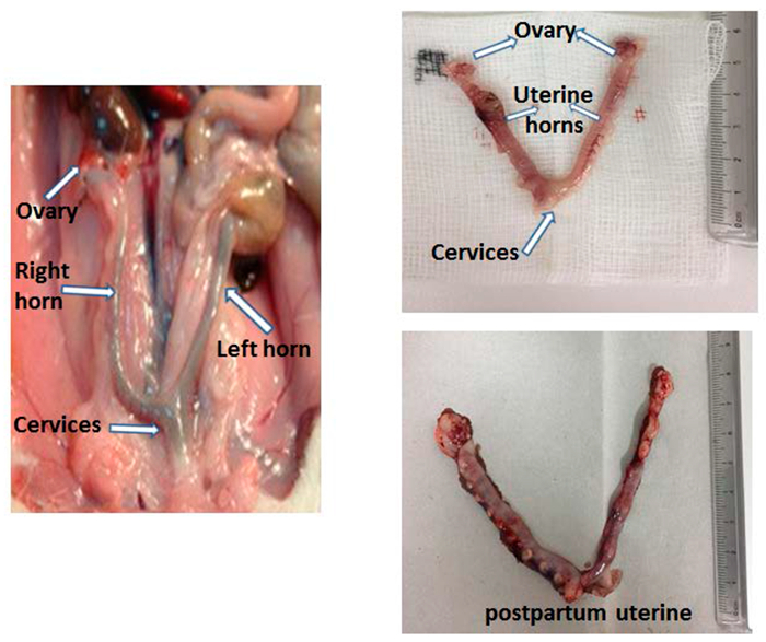
Figure 1: Anatomy Please click here to view a larger version of this figure.
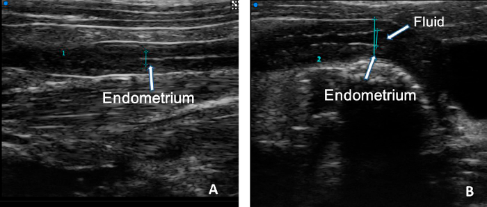
Figure 2: Measurement of the endometrial thickness (B-mode). The thickness of the endometrium (blue line) (A). The antero-posterior uterine horn diameters (large blue line) and thickness of the endometrium (short blue line) during estrus cycle (B). Please click here to view a larger version of this figure.
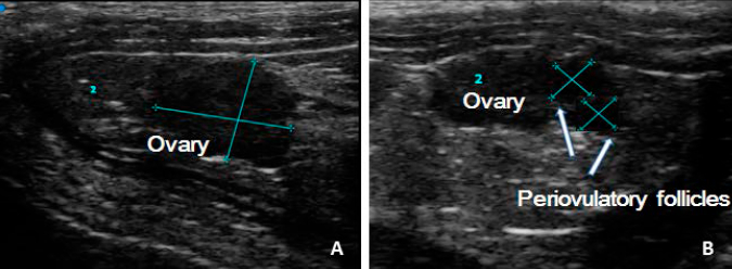
Figure 3: (A) Measurement of the diameter of the left ovary; (B) Ovary and follicles during estrus phase.
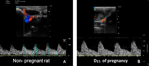
Figure 4: Measurement of uterine artery blood flow. (A) Uterine artery blood flow in non-pregnant rats. (B) Uterine artery blood flow in 15 th day of pregnant rats.
(P >0.05, no significant difference in each group).SD: Standard DesviationPSV: peak systolic velocityEDV: end-diastolic velocityS/D: Systolic to diastolic ratioRI: resistance index. ((RI)=[PSV–EDV]/PSV)
Variable (mm±SD) |
Left side | Right side | P. value |
Horn diameter (mm) | 1.78±0.24 | 1.73±0.28 | 0.626 |
Endometrium thickness (mm) | 0.75±0.06 | 0.76±0.05 | 0.752 |
Ovary diameter (mm) | 3.69±0.52 | 3.62±0.32 | 0.107 |
Follicle size (mm) | 1.68±0.31 | 1.74±0.29 | 0.859 |
PSV (mm/s) | 91.52±17.91 | 93.07±22.87 | 0.055 |
EDV (mm/s) | 34.18±9.36 | 36.67±11.14 | 0.178 |
S/D | 2.78±0.59 | 2.62±0.50 | 0.294 |
RI | 0.62±0.08 | 0.60±0.08 | 0.876 |
(P >0.05, no significant difference in each group). | |||
SD: Standard Desviation | |||
PSV: peak systolic velocity | |||
EDV: end-diastolic velocity | |||
S/D: Systolic to diastolic ratio | |||
RI: resistance index. ((RI)=[PSV–EDV]/PSV) |
Table 1: The differences in the left and right uterine horn and ovary.
Variable (mm±SD) |
Estrus phase (Group 1) |
Non-estrus phase (Group 2) |
P.value |
Horn diameter (mm) | 1.71±0.18 | 1.83±0.23 | 0.433 |
Endometrium thickness (mm) | 0.78±0.04 | 0.72±0.05 | 0.168 |
Ovary diameter (mm) | 3.71±0.56 | 3.66±0.47 | 0.515 |
PSV (mm/s) | 92.05±17.93 | 94.15±20.62 | 0.886 |
EDV (mm/s) | 37.81±9.64 | 34.72±5.38 | 0.096 |
S/D | 2.61±0.58 | 2.77±0.44 | 0.249 |
RI | 0.60±0.08 | 0.63±0.06 | 0.232 |
(P >0.05, no significant difference in each group). | |||
SD: Standard Desviation | |||
PSV: peak systolic velocity | |||
EDV: end-diastolic velocity | |||
S/D: Systolic to diastolic ratio | |||
RI: resistance index. ((RI)=[PSV–EDV]/PSV) |
Table 2: The differences between different estrous cycle phases in non-pregnant rats.
Variable | D9 | D15 | D18 | P value |
PSV(mm/s) | 111.08±5.93a,b | 122.64±7.49c | 131.91±3.50 | <0.05 |
EDV(mm/s) | 38.80±3.37d,e | 56.43±3.10f | 79.29±5.47 | <0.05 |
S/D | 2.87±0.12g,h | 2.17±0.16i | 1.67±0.14 | <0.05 |
RI | 0.65±0.02 j,k | 0.54±0.04L | 0.39±0.05 | <0.05 |
PSV= Peak systolic velocity | ||||
EDV=End-diastolic velocity. | ||||
S/D=Systolic to diastolic ratio (PSV/EDV). | ||||
RI=Resistance index ((RI)= ([PSV–EDV]/PSV). | ||||
D9= Day 9 of gestation | ||||
D15= Day 15 of gestation | ||||
D18=Day 18 of gestation | ||||
SD:The errors indicate Standard Deviation (±). | ||||
(P<0.05, no significant difference in each group) | ||||
P value: D9 vs D15 a=0.03; d=0.001; g=0.01; j=0.01. D9 vs D18 b=0.003; e=0.001; h=0.01; k=0.001. D15 vs D18 c=0.03; f=0.001; i=0.03; L=0.04. |
Table 3: The differences in uterine artery blood flow in pregnant rats.
Due to the procedural modifications and troubleshooting that was required in this study, despite our aim of identifying all the phases of the estrus cycle in rats using ultrasound, we were unable to find any significant differences. We hypothesize that these difficulties may be because the estrus cycle only lasts a few days in rats, unlike the cycle in women. We are sure that all measurements were done at exactly the right time to determine any differences. Therefore, we regrouped the estrus cycle phases into only two phases to evaluate for any differences, but none were observed. As expected, we found no significant difference between the measurements of each uterine horn, which will allow us to choose either horn to perform measurements in the future. Studies on the effects of isoflurane use on rodents are contradictory. It may be teratogenic, but only if administered at anesthetic concentrations for many hours or several days. In our experiment, the examination time in the pregnant rats was less than 30 min, so we did not find any adverse effects in any pregnant rats or their offspring.
We would like to have found more similarities to ultrasound evaluation of the reproductive tract in women, in which there are clear echogenic differences between the follicular phase, periovulatory and secretory phases, but such changes were not seen in the rat model. This fact can be explained because the rat estrus cycle phases only last four to five days. Short estrus cycle phases and gestation periods make the rat an ideal animal for research on reproduction5. Nevertheless, this fact may be the reason why there are no significant differences in the diameter of the uterine horn, endometrial thickness and ovarian diameter in the estrus and non-estrus phases. It is difficult to take the measurements at exactly the right time to ascertain differences, and despite taking measurements every day, we did not find significant changes.
Despite the limitations above, we propose the use of ultrasonography to monitor changes in the reproductive system, including thickness and morphology of reproductive organs. We can affirm this use of ultrasonography because the thickness of the endometrium in 8-week-old Sprague-Dawley rats as measured histopathologically (one layer 359.13 ± 49.70 mm) by Jing et al.7 is similar to the results here. In spite of the various techniques for measuring endometrial thickness histopathologically and sonographically, we obtained similar results. Though power Doppler and color Doppler have been used on pregnant rats for several years8,9, there have been few investigations measuring uterine artery blood flow in non-pregnant rats. Now with the development of the ultrasonography, we can take advantage of this technique for monitoring changes in the reproductive tract during each different stage, even at the very beginning of pregnancy.
Future applications of ultrasound may include exploration of the mechanism of embryo implantation and treatment of thin endometrium in rat animal models. Also, by monitoring the characteristics of developing follicles, we can obtain more comprehensive knowledge about their function in an ovarian transplantation model. Currently, there are few investigations using 3D imaging and molecular ultrasound imaging of the reproductive system of rats10, and we will apply this technique on a thin endometrium model in the future.
We can draw the conclusion that the rat is a suitable model for the study of reproductive organ dynamics using transcutaneous ultrasound bio-microscopy, which does not require sacrifice of the animal.
The authors have nothing to disclose.
This research was supported by Animal Experimentation Section of Murcia University and the Obstetrics and Gynecology Department of Murcia University. We thank all the technicians working at the CEIB (Centro Experimental en Investigaciones Biomédicas), the section of animal experimentation of the University of Murcia, who have collaborated on this project.
Vevo3100 high-resolution in vivo micro-imaging system* | Visual Sonics, inc. | www.visualsonics.com/products | |
Vevo integrated rail system including physiological monitoring unit. | Visual Sonics, inc. | www.visualsonics.com/products | |
MX400 Transducter | Visual Sonics, inc. | www.visualsonics.com/products | |
Vevo Lab Software | Visual Sonics, inc. | www.visualsonics.com/products | |
HSD: Sprague Dawley SD | Envigo, inc. | Rat strain | |
Lubricating Gel | General Supply | ||
CIBERTEC CA-EAC20 Anesthesia Trolley System | Cibertec S.A | Anesthesia Machine | |
Ecogel 100 ultrasound gel | Eco-Med Pharmaceuticals Inc. | ||
Hair removal lotion (Nair) | General Supply | ||
Isoflurane | Esteve Veterinaria | Inhalatory anesthesia | |
* Required software is Vevo software including B-Mode application, pulse wave Doppler application, and vascular strain analysis tools package. |