Summary

巨大リポソームの膜カーボンナノ チューブを引っ張ってください。

Published: December 07, 2017
doi:

Summary

多くのタンパク質の細胞感覚し、膜の曲率を誘発します。湾曲した膜体外タンパク質または任意曲率活性分子の相互作用を研究する脂質小胞から膜ナノチューブをプルする方法について述べる。

Abstract

細胞膜の再編は、エンドサイトーシス、人身売買、糸状等の形成など多くの細胞現象の不可欠な部分です。多くの異なった蛋白質に関連付ける曲面膜感覚または膜の曲率を誘発する能力のため。通常、これらのプロセスは複雑すぎるために、細胞の定量的研究し蛋白質の多数を含みます。エンドサイトーシスの首などの曲線の細胞構造を模倣した、曲線状の膜の in vitro再構成するプロトコルについて述べる。巨大リポソーム (GUV) 小嚢は、ピペット吸引がその内部圧力と表面張力を制御、細胞膜のモデルとして使用されます。光ピンセットによる旦那のポイント引き力を適用するには、平膜に接続されている高曲率のカーボンナノ チューブが作成されます。このメソッドは、曲げ剛性などの脂質膜の基本的な機械的性質を測定する伝統的に使用されています。近年、タンパク質が膜の湾曲や形状および膜の力学に影響する方法と対話する方法の研究に展開されています。マイクロマニピュレーション、マイクロインジェクション、光ピンセットと共焦点顕微鏡を組み合わせたシステムは、膜の曲率、膜張力および蛋白質の表面密度の測定を同時にできます。これらの測定からタンパク質膜システムの多くの重要な機械的および形態学のプロパティを推測できます。さらに、我々 は生理的塩濃度および分類された蛋白質と脂質の蛍光強度から膜タンパク質の表面密度を定量化する手法の存在下で膜を作成するプロトコルをレイアウトします。

Introduction

エンドサイトーシス、人身売買、糸状、感染症などの形成など多くの細胞プロセスは細胞膜1,2という形で劇的な変化を伴っています。セルで、いくつかの蛋白質は膜にバインドし、その形状を変更するこれらのプロセスに参加します。最も顕著な例は、本質的にドメイン3,4,5,6,7バー曲線特性を含む箱/アンフィフィシン/Rv (バー) 蛋白質家族のメンバーです。通常、彼らは表面と、多くの場合、両親媒性ヘリックスを 2 層も浅く挿入バー ドメインを付着する膜と対話します。形状、サイズ、および両親媒性ヘリックスの数と共に BAR ドメインの料金を決定します: (1) 膜の湾曲の方向 (すなわち、かどうか彼らは陥入や突起を誘発するが)、(2) 膜の大きさ曲率5,8。注記のうち、ここで正の曲率は曲線状の膜、すなわち、相互作用する粒子と負の膨らみのそれ以外の凸側として定義されます。さらに、蛋白質バーの定量的研究は、膜への影響は物理的なパラメーターのセットによって異なりますを明らかにした: 表面蛋白質、膜張力膜形状 (フラット鋼管球の密度図形)7。タンパク質バーこれらのパラメーターに応じてすることができます: (1) 膜の曲率のセンサーとしての機能、(2) 膜を曲げたり、(3) 膜切断7を誘発します。

膜再編のセルで、エンドサイトーシスなどの現象の定量的側面を勉強に関連するコンポーネントの数により生体内では非常に挑戦的です。セルに湾曲した膜を模倣した最小限のコンポーネントのin vitro再構成は、どのように膜カービング タンパク質の動作機構の理解を得るための手段を提供します。この記事では、膜カーボンナノ チューブ体外顕微操作、共焦点顕微鏡、光ピンセットを使用して再構成するプロトコルについて説明します。アプローチは、タンパク質、脂質、または小さい分子が湾曲した膜と対話する方法を定量的な方法で勉強する使用できます。脂質膜は、曲率が無視できる膜カービング結合分子のサイズと比較して、細胞膜のモデルとして使用されます。彼らは、脂質膜の水和と交流電流 (AC)10の下でそれを膨潤膜に小胞を形成する electroformation メソッド9を使用して準備されます。最も一般的な基質 Guv が栽培されているインジウムの錫の酸化物 (ITO) のいずれか半導べ電性皮膜や白金線 (Pt 線)11。この作品は、このメソッドは、バッファー12に塩の存在下で膜を作るの代替よりもはるかに良い動作するように示されている、Guv、Pt ワイヤ上育ちます。Electroformation プロトコルここでそれを再現するのに十分な詳細に説明しています、我々 は詳細13,14に、膜を作るのと同様、他の方法を記載されている以前の記事へ読者を参照します。私たちの手で Pt ワイヤ上 electroformation 正常にもたらした膜合成脂質のミックスや天然脂質から 〜 100 mM の NaCl を含むバッファーで抽出。さらに、成長過程における膜内タンパク質をカプセル化することが可能だったも。図 1 a; での例が electroformation 商工会議所を表示します。両側からす coverslips に封印できるポリテトラフルオロ エチレン (PTFE) から作られたホルダーに挿入 2 ~ 10 cm ロング Pt ワイヤーが装備されている 1 〜 2 cm 離れて (図 1 a)。

Figure 1
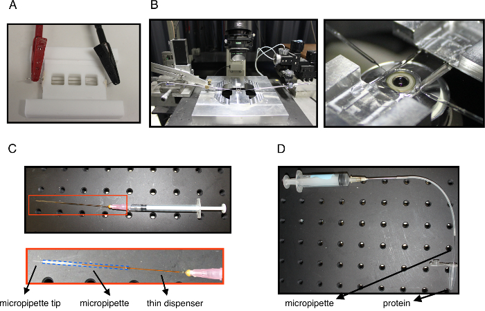
図 1: 実験的セットアップします。白金線に接続された電気コネクタの旦那 electroformation (A) 室。(B) 左: 顕微鏡を示す実験的システム、上記の目的は、2 つのマイクロ ピペット (左と右) 実験室マニピュレーターに取り付け、チューブを引っ張るとタンパク質の実験室に挿入インジェクション。右: 実験室のクローズ アップ ビューを吸引し挿入注入マイクロ ピペットの先端を示す目的上マウント。(C) A の注射器そのバックエンドでマイクロ ピペットに挿入される細いディスペンサーを装備しました。下部は、青の点線のアウトライン、ピペットでマイクロ ピペット内ディスペンサーのクローズ アップ ビューです。このシステムは、カゼイン ガラス表面にまた必要なとき鉱物油で塗りつぶしをバックアップすると、マイクロ ピペットを埋めるためです。(D) A システム μ L タンパク質溶液の量を吸引するために使用します。針は、注射器と注入マイクロ ピペットに接続されているチューブに接続されているです。マイクロ ピペット チップは慎重にタンパク質溶液に浸漬、マイクロ ピペットのチップを記入するので吸気します。マイクロ ピペットがパネル c. に示すシステムを使用して鉱物油で再び埋められますこの図の拡大版を表示するのにはここをクリックしてください

7 からの半径に至るまで膜ナノチューブ nm ~ 数百 nm は、外部の力が、旦那から引っ張ることができます。このメソッドの細胞膜と小胞、曲げ剛性15,16などの弾性的性質を測定するために設計されました。最も最近の作品の方法は microinjecting プル カーボンナノ チューブ7,17近く蛋白質による曲面膜とタンパク質の相互作用を研究に拡張されました。他の方法は蛋白質の膜カービングを勉強するために開発されています。1 つの方法で蛋白質は不動態化された表面につながれた異なるサイズのリポソームと孵化させます。共焦点の顕微鏡を使用して、曲率による並べ替え18,19を示すことができます, リポソーム径の関数として蛋白質の結合を測定します。別の方法で蛋白質は尿細管20,21を自発的に誘発する能力を測定するマイクロ吸気 GUV 近く注入されます。このプロトコルで説明する方法は、エンドサイトーシスに関与する膜カービング蛋白質を研究に適してほとんどの蛋白質が通常の貨物を含む細胞膜の陥入を接続する前もって形成された膜ナノチューブに遭遇、平膜を基になります。さらに、このメソッドとは異なり、つなぎ縄でつながれた小さなリポソームを用いる試金で膜カーボンナノ チューブは継続的に接続されて膜;したがって、それは GUV、状況が予想される生体内力学的平衡です。したがって、基本的な膜の物理が適用され、我々 は我々 の測定22,23,24から機械的性質の茄多を推測できます。

このメソッドの完全な実装、必要な機器は、共焦点顕微鏡、光ピンセット、水タンク (図 1 b) に接続されている 1 つまたは 2 つのマイクロ ピペットに含まれます。すべての 3 つを組み合わせて、同時に力25をチューブの膜、膜の曲率、蛋白質の表面密度を測定し不可能です。マイクロ ピペット吸引は不可欠ですし、静水圧による吸引圧26を制御する水タンクに接続されているホルダーにガラス ピペットを挿入することによって簡単に構築します。マイクロ ピペットとホルダーは高精度動きの圧電アクチュエータを用いたマイクロマニピュレーターと、理想的には、一方向に制御されます。カーボンナノ チューブ、旦那は微小ビーズを引いて離れて、カーボンナノ チューブの作成にこだわった簡単に microaspirated を引く。この実装では、ビーズが27公開されたプロトコルに従うことによって構築することができます光ピンセットを用いて行われます。正確な力の測定を犠牲にして、光ピンセットとプル カーボンナノ チューブのさまざまな方法で調剤することが可能です。光トラップを構築するも困難な場合、または力の測定は必須ではありませんなど単に湾曲した膜蛋白質の好みをチェックしたい場合、2 番目のマイクロ ピペット28の先端に吸引ビーズを使用してチューブを引っ張ることが。また、重力29を使用してチューブを引くか、30,31をフローすることが可能です。さらに、共焦点顕微鏡必須ではありません。しかし、それは蛋白質の表面密度を測定するので勧めします。また、脂質膜力と緊張とは関係なくこうして、管内の蛍光強度からカーボンナノ チューブの半径を測定できます。蛍光から推論のチューブの半径は膜付着蛋白質25の存在のため確立された方程式から逸脱するこれらの量の間の関係は特に重要です。重要なは、1 つは管の曲率を測定するそれはできない、光学トラップと共焦点顕微鏡のはやれない。

このプロトコルで説明するメソッドは、様々 な末梢膜タンパク質ナノチューブ バー家族25,32,33,34 からの大抵それらの曲率による並べ替えを研究に使用されています.また KvAP に濃縮されて円錐形の貫通型カリウム チャネルは、タンパク質35バーとしてカーボンナノ チューブを同じ方法で湾曲したが示されました。膜内タンパク質をカプセル化する方法を最適化することによって負の曲率とタンパク質の相互作用は、最近よく36として研究されています。タンパク質足場25,37の形成を明らかにし、どちらかライン テンション38, タンパク質ダイナミン39、またはバーの膜切断機構の研究にさらに、このメソッドが使用されていますタンパク質40,41。蛋白質に加えて小さな分子やイオンで曲率も誘導できます。このメソッドを使用すると、カルシウム イオンは塩無料条件42の下で正の曲率を誘導するために示されていた。興味深いことに、脂質が demixing ポイント43,44近くにある組成のだけが並べ替え、曲率を受けることができることはまた示します。合計では、メソッドは、方法のいずれかの積分膜成分 (例えば脂質や膜貫通タンパク質) の調査に興味を持って研究によって使用することができますまたは末梢分子をバインド (または内部または外膜) の対話開放円筒曲面膜、機械的かつ定量的視点から。それはまた22,23,45膜自体の機械的性質の測定に興味のある方のものは。

Protocol

1 Pt 線の Electroformation によって膜の作製 Electroformation の洗浄 (導入および図 1A参照) と Pt-ワイヤ エタノールや脂質を洗い流すためアセトンなどの有機溶剤と水で塩を洗い流す。注: 私達は徹底的にエタノールに浸したティッシュでふき取り、アセトン、エタノール、水、5 分ごとで sonicating を提案します。 1 mg/mL のクロロホルムで必要な脂?…

Representative Results

チューブを抜く実験膜に関する重要な力学的情報を与えることができます。旦那50,51 から引き出されたチューブ式のタンパク質や他の分子膜膜曲率を持つカップルを強制的に、Canham Helfrich ハミルトニアンを用いた膜張力チューブの半径を関連付けることができますがない場合 <i…

Discussion

膜からチューブを抜く方法、膜蛋白質システムに豊富な情報と、膜の基本的な機械的性質を測定するための手段だけではないが、タンパク質や膜間の結合に光を当てること曲率。サブミクロン リポソーム表面パッシベーションされた18,19につながれたと蛋白質をインキュベートするか観察することによって、タンパク質の膜カーブの効果を測定す?…

Disclosures

The authors have nothing to disclose.

Acknowledgements

著者は、ブノワ ・ ソール、ダミアン キュヴリエ、ピエール ・ Nassoy、フランソワ Quemeneur、グループでカーボンナノ チューブ法を確立するギル Toombes 彼らの本質的な貢献のためにありがとうございます。脱法属する CNRS コンソーシアム CellTiss、Labex CelTisPhyBio (ANR-11-LABX0038)、およびパリ科学 et レトル (ANR-10-アイデックス-0001-02)。F. C. ツァイ エンボ長期フェローシップ (ALTF 1527-2014 年)、マリー キュリー アクション (H2020 MSCA-場合 2014、プロジェクト膜・ エズリン アクチン) によって資金を供給されました。修士はシモンズの仲間社会短期やつです。

Materials

1,2-Dioleoyl-sn-glycero-3-phosphatidylcholine (DOPC) Avanti 850375 Example lipid used in data for Figure 3
1,2-distearoyl-sn-glycero-3-phosphoethanolamine-N-[biotinyl(polyethyleneglycol)-2000] [DSPE-PEG(2000)-biotin] Avanti 880129 biotinylated lipid
BODIPY-TR-C5-ceramide Molecular Probes (ThermoFisher) D7540 fluorescent lipid
BODIPY- FLC5-hexadecanoyl phosphatidylcholine (HPC*) Molecular Probes (ThermoFisher) fluorescent lipid for protein density calibration
egg L-α-phosphatidylcholine (eggPC) Avanti 840051 used for calibrating the tube radius constant
β-casein from bovine milk (>99%) Sigma Aldrich C6905 used for passivating the micropipette and the experimental chamber
Sucrose Sigma Aldrich S7903
D(+) glucose Sigma Aldrich G7021
NaCl Sigma Aldrich S7653
Tris Sigma Aldrich 10708976001
osmometer Loser n/a
Pt-wires, 0.5 mm diameter, 99.99% pure Goodfellow USA PT005139 used for GUV electroformation
function generator n/a used to create current for GUV electroformation
putty sealant Vitrex (from CML France) CRIT 140013 used to seal the electroformation chamber
bath sonicator n/a useful to clean the electroformation chamber, but not crucial
Nikon TE2000 inverted microscope, eC1 confocal system (Nikon), with two laser lines (λ = 488 nm and 543 nm); optical tweezers induced by a 5 W ytterbium fiber continuous wave laser (λ > 1070 nm; IPG GmBH Germany) an example of a confocal microscopy system equipped with optical tweezers
micromanipulators n/a
borosilicate capillaries (with internal and external radii of 0.78 mm and 1 mm, respectively) Harvard apparatus 30-0036
micropipette puller Sutter Instrument P-2100
microforge Narishige MF-800
piezoelectric actuator Physik Instrumente n/a
polystyrene streptavidin coated beads (diameter 3.2 µm) Spherotech SVP-30-5

References

  1. McMahon, H. T., Gallop, J. L. Membrane curvature and mechanisms of dynamic cell membrane remodelling. Nature. 438 (7068), 590-596 (2005).
  2. Johannes, L., Wunder, C., Bassereau, P. Bending “on the rocks”–a cocktail of biophysical modules to build endocytic pathways. Cold Spring Harb Perspect Biol. 6 (1), (2014).
  3. Dawson, J. C., Legg, J. A., Machesky, L. M. Bar domain proteins: a role in tubulation, scission and actin assembly in clathrin-mediated endocytosis. Trends Cell Biol. 16 (10), 493-498 (2006).
  4. Mim, C., Unger, V. M. Membrane curvature and its generation by BAR proteins. Trends Biochem Sci. 37 (12), 526-533 (2012).
  5. Qualmann, B., Koch, D., Kessels, M. M. Let’s go bananas: revisiting the endocytic BAR code. EMBO J. 30 (17), 3501-3515 (2011).
  6. Rao, Y., Haucke, V. Membrane shaping by the Bin/amphiphysin/Rvs (BAR) domain protein superfamily. Cell Mol Life Sci. 68 (24), 3983-3993 (2011).
  7. Simunovic, M., Voth, G. A., Callan-Jones, A., Bassereau, P. When Physics Takes Over: BAR Proteins and Membrane Curvature. Trends Cell Biol. 25 (12), 780-792 (2015).
  8. Safari, F., Suetsugu, S. The BAR Domain Superfamily Proteins from Subcellular Structures to Human Diseases. Membranes (Basel). 2 (1), 91-117 (2012).
  9. Angelova, M. I., Soléau, S., Méléard, P., Faucon, F., Bothorel, P., Helm, C., Lösche, M., Möhwald, H. . Progress in Colloid and Polymer Science Vol. 89: Trends in Colloid and Interface Science VI. 89, 127-131 (1992).
  10. Meleard, P., Bagatolli, L. A., Pott, T. Giant unilamellar vesicle electroformation from lipid mixtures to native membranes under physiological conditions. Methods Enzymol. 465, 161-176 (2009).
  11. Montes, L. R., Alonso, A., Goni, F. M., Bagatolli, L. A. Giant unilamellar vesicles electroformed from native membranes and organic lipid mixtures under physiological conditions. Biophys J. 93 (10), 3548-3554 (2007).
  12. Mathivet, L., Cribier, S., Devaux, P. F. Shape change and physical properties of giant phospholipid vesicles prepared in the presence of an AC electric field. Biophys J. 70 (3), 1112-1121 (1996).
  13. Collins, M. D., Gordon, S. E. Giant liposome preparation for imaging and patch-clamp electrophysiology. J Vis Exp. (76), (2013).
  14. Garten, M., Aimon, S., Bassereau, P., Toombes, G. E. Reconstitution of a transmembrane protein, the voltage-gated ion channel, KvAP, into giant unilamellar vesicles for microscopy and patch clamp studies. J Vis Exp. (95), e52281 (2015).
  15. Hochmuth, R. M., Evans, E. A. Extensional flow of erythrocyte membrane from cell body to elastic tether. I. Analysis. Biophys J. 39 (1), 71-81 (1982).
  16. Hochmuth, R. M., Wiles, H. C., Evans, E. A., McCown, J. T. Extensional flow of erythrocyte membrane from cell body to elastic tether. II. Experiment. Biophys J. 39 (1), 83-89 (1982).
  17. Bassereau, P., Sorre, B., Levy, A. Bending lipid membranes: experiments after W. Helfrich’s model. Adv Colloid Interface Sci. 208, 47-57 (2014).
  18. Hatzakis, N. S., et al. How curved membranes recruit amphipathic helices and protein anchoring motifs. Nature chemical biology. 5 (11), 835-841 (2009).
  19. Bhatia, V. K., et al. Amphipathic motifs in BAR domains are essential for membrane curvature sensing. EMBO J. 28 (21), 3303-3314 (2009).
  20. Shi, Z., Baumgart, T. Membrane tension and peripheral protein density mediate membrane shape transitions. Nat Commun. 6, 5974 (2015).
  21. Chen, Z., Shi, Z., Baumgart, T. Regulation of membrane-shape transitions induced by I-BAR domains. Biophys J. 109 (2), 298-307 (2015).
  22. Cuvelier, D., Derenyi, I., Bassereau, P., Nassoy, P. Coalescence of membrane tethers: experiments, theory, and applications. Biophys J. 88 (4), 2714-2726 (2005).
  23. Derenyi, I., Julicher, F., Prost, J. Formation and interaction of membrane tubes. Phys Rev Lett. 88 (23), 238101 (2002).
  24. Dimova, R. Recent developments in the field of bending rigidity measurements on membranes. Adv Colloid Interface Sci. 208, 225-234 (2014).
  25. Sorre, B., et al. Nature of curvature coupling of amphiphysin with membranes depends on its bound density. Proc Natl Acad Sci U S A. 109 (1), 173-178 (2012).
  26. Evans, E., Rawicz, W. Entropy-driven tension and bending elasticity in condensed-fluid membranes. Phys Rev Lett. 64 (17), 2094-2097 (1990).
  27. Neuman, K. C., Nagy, A. Single-molecule force spectroscopy: optical tweezers, magnetic tweezers and atomic force microscopy. Nat Methods. 5 (6), 491-505 (2008).
  28. Capraro, B. R., Yoon, Y., Cho, W., Baumgart, T. Curvature sensing by the epsin N-terminal homology domain measured on cylindrical lipid membrane tethers. J Am Chem Soc. 132 (4), 1200-1201 (2010).
  29. Bo, L., Waugh, R. E. Determination of bilayer membrane bending stiffness by tether formation from giant, thin-walled vesicles. Biophys J. 55 (3), 509-517 (1989).
  30. Rossier, O., et al. Giant Vesicles under Flows: Extrusion and Retraction of Tubes. Langmuir. 19 (3), 575-584 (2003).
  31. Borghi, N., Rossier, O., Brochard-Wyart, F. Hydrodynamic extrusion of tubes from giant vesicles. EPL (Europhysics Letters). 64 (6), 837 (2003).
  32. Zhu, C., Das, S. L., Baumgart, T. Nonlinear sorting, curvature generation, and crowding of endophilin N-BAR on tubular membranes. Biophys J. 102 (8), 1837-1845 (2012).
  33. Ramesh, P., et al. FBAR syndapin 1 recognizes and stabilizes highly curved tubular membranes in a concentration dependent manner. Sci Rep. 3, 1565 (2013).
  34. Knorr, R. L., et al. Membrane morphology is actively transformed by covalent binding of the protein Atg8 to PE-lipids. PLoS One. 9 (12), e115357 (2014).
  35. Aimon, S., et al. Membrane shape modulates transmembrane protein distribution. Dev Cell. 28 (2), 212-218 (2014).
  36. Prévost, C., et al. IRSp53 senses negative membrane curvature and phase separates along membrane tubules. Nat Commun. , (2015).
  37. Simunovic, M., et al. How curvature-generating proteins build scaffolds on membrane nanotubes. Proc Natl Acad Sci U S A. 113 (40), 11226-11231 (2016).
  38. Allain, J. M., Storm, C., Roux, A., Ben Amar, M., Joanny, J. F. Fission of a multiphase membrane tube. Phys Rev Lett. 93 (15), 158104 (2004).
  39. Morlot, S., et al. Membrane shape at the edge of the dynamin helix sets location and duration of the fission reaction. Cell. 151 (3), 619-629 (2012).
  40. Renard, H. F., et al. Endophilin-A2 functions in membrane scission in clathrin-independent endocytosis. Nature. 517 (7535), 493-496 (2015).
  41. Simunovic, M., et al. Friction Mediates Scission of Tubular Membranes Scaffolded by BAR Proteins. Cell. 170 (1), 172-184 (2017).
  42. Simunovic, M., Lee, K. Y., Bassereau, P. Celebrating Soft Matter’s 10th anniversary: screening of the calcium-induced spontaneous curvature of lipid membranes. Soft Matter. 11 (25), 5030-5036 (2015).
  43. Sorre, B., et al. Curvature-driven lipid sorting needs proximity to a demixing point and is aided by proteins. Proc Natl Acad Sci U S A. 106 (14), 5622-5626 (2009).
  44. Heinrich, M., Tian, A., Esposito, C., Baumgart, T. Dynamic sorting of lipids and proteins in membrane tubes with a moving phase boundary. Proc Natl Acad Sci U S A. 107 (16), 7208-7213 (2010).
  45. Dasgupta, R., Dimova, R. Inward and outward membrane tubes pulled from giant vesicles. Journal of Physics D: Applied Physics. 47 (28), 282001 (2014).
  46. Vitkova, V., Genova, J., Mitov, M. D., Bivas, I. Sugars in the aqueous phase change the mechanical properties of lipid mono- and bilayers. Molecular Crystals and Liquid Crystals. 449, 95-106 (2006).
  47. Bouvrais, H. . Mechanical properties of giant vesicle membranes investigated by flickering technique. , (2011).
  48. Koster, G., Cacciuto, A., Derenyi, I., Frenkel, D., Dogterom, M. Force barriers for membrane tube formation. Phys Rev Lett. 94 (6), 068101 (2005).
  49. Kwok, R., Evans, E. Thermoelasticity of large lecithin bilayer vesicles. Biophys J. 35 (3), 637-652 (1981).
  50. Helfrich, W. Elastic properties of lipid bilayers: theory and possible experiments. Z Naturforsch C. 28 (11), 693-703 (1973).
  51. Canham, P. B. The minimum energy of bending as a possible explanation of the biconcave shape of the human red blood cell. J Theor Biol. 26 (1), 61-81 (1970).
  52. Marcerou, J. P., Prost, J., Gruler, H. Elastic Model of Protein-Protein Interaction. Nuovo Cimento Della Societa Italiana Di Fisica D-Condensed Matter Atomic Molecular and Chemical Physics Fluids Plasmas Biophysics. 3 (1), 204-210 (1984).
  53. Leibler, S. Curvature Instability in Membranes. J Phys-Paris. 47 (3), 507-516 (1986).
check_url/56086?article_type=t

Play Video

Cite This Article
Prévost, C., Tsai, F., Bassereau, P., Simunovic, M. Pulling Membrane Nanotubes from Giant Unilamellar Vesicles. J. Vis. Exp. (130), e56086, doi:10.3791/56086 (2017).

View Video