Here, we present a protocol for immunofluorescence staining to observe the endothelial cells of the mouse aorta directly. This technique is useful when studying the cellular and molecular phenotype of endothelial cells in different flow patterns and in the development of atherosclerosis.
Aberrant changes in endothelial phenotype and morphology are considered to be initial events in the pathogenesis of atherosclerosis. Direct observation of the intact endothelium will provide valuable information for understanding the cellular and molecular events in the dysfunctional endothelial cells. Here, we describe a modified en face immunofluorescence staining technique which enables scientists to obtain clear images of the intact endothelial surface and analyze the molecule expression patterns in situ. The method is simple and reliable for observing the entire endothelial monolayer at different sites of the aorta. This technique may be a promising tool for understanding the pathophysiology of atherosclerosis, especially at an early stage.
The early changes in the vasculature primarily initiate in the endothelium, which functions as a selective barrier between the blood and the vessel wall with its intercellular tight junctional complexes1. Substantial evidence points to a critical role for the mechanical effects of blood flow in modulating endothelial function2. Fluid shear stress, a frictional force generated by blood flow, differentially shapes endothelial cell morphology and function, depending on the specific flow paradigms at different vascular sites2,3. Atherosclerotic lesions preferentially occur at sites of disturbed blood flow (d-flow), such as vessel curvatures, flow dividers, and branch points, as compared to regions of steady flow (s-flow), such as the straight segment of the artery. Therefore, direct observation of endothelial morphology and molecule expression patterns should provide important insights into the structural and functional phenotypes of endothelial cells under varying flow paradigms.
Cultured endothelial cells may not express the actual phenotype as they do in vivo partly due to the loss of impact of fluid shear stress, surrounding cytokines, and cell-cell or cell-extracellular matrix interactions. To aid this, the intact endothelial cell monolayer can be studied on transverse sections using classical immunohistochemistry. However, the endothelial monolayer is so thin and fragile that it usually cannot be observed clearly. En face immunohistochemistry has been used to observe the inner surface of the endothelium but is either complicated or erratic in its results because the endothelium is easily stripped from the underlying tissue, or just part of the arterial wall of rats or rabbits, whose walls are thick, is mounted4,5.
Mouse models have considerable advantages over other animals in many respects. Here, we employ a modified en face immunofluorescence technique to analyze endothelial cells of the aortic arch and thoracic aorta in C57BL/6 mouse. Such a technique has been widely used to study the endothelial pathophysiology in different flow patterns and in the development of atherosclerosis6,7,8,9,10. This method allows scientists to observe the entire surface of the endothelium clearly and to compare the expression patterns of a given protein in regions under different fluid shear stress.
All animal experiments were conducted in accordance with experimental protocols approved by the Committee on Animal Resources of Shanghai Jiao Tong University.
1. Perfusion of the mouse aorta
2. Dissection and longitudinally opening of the aorta
3. Pretreatment and immunostaining of the aorta
4. Mounting of the aorta on the glass slide
5. Observation of the aorta
A 12-week-old C57BL/6 mouse was euthanized and perfused with normal saline containing 40 units/mL heparin and, then, prechilled 4% paraformaldehyde. The mouse aorta was exposed under a dissecting microscope (Figure 1), dissected, and cut open longitudinally (Figure 2). En face immunofluorescence staining of the vascular endothelial cells was performed as illustrated in Figure 3 and Table 1. En face immunofluorescence of the vascular cell adhesion protein-1 (VCAM-1) expression with VE-cadherin as endothelial marker was shown under varying flow patterns from different regions of the mouse aorta. DAPI was also counterstained to show the cell nuclei for better visualization. The endothelial and smooth muscle cells can be easily distinguished from the morphology of the cell nuclei when looking through the z-stacks under the microscope since the endothelial cell nuclei are oval shaped and bigger than the spindle-shaped smooth muscle cell nuclei. The representative en face images are shown in Figure 4. The aorta was examined by the LSM 710 Laser Scanning Microscope (Table of Materials) with a FLUAR 40x/1,3 oil lens. From the en face immunofluorescence staining, we can clearly and directly observe that the expression of VCAM-1 was more abundant in regions under disturbed flow (lesser curvature of the aorta arch) than in those under steady flow (greater curvature of the aorta arch and the thoracic aorta).
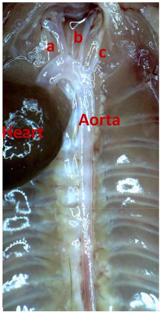
Figure 1: Exposing the mouse aorta under a dissecting microscope. Remove the connective tissues along the aorta as cleanly as possible. a = innominate artery; b = left common carotid artery; c = left subclavian artery. Please click here to view a larger version of this figure.
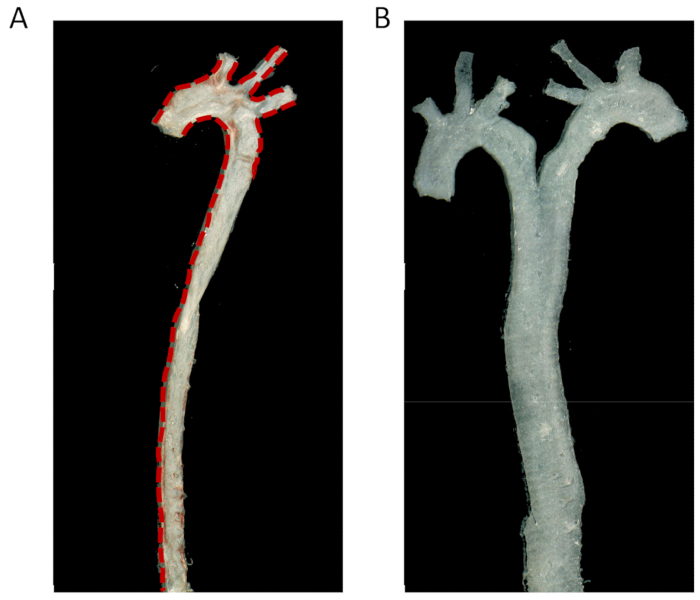
Figure 2: Dissection of the mouse thoracic aorta and cutting it open longitudinally. (A) The thoracic aorta from the heart to the celiac trunk was cut open along the lesser curve longitudinally, and along the greater curve, until the straight segment was met. The three branches of the aortic arch, including the innominate, left common carotid, and left subclavian artery, were also cut open with microscissors. Red dashed lines indicate the cutting line. (B) The aorta was opened and spread flat on the glass slides. Please click here to view a larger version of this figure.
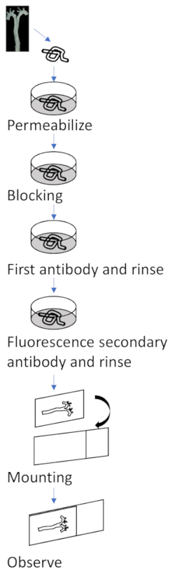
Figure 3: Schema for en face immunofluorescence staining of vascular endothelial cells. First, permeabilize the dissected aorta with 0.1% polyoxyethylene octyl phenyl ether in PBS for 10 min and block it with 10% normal goat serum in TBS containing 2.5% polysorbate 20 for 1 h at room temperature in a 12-well cell culture plate. Next, incubate the aorta with primary antibody in the blocking buffer overnight at 4 °C, and rinse it three times with washing solution (TBS containing 2.5% polysorbate 20). Then, apply fluorescence-conjugated secondary antibodies for 1 h at room temperature and rinse three times. Counterstain the aorta with DAPI for 10 min and rinse it three times in the washing solution. At last, mount the aorta on a glass slide and observe the aorta by a laser-scanning confocal microscope. Please click here to view a larger version of this figure.
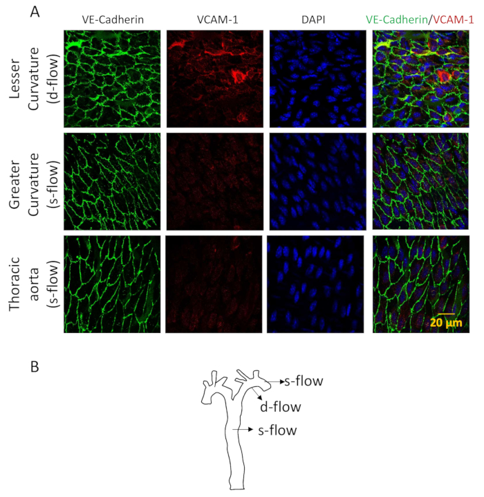
Figure 4: Representative en face staining results. (A) En face immunofluorescence analysis of the lesser curvature of a mouse aorta (d-flow areas) and the greater curvature or thoracic aorta (s-flow areas) to compare the endothelial VCAM-1 expression under different flow paradigms. Endothelial cell morphology is shown by VE-cadherin staining. (B) Representative d-flow areas (lesser curvature) and s-flow areas (greater curvature and thoracic aorta) are indicated by arrows. Please click here to view a larger version of this figure.
Step | Process | Buffer | Temperature | Time |
1 | Permeabilize | 0.1% polyoxyethylene octyl phenyl ether in PBS | Room temperature | 10 min |
2 | Blocking | 10% normal goat serum in Tris-buffered saline (TBS) containing 2.5% polysorbate 20 | Room temperature | 1 h |
3 | First antibody (5 g/mL) | Blocking buffer | 4 °C | Overnight |
4 | Rinse | TBS containing 2.5% polysorbate 20 | Room temperature | 5 min, 3 times |
5 | Fluorescence secondary antibody (1:1000) | Blocking buffer | Room temperature | 1 h |
6 | Rinse | TBS containing 2.5% polysorbate 20 | Room temperature | 5 min, 3 times |
Table 1: Detailed information about the en faceimmunofluorescence staining procedure.
The endothelium is exposed to numerous proatherogenic factors, including lipids, inflammatory mediators, and fluid shear stress1,11,12. Direct observation of endothelial cells in situ provides the special advantages to analyze changes in cell morphology, intercellular junctions, and molecule expression patterns in response to the injury stimuli.
Previous studies have provided two different en face immunohistochemical techniques to observe the endothelium of the arterial wall4,5. One is to obtain an endothelial monolayer by stripping the endothelium from the vessel wall by a special technique, which is difficult to master and could result in the loss of much endothelium at the branching sites of the artery4. The other is mounting the whole arterial wall as we described. Whole-mount preparation is easy to perform and can maintain the whole endothelial monolayer in rabbit and rats5,13. However, spreading the whole mouse aortic specimen flat is not easy, and failure to do so may largely affect the quality of the observation. The preparation of the aortic specimen as described here, with the curvatures cut open and the luminal surface faced downward on the coverslip, makes it easier to spread the aortic specimen flat on the slide glass. Moreover, a laser-scanning confocal microscope is required to better focus on the thin endothelial cell monolayer lining the blood vessel.
A key point of this technique is to stretch the aortic specimen as flat as possible to get a better observation of the aorta. In addition, heparin should be added to normal saline before perfusion, and the aortic specimen should be treated gently throughout the process. Paraformaldehyde should be freshly prepared and kept at 4 °C until usage.
There are limitations to this technique. First, the capacity of en face immunofluorescence to detect subendothelial substance, such as deposited low-density lipoprotein, is limited when compared to regular immunohistochemistry of tissue sections. Second, the fluorescent signals are readily bleached after a scan by the confocal microscope, especially when performing z-axis analysis. Moreover, the expression of the target molecule can only be semiquantified, according to its fluorescent intensity.
In summary, this modified en face immunofluorescence staining technique provides an easy way to analyze the morphology and protein expression pattern of vascular endothelial cells in regions under different blood flow paradigms6,7 and in the pathogenesis of atherosclerosis.
The authors have nothing to disclose.
This study was supported by the National Natural Science Foundation of China (Grant No. 81670451, 81770430), the Shanghai Rising-Star Program (Grant No. 17QA1403000), and the Science Technology Committee of the Shanghai Municipal Government (Grant No. 14441903002, 15411963700).
Antifade mountant | Servicebio | G1401 | |
Delicate Forceps | RWD Life Science | F11001-11 | |
Delicate Scissors | RWD Life Science | S12003-09 | |
Dissecting Forceps | RWD Life Science | F12005-10 | |
Mciro Spring Scissors | RWD Life Science | S11001-08 | |
Polyoxyethylene octyl phenyl ether (Triton X-100) | Amresco | M143 | |
Polysorbate 20 (Tween 20) | Amresco | 0777 | |
VCAM-1 antibody | Abcam | ab134047 | |
VE-Cadherin antibody | BD Biosciences | 555289 | |
Alexa Fluor 555 labeled anti-rabbit IgG | invitrogen | A-31572 | |
Alexa Fluor 488 labeled anti-rat IgG | invitrogen | A-21208 | |
Laser Scanning Microscope | Carl Zeiss |