Here we describe a method to visualize endoplasmic reticulum-associated mRNAs in mammalian tissue culture cells. This technique involves the selective permeabilization of the plasma membrane with digitonin to remove cytoplasmic contents followed by fluorescent in situ hybridization to detect either bulk poly(A) mRNA or specific transcripts.
In eukaryotes, most of the messenger RNAs (mRNAs) that encode secreted and membrane proteins are localized to the surface of the endoplasmic reticulum (ER). However, the visualization of these mRNAs can be challenging. This is especially true when only a fraction of the mRNA is ER-associated and their distribution to this organelle is obstructed by non-targeted (i.e. “free”) transcripts. In order to monitor ER-associated mRNAs, we have developed a method in which cells are treated with a short exposure to a digitonin extraction solution that selectively permeabilizes the plasma membrane, and thus removes the cytoplasmic contents, while simultaneously maintaining the integrity of the ER. When this method is coupled with fluorescent in situ hybridization (FISH), one can clearly visualize ER-bound mRNAs by fluorescent microscopy. Using this protocol the degree of ER-association for either bulk poly(A) transcripts or specific mRNAs can be assessed and even quantified. In the process, one can use this assay to investigate the nature of mRNA-ER interactions.
In eukaryotes, mRNA encoding secreted and membrane proteins can be targeted to the ER co-translationally by the signal recognition particle1,2 and can be maintained on the ER via the direct interactions between ribosomes and translocons during the translation3,4. However, whether mRNAs can be targeted and maintained on the ER independent of either ribosomes or translation was unclear until very recently. Previous studies attempted to address whether there is translation-independent mRNA association with the ER using cellular fractionation techniques. Because harsh chemical conditions were required to disassociate ribosomes from ER derived vesicles, which might disrupt the potentially delicate mRNA-ER association, these studies were inconclusive, providing evidence for5-8 and against9,10 ribosomal-independent anchoring of mRNA to the ER.
To circumvent these problems we have developed a protocol to isolate and image ER-bound mRNAs. This procedure involves a mild extraction treatment, which effectively removes all the cytoplasmic content of the cell (including non ER-bound mRNAs) while simultaneously preserving the ER morphology and all of its associated molecules. Using this protocol we have demonstrated that a subset of mRNAs are targeted and then maintained on the ER independently of ribosomes or translation11.
1. Preparation of Materials for Extraction
2. Digitonin Extraction
3. FISH Staining
4. Imaging and Quantification
To determine the percentage of mRNA that is ER-associated, COS-7 cells that were transfected with plasmids that contained the placental alkaline phosphatase (ALPP) or cytochrome P450-8B1 (CYP8B1) were either extracted with digitonin and then fixed, or directly fixed (see Figure 1, compare “Unextracted Ctrl” to “Extracted Ctrl”). The non-nuclear fluorescence was quantified in both samples and the fraction of ER-associated mRNA was calculated (Figure 2). Our data clearly shows that for both ALPP and CYP8B1, about 60% of the cytoplasmic mRNA is associated with the ER. Since both of these mRNAs encode proteins that are processed in the ER-lumen, our results are consistent with the idea that such mRNAs are translated on the surface of the ER.
To monitor the ER-association of these transcripts after ribosome dissociation, transfected COS-7 cells were treated with control media, puromycin or HHT for 30 min then extracted, fixed and stained using specific FISH probes directed against each mRNA (for representative images see Figure 1, for quantification of ER- and nuclear-associated mRNA see Figure 3). Note that only ALPP, and not CYP8B1 mRNA, is maintained on the ER after ribosomes are disassembled with puromycin or HHT (Figures 2-3). Importantly the level of nuclear mRNA did not change between the variously treated samples for either mRNA (Figure 3). This constant nuclear FISH signal indicates that the changes in fluorescence intensity in the ER fraction are not due to alterations in mRNA expression or variations in FISH staining.
From these results we conclude that a subset of ER-targeted mRNAs, such as the ALPP transcript, is maintained on the surface of this organelle after ribosome disassembly.
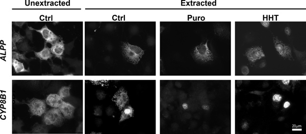
Figure 1. ALPP, but not CYP8B1 mRNA remains associated with the ER independently of ribosomes and translation. COS-7 cells were transfected with plasmids containing either the ALPP (top row), or CYP8B1 (bottom row) genes and allowed to express mRNA for 18-24 hr. The cells were then treated with DMSO control medium (“Ctrl”), puromycin (“Puro”) or HHT for 30 min. Cells were either directly fixed (“unextracted”) or first extracted then fixed. Note that while the control and HHT-treated cells were extracted with digitonin alone, Puro-treated cells were extracted with digitonin and EDTA. Cells were stained for mRNA using specific FISH probes, and imaged. Scale bar = 20 μm.
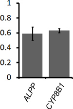
Figure 2. Quantification of the level of ER-association for ALPP and CYP8B1 mRNA. Transfected COS-7 cells were either directly fixed to determine the total level of mRNA in the cytoplasm (see Figure 1, “Unextracted Ctrl” cells) or first extracted and then fixed to determine the amount of ER-associated mRNA (see Figure 1, “Extracted Ctrl” cells). For each experiment the average ER-associated fluorescence of 30-40 extracted cells was divided by the average cytoplasmic fluorescence of 30-40 unextracted cells, to give the fraction of ER-bound mRNA (y-axis). Each bar represents the average value and standard error of three independent experiments.
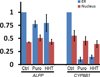
Figure 3. Quantification of the ER-association of ALPP and CYP8B1 mRNA after ribosome dissociation. Transfected COS-7 cells were treated with control medium or various translation inhibitors, extracted, fixed, stained for mRNA using specific FISH probes and imaged (see Figure 1, “Extracted” cells). The fluorescence intensities of mRNA in the ER and nucleus were quantified. Each bar represents the average and standard error of three independent experiments, each consisting of the average integrated intensity of 30 cells over background and normalized to the fluorescence in the ER of control-treated cells (y-axis). Note that although ribosome disruption caused CYP8B1 mRNA to dissociate from the ER, the levels of nuclear mRNA were unaffected.
The localization of mRNAs to various subcellular sites, through the interaction of transcripts with mRNA localization proteins, is a widespread phenomenon important for the proper sorting of proteins to their final destination, and for the fine tuning of gene expression to the local requirements of a subcellular region19,20.
Using the assay described here, we revisited the question of whether mRNAs that encode secretory proteins can be maintained on the ER in the presence or absence of ribosomes or translation. Indeed, we found that a subset of these mRNAs have this activity11. For example, using this extraction protocol, we determined that both ALPP and CYP8B1 mRNAs are localized on the surface of the ER in control treated COS-7 cells (Figures 1-2). However, after treating the cells with translation inhibitors puromycin or HHT, only ALPP, but not CYP8B1 mRNA, remained associated with the ER in the absence of functional ribosomes and translation (Figures 1, 3). In addition we previously found that ALPP mRNA remains ER-associated in COS-7 cells treated with pactamycin11. Since these various treatments act through divergent mechanisms to clear mRNA of associated ribosomes, it is unlikely that the ER-association of ALPP is the result of some indirect cellular response to any particular drug. To confirm that these translation inhibitors are effective, it is also helpful to test whether they inhibit the incorporation of 35S-methionine into newly synthesized proteins. For example, we have found that MDMP-treatment (100 μM, 15 min) can only inhibit 40-60% of all protein synthesis in COS-7 cells (X.A. Cui and A.F. Palazzo, unpublished observations). As a result, this drug does not efficiently disrupt the ER-localization of mRNAs that require translation for their targeting and maintenance (X.A. Cui and A.F. Palazzo, unpublished observations).
To further ensure that ER-targeting of the mRNA is not dependent on encoded signal sequence or transmembrane domains, one can alter the exogenous transcript so that it encodes a cytoplasmic protein. Indeed, we previously demonstrated that a version of the ALPP mRNA that lacks both signal sequence and transmembrane coding regions is still able to localize to the ER11.
It is also worth noting that the method presented here when combined with other assays used to analyze mRNA nuclear export21, can be used to define the kinetics and requirements of mRNA transport from one compartment to another (e.g. from the nucleus to the surface of the ER). Indeed, by microinjecting plasmids that contain the ALPP gene into the nuclei of COS-7 cell that were pretreated with translation inhibitors, we previously demonstrated that newly made ALPP mRNAs are targeted to the ER independently of translation or ribosome-association11.
Although we do not fully understand how these mRNAs are targeted and anchored to the ER, it is likely that this organelle contains mRNA receptors. Indeed we recently identified p180, a large positively-charged membrane-bound protein, as being required for the ability of ALPP mRNA to be maintained on the ER independently of translation11. However, it is likely that other mRNA receptors must be present on the surface of the ER11. With the help of the technique outlined here, we hope to identify and dissect many of the molecular mechanisms that help regulate mRNA localization in mammalian cells.
The authors have nothing to disclose.
This work was supported by grants from the National Science and Engineering Research Council of Canada to X.A.C. and A.F.P.