In this method, we use photopolymerization and click chemistry techniques to create protein or peptide patterns on the surface of polyethylene glycol (PEG) hydrogels, providing immobilized bioactive signals for the study of cellular responses in vitro.
There are many biological stimuli that can influence cell behavior and stem cell differentiation. General cell culture approaches rely on soluble factors within the medium to control cell behavior. However, soluble additions cannot mimic certain signaling motifs, such as matrix-bound growth factors, cell-cell signaling, and spatial biochemical cues, which are common influences on cells. Furthermore, biophysical properties of the matrix, such as substrate stiffness, play important roles in cell fate, which is not easily manipulated using conventional cell culturing practices. In this method, we describe a straightforward protocol to provide patterned bioactive proteins on synthetic polyethylene glycol (PEG) hydrogels using photochemistry. This platform allows for the independent control of substrate stiffness and spatial biochemical cues. These hydrogels can achieve a large range of physiologically relevant stiffness values. Additionally, the surfaces of these hydrogels can be photopatterned with bioactive peptides or proteins via thiol-ene click chemistry reactions. These methods have been optimized to retain protein function after surface immobilization. This is a versatile protocol that can be applied to any protein or peptide of interest to create a variety of patterns. Finally, cells seeded onto the surfaces of these bioactive hydrogels can be monitored over time as they respond to spatially specific signals.
There are many stimuli that influence cell behavior. Generally, typical cell culturing techniques rely on soluble factors to elicit cellular responses; however, there are limitations to this approach. These methods are unable to accurately display all signaling motifs commonly found in vivo. Such signaling mechanisms include sequestered growth factors, cell-cell signaling, and spatially-specific biochemical cues.Furthermore, substrate stiffness can play an important role in cell behavior and stem cell differentiation and is not easily manipulated using common cell culturing practices1,2. Biomaterial approaches offer a new platform to begin exploring these signaling mechanisms. In particular, hydrogels are excellent candidates for tuning substrate stiffness3,4, immobilizing proteins and peptides5,6, and creating spatially specific patterns7,8.
Hydrogels are commonly used as scaffolds in tissue engineering due to their biophysical and biochemical commonalities with the extracellular matrix (ECM)9,10. Natural polymers are common choices for scaffolds, as they are biocompatible and are found in many tissues of the body. The limitation of using natural polymers as substrates is that they lack easily manipulated chemical moieties for bioconjugation. On the other hand, synthetic hydrogels, as such as PEG, are excellent platforms for targeted chemistries11,12. Additionally, PEG hydrogels do not elicit a cellular response and therefore are used as inert backbones for creating bioactive scaffolds.
To create bioactive hydrogels, both photopolymerization and thiol-ene click chemistry reactions are employed. These photoreactions require a photoinitiator and a UV light source. When photoinitiators are introduced to UV light, bonds break to form radicals. Theses radicals are necessary for initiating the reaction but can negatively affect protein bioactivity12,13. Therefore, it is crucial to optimize photoinitiator and UV exposure times to maintain protein bioactivity.
In this method, hydrogels are synthesized through acrylate-acrylate chain growth photopolymerization. PEG-diacrylate (PEGDA) monomers react with each other to form branched polymer networks responsible for the structure of the hydrogel. The concentration of PEGDA monomers within the gel precursor solution will control the substrate stiffness. Due to the small pore size of the hydrogel, ECM proteins such as fibronectin can be easily incorporated within the hydrogel for the purpose of cell attachment. Finally, these hydrogels can be surface-patterned with bioactive peptides or proteins via thiol-ene click chemistry reactions. Here, unreacted free acrylates within the hydrogel system will react with free thiols located on the protein or peptide when exposed to UV light. After the proteins or peptides are immobilized on the hydrogel surface, the hydrogel can be stored at 4 °C for several weeks without losing bioactivity. This offers convenience, flexible experimental planning, and the possibility for collaboration between labs. Overall, this platform allows for biomechanical and spatial biochemical control, independent of each other, for the opportunity to influence cellular behavior.
1. Preparation of Materials for Hydrogel Synthesis
2. Modifying Proteins with a Free Thiol
3. Hydrogel Formation
4. Hydrogel Stiffness Measurements
5. Protein Patterning
6. Preparing Hydrogels for Cell Seeding
7. Cell Seeding on Hydrogels
8. Evaluation of Bioactivity
The protocol to create bioactive patterns on the surface of PEG hydrogels is illustrated in Figure 1. A spreadsheet was developed to calculate the volume and concentration for each stock solution (Table 1A). Proteins to be immobilized onto the surface of the hydrogel are modified with 2-iminothiolane (Figure 1B). This reaction is performed using the volumes from Table 1B. The precursor hydrogel solution is prepared with 10% weight/volume of PEGDA with LAP (Figure 1A). Various precursor PEGDA concentrations can be used to yield the desired substrate stiffness (Figure 2A). Fibronectin is included within this precursor solution for cell attachment purposes. After thorough mixing, this solution is pipetted into the prepared mold and exposed to UV light (Figure 1C). UV light exposure should be minimized; exposure should be just enough to produce a hydrogel. Hydrogel samples are punched out to the appropriate diameter for the desired well plate (Figure 1C). For surface patterning, modified protein solution is pipetted onto the surface of a hydrogel and spread evenly. Minimal volume should be used; protein volume should be just enough to cover the entire surface of the hydrogel. The predesigned photomask is placed directly onto the hydrogel surface; air bubbles between the mask and the hydrogel should be avoided. A second round of UV light is used to covalently conjugate UV-exposed proteins to the hydrogel. Hydrogel samples are rinsed to remove unreacted proteins and reveal the immobilized protein pattern (Figure 1D).
It is important to minimize photoinitiator concentration and UV exposure time when proteins are present. Using lysozyme bioactivity as an indicator, we found that the LAP photoinitiator concentration should be less than 2 mM (Figure 2B) and the UV exposure time should total less than 2 min (Figure 2C) to retain a protein bioactivity greater than 80%.
UV exposure time during hydrogel formation and protein patterning are both important parameters for developing a successful protocol (Figure 3). First of all, minimizing UV exposure during hydrogel formation is critical to maintaining free acrylate functional groups for subsequent protein immobilization reactions (Figure 3A). Hydrogels exposed to UV light for longer than 2 min are unable to create immobilized protein patterns. Additionally, as the UV exposure to the protein pattern increases, more proteins react to the surface (Figure 3B).
Finally, cells can be cultured onto these patterned hydrogel substrates to manipulate cell behavior. To show the potential of immobilized patterns on hydrogels, we patterned VEGF, a growth factor important for endothelial cells, and cultured HUVECs on the surface using basal EGM-2 medium (Figure 4). HUVECs were uniformly seeded onto the surface of VEGF-patterned PEG hydrogels (Figure 4A). Two days after seeding, HUVECs were observed to migrate towards the spatial regions of the hydrogel that contained immobilized VEGF (Figure 4B, C). This is one example of a bioactive protein pattern on PEG hydrogels being used to influence cell behavior.
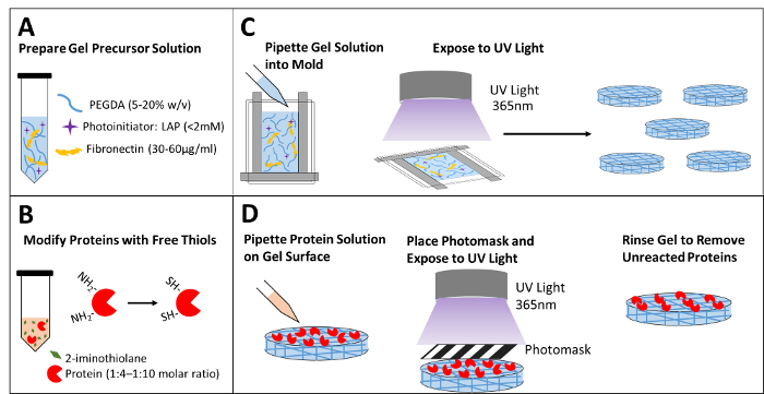
Figure 1: Schematic of hydrogel formation and protein patterning. (A) Prepare precursor hydrogel solution with PEGDA monomers, photoinitiator, and extracellular protein for cell attachment. (B) Modify the proteins with free thiol groups by reacting with 2-iminothiolane. (C) Pipette the precursor solution into the prepared mold and expose it to UV light to form the hydrogel. Punch out hydrogel samples of the desired size. (D) Pipette the modified protein solution onto the surface of the hydrogel, place a photomask on the surface, and expose it to a second round of UV light. Rinse the gel to remove unreacted species prior to imaging and cell seeding. Please click here to view a larger version of this figure.
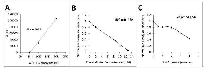
Figure 2: Modulating stiffness and protein bioactivity. (A) Changes in the concentration of PEGDA monomers within the precursor gel solution alter hydrogel stiffness. (B) Increasing the concentration of LAP photoinitiator lowers protein function after 1 min of UV exposure. (C) Increasing UV exposure time lowers protein function with 2 mM LAP. All error bars represent standard deviation of replicates. Please click here to view a larger version of this figure.
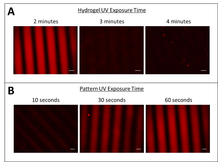
Figure 3: Pattern and hydrogel UV exposure times optimized for protein patterning. (A) Minimizing UV exposure times for hydrogel formation allows for surface patterning. (B) Increasing UV exposure times for surface patterning increases pattern strength. Scale bars = 200 µm. Please click here to view a larger version of this figure.
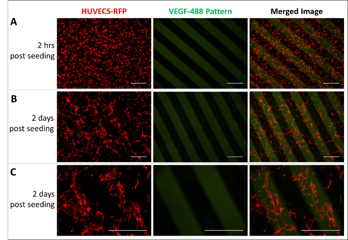
Figure 4: Endothelial cells responding to a VEGF pattern. (A) Uniform HUVECs seeded on hydrogels. (B and C) HUVECs sense the VEGF pattern and migrate towards immobilized VEGF. Images were taken two days after seeding in (B) 4X and (C) 10X magnification. Scale bars = 500 µm. HUVEC-RFPs (red) and VEGF-488 pattern (green) were captured with excitation and emission filters at 528/553 and 465/495, respectively. Please click here to view a larger version of this figure.
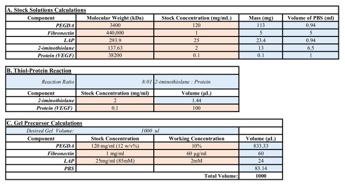
Table 1: Calculations for stock and gel precursor solutions. The red box indicates user-defined values, such as molecular weight, desired stock concentrations, and weighed-out masses. Blue boxes represent values that have been calculated based on user-defined values.
This protocol provides a method for creating bioactive protein patterns for biological applications. There are several modifications that can be made to adapt this protocol for different experiments. First, cell attachment requirements will vary for different cell types. If poor cell attachment to the gels is initially observed, increasing the concentration of the ECM protein within the precursor solution is advised. Other ECM proteins can be used instead of fibronectin, including different types of collagen, laminin, or a combination thereof. For each new cell type, cell attachment should be optimized prior to hydrogel patterning. This protocol also allows for user-designed photomasks. Based on the desired application, photomasks can be produced with various feature sizes, shapes, and overall patterns. Uniform immobilization can be achieved in the absence of a photomask.
This protocol has certain limitations. As highlighted in the Representative Results, this method is sensitive to the amount of UV light at each step. Overexposure during the hydrogel formation step limits the available acrylate groups for subsequent surface bioconjugation. Therefore, a key step in this protocol is well-managed UV light exposure times for each step. Also, this protocol requires a high concentration protein stock for successful patterning. Low concentrations of protein will result in poor surface patterns. Additionally, photomasks are also limiting in that they can only produce discrete patterns. More complex patterns can be achieved with similar approaches but require a more advanced methodology.
This protocol is significant to existing methods as it provides a simple method for adjusting substrate stiffness and protein patterning. Using acrylate chemistry for the hydrogel formation allows for a magnitude range of substrate stiffness within the physiological range. Simply adjusting the concentration of PEGDA within the precursor solution gives control over the hydrogel stiffness. Additionally, the use of click chemistry for protein patterning allows for rapid conjugation between hydrogel substrate and thiolated modified protein. This is a key design feature, as it allows this protocol to be applicable to any protein or peptide of interest.
PEG hydrogels are promising biomaterials that can be used to explore new platforms for displaying biochemical cues to biological systems. Whether uniform surface immobilization or spatially specific patterns, these techniques provide novel ways to control cell behavior. Moving forward, the advancement of biomaterial technology will provide new insights into cell behavior and further our abilities to recapitulate in vivo signaling motifs within in vitro systems. This can be beneficial for stem cell differentiation and modeling developmental signaling within a controlled experimental system.
The authors have nothing to disclose.
This study was mainly supported by grants from the American Heart Association Scientist Development Grant (12SDG12050083 to G.D.), the National Institutes of Health (R21HL102773, R01HL118245 to G.D.) and the National Science Foundation (CBET-1263455 and CBET-1350240 to G.D.).
PEG-diacrylate (PEGDA) | Laysan Bio | ACRL-PEG-ACRL-3400 | Can also be synthesized or purchased through other venders. Different molecular weights can be used. |
Lithium Phenyl-2,4,6-trimethylbenzoylphosphinate (LAP) | Synthesized in lab | ||
Fibronectin | Corning | 356008 | Other cell attachment proteins can be used, such as laminin, matrigel |
Phosphate-buffered saline (PBS) | Sigma | D8537-500ML | |
Photomask | FineLine Imaging | n/a | Custom prints on transparent sheets with high resolution DPI. |
Binder Clips | Various Vendors | ||
Compact UV Light Source (365nm) | UVP | UVP-21 | Other UV light sources can be used, calibration of power is required. |
2-iminothiolane (Pierce Traut’s Reagent) | Thermo Sci. | 26101 | |
Ellman’s Reagent: DTNB; 5,5-dithio-bis(2-nitrobenzoic acid) | Thermo Sci. | 22582 | |
human umbilical vein endothelial cells (HUVECs) | Lonza | passage number between 6- 10 | |
EGM-2 Media | Lonza | CC31-56, CC-3162 | EGM-2 without growth factors was used in experiments. Full EGM-2 media was used for cell maintainance |
0.25% Trypsin EDTA | Life Tech | 25200-056 | |
Trypsin Neutralizer | Life Tech | R-002-100 | |
Centrifuge | Various Venders | ||
Hemocytometer | Hausser Sci. Bright-line | ||
Ethylenediaminetetraacetic acid (EDTA) | Sigma Aldrich | E6758 | |
0.22µm filter | Cell Treat | 229743 | |
1mL Syringe | |||
Glass Microscope Slides | Fisher Sci. | 12-550C | |
Plastic spacers | Various Venders | 0.5mm thickness | |
70% Ethanol | BICCA | 2546.70-1 | |
Bio-shield | Bio-shield | 19-150-0010 | |
Bradford Reagent | BIO-RAD | ||
Desalting Resin – Sephadex G-25 | GE Healthcare | 95016-754 | |
Microspin Columns | Thermo Sci. | PI69725 | |
AR-G2 rehometer | TA Instruments |